




High-performance self-powered X-ray detection materials based on KTiOAsO4 polar crystals
 0
Abstract
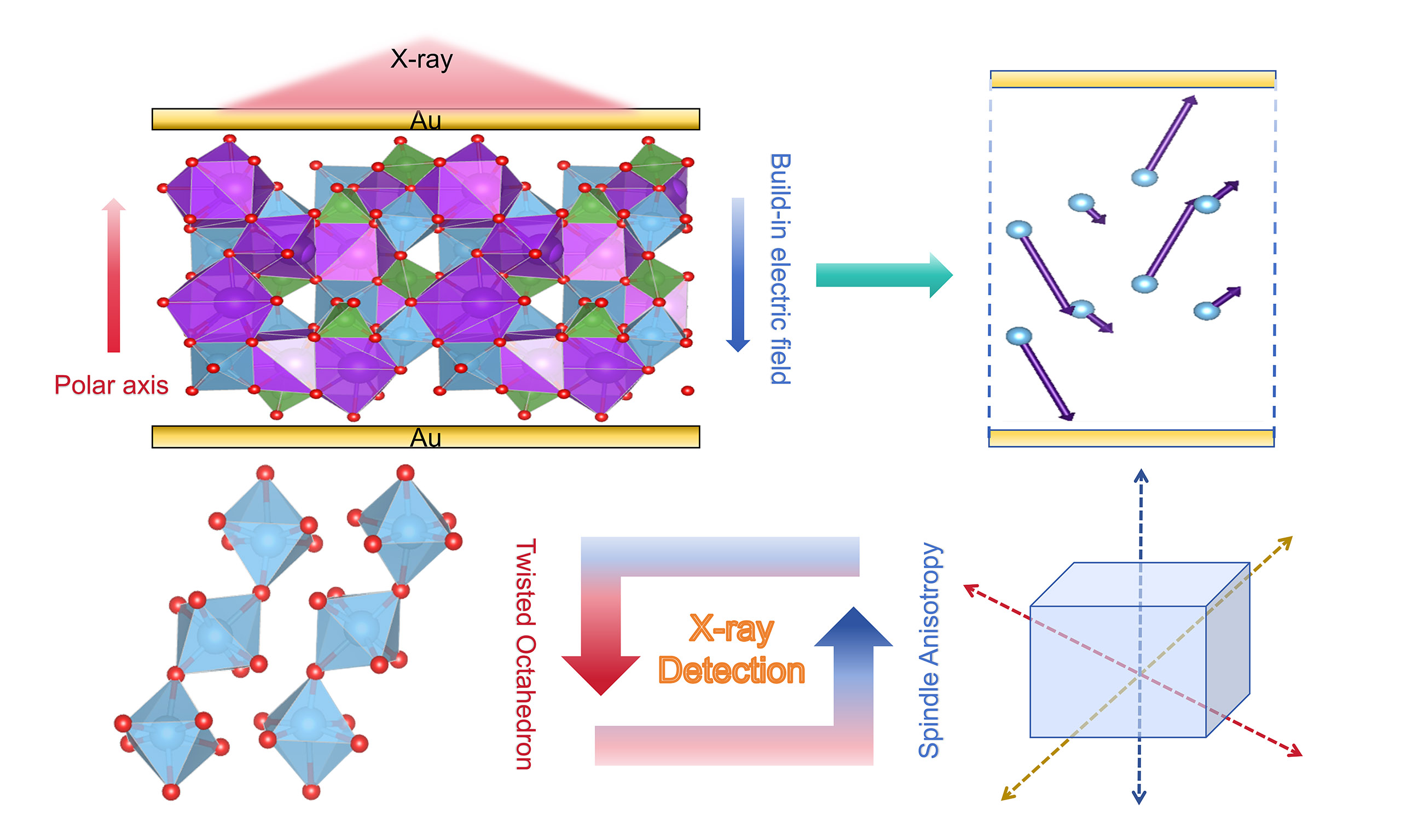
Self-powered X-ray detectors can perform X-ray inspections without requiring an external power supply, enabling the design of highly portable X-ray detection devices. As a result, they hold broad application prospects in the fields of industrial non-destructive testing and medical examinations. This paper reports a self-powered X-ray detector based on a KTiOAsO4 polar crystal, which possesses a strong built-in electric field (approximately
Keywords
INTRODUCTION
By interacting with electrons in the object being examined, X-rays can carry information about that object through changes in signal intensity[1,2]. Thus, direct detection of changes in X-rays allows accurate retrieval of information about the subject[3-5]. The two most widely used types of X-ray detectors are direct and indirect detectors. Indirect X-ray detectors consist of two main parts: the scintillator material converts the incident X-rays into photons, and the photoelectric sensor converts the optical signal into an electrical signal[6]. Direct X-ray detectors directly convert X-ray signals into electrical signals. They effectively reduce signal attenuation and, compared to indirect detectors, offer higher spatial resolution and superior image quality, thereby demonstrating significant technical advantages[7-12]. Currently, most high-performance direct X-ray detectors are made of inorganic semiconductor materials, such as silicon, germanium, selenium, cadmium-zinc-tellurium, and chalcogenides[13]. These detection materials demonstrate exceptional performance in X-ray detection; however, their application is constrained by a number of limitations, primarily including severe X-ray attenuation, low damage thresholds, high operating voltages, significant noise levels, severe baseline drift, high costs, and contamination issues during the manufacturing process[14-18]. In addition, significant progress has been made in organic and inorganic materials, such as MAPbI3, MAPbBr3, CsPbBr3, and MA3Bi2[19-21]. These materials contain high-Z elements such as lead (Pb), bismuth (Bi), iodine (I), and bromine (Br), and therefore possess large X-ray absorption cross-sections and excellent carrier transport properties[22-24]. However, the vast majority of them contain toxic elements, are prone to deliquescence, and are susceptible to decomposition or phase transitions under high-temperature conditions[22-24].
Compared with conventional inorganic X-ray detection materials, oxide crystals can be grown using the flux method, which is favorable for obtaining high-quality bulk single crystals[25,26]. Compared with organic-inorganic composites, oxide crystals have wider band gaps and stable physicochemical properties, especially their unparalleled high-temperature stability, which gives them a wide operating temperature range and allows them to be used under extreme conditions[25-28]. Previous studies based on oxide X-ray detection materials have provided important avenues to address these issues. KTiOAsO4 (KTA) is an oxide crystal with excellent physicochemical stability. Its elemental composition includes elements with medium atomic numbers, such as arsenic (As) and titanium (Ti), giving it appreciable X-ray absorption capacity. Furthermore, KTA crystals can be grown into large, low-defect single crystals using the well-established flux method. Although KTA contains arsenic and requires appropriate protocols during growth and processing to ensure safety, it does not contain highly toxic heavy metals such as lead and cadmium, which are commonly found in many perovskite-based detectors. Furthermore, once crystallized, it is chemically stable and non-toxic during routine use, thereby reducing the risks associated with its manufacture and use. Additionally, KTA exhibits excellent thermal and chemical stability and resistance to phase transitions or decomposition. It is expected to overcome the application limitations of traditional detector materials under extreme conditions, offering a new candidate material for high-performance, environmentally friendly direct X-ray detectors.
This study systematically investigated the X-ray detection performance of KTA polar crystals, evaluating key metrics including the carrier mobility-lifetime product (μτ), sensitivity, signal-to-noise ratio, and detection limit under both biased and self-powered operating modes. The anisotropic response behavior and its structural origins were also comprehensively analyzed. The results demonstrate that KTA crystals, leveraging their unique polar structure, strong built-in electric field, and excellent physicochemical stability, deliver competitive X-ray detection performance, with notable self-powered capability and effective suppression of ion migration. Its crystal structure and the arrangement of TiO6 octahedra provide insights into how polar crystal symmetry governs the trade-off between sensitivity and detection limit. These characteristics establish KTA as a promising candidate material for portable, low-dose X-ray detection applications in domains including medical diagnostics, safety inspections, and industrial non-destructive testing.
MATERIALS AND METHODS
Materials
KTA single crystals serving as the core material were grown by our research group. The samples measured 6 × 6 × 2 mm3, and no obvious defects were observed. The samples were numbered KTA-X, KTA-Y, and KTA-Z according to the crystal axis perpendicular to the test plane.
Detector preparation
Au electrodes were used for detector fabrication. A physical mask was closely bonded to the KTA crystal, and an Au target with 99.999% purity was used as the sputtering source. The KTA crystal was placed in a vacuum chamber, and sputtering was initiated once the pressure fell below 10 Pa. Using ion sputtering and a physical mask, a 100-nm-thick, 2 × 2 mm2 gold electrode was deposited onto the crystal surface to fabricate a single-crystal detector. The detector exhibits an Au/KTA/Au structure. This fabrication method is simple to perform and offers good reproducibility.
Detector performance characterization
The X-ray source was an X-ray tube (E7252X, Toshiba, Japan) operated at 40-150 keV; the tube current was adjusted from 10 mA to 100 mA to vary the X-ray dose rate. Current-voltage (I-V) and current-time (I-t) curves were recorded using a 6517B high-precision electrometer (Keithley, USA). Measurements were conducted under optically isolated conditions (in the dark) to ensure that the recorded current originated entirely from X-ray excitation rather than from photo-generated carriers. In X-ray detection, the effective device area (4 mm2) is defined as the metal area on the device surface in contact with the probe. All detector performance tests were conducted on unpackaged devices at room temperature.
X-ray imaging performance
The X-ray imaging performance of KTA X-ray detectors was measured on a custom-built platform. The X-ray detector sample used for imaging was coated with a gold electrode with an area of 6 × 6 mm2 on one side and a planar array electrode on the other side. The area of each small electrode was 0.5 × 0.5 mm2, and the distance between the electrodes was 0.1 mm. The imaging object was placed between a planar array X-ray detector and an X-ray beam, and the electrical signals of the detector were recorded at different positions. The imaging object was chosen to be smaller than the planar array detector so that the whole object could be imaged in a single acquisition without moving the detector, thereby avoiding unwanted errors.
RESULTS AND DISCUSSION
Crystal properties
The KTA crystal belongs to the orthorhombic crystal system with space group Pna21, and its polar axis extends along the crystallographic Z-axis. Figure 1A shows KTA samples (polished on both sides) along three principal axis directions, which were used for the fabrication of X-ray detectors. The device configuration is displayed in Figure 1B. We further compared the X-ray absorption coefficients of KTA with those of some previously reported X-ray detection materials [Figure 1C]. Within the typical detection range, the coefficient of KTA crystals is lower than that of Se, Ga2O3, and CdZnTe due to the smaller atomic numbers of K, As, Ti, and O. Specifically, at 40 keV, a 2-mm-thick KTA crystal absorbs nearly 100% of the incident photons [Figure 1D]. Resistivity is a key parameter affecting X-ray detection performance. It determines the dark current level of the material, which in turn influences the detector’s sensitivity. To evaluate the resistivity of KTA crystals, we employed the two-probe method to measure the resistivity of KTA samples along different crystal orientations at room temperature. The results reveal that the resistivities along the X, Y, and Z axes are 4.02 × 10⁹, 7.69 × 10⁹, and 5.15 × 10⁶ Ω·cm, respectively, exhibiting significant anisotropy [Figure 1E and F]. This anisotropy stems from differences in atomic bonding patterns within the crystal structure and the directionality of carrier transport paths. Specifically, the resistivity along the Z-axis (polar axis) is approximately three orders of magnitude lower than that along the X- and Y-axes, indicating that the migration resistance of charge carriers in the Z-axis is lower. However, when the resistivity is too low, it leads to excessively high dark current, and photocurrent shielding effects arise, significantly reducing the detector’s sensitivity and signal-to-noise ratio. This hinders accurate measurements and practical applications [Supplementary Figure 1]. Therefore, polar-axis orientation is not an ideal choice for X-ray detection. To meet the requirements for high signal-to-noise ratio and high sensitivity in practical applications, the non-polar axis directions are the most appropriate choice.
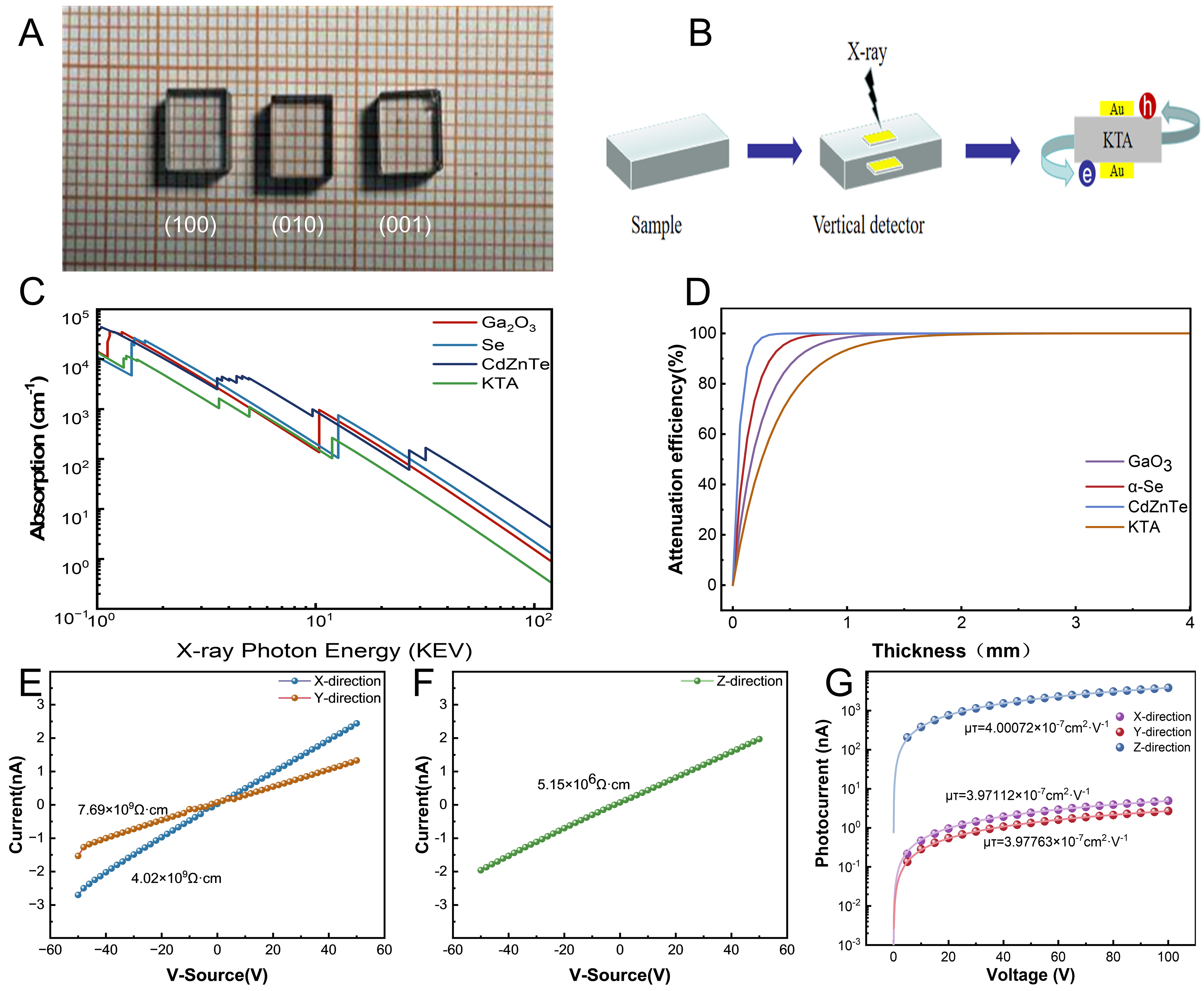
Figure 1. (A) Photograph of the KTA samples (original image by the authors). (B) Schematic of the X-ray detector configuration. (C) Comparison of X-ray absorption coefficients between KTA and other materials[33]. (D) Comparison of X-ray attenuation efficiency between KTA and other materials[33]. (E) Resistivity of the KTA crystal along the X and Y axes. (F) Resistivity of the KTA crystal along the Z axis. (G) Carrier mobility-lifetime product of the KTA crystal along the three principal axes.
As a key parameter characterizing charge transport capability and carrier extraction efficiency, the carrier mobility-lifetime product (μτ) directly determines the ultimate responsivity and quantum efficiency of photodetectors. To quantitatively evaluate the carrier transport properties of KTA crystals, we systematically investigated their voltage-dependent photoconductive behavior by applying a linearly swept DC bias from 0 to 100 V at an X-ray dose rate of 55.0374 μGyair·s-1 (120 keV). Additionally, the dynamic photocurrent response was simultaneously monitored under dark and X-ray irradiation conditions to determine the field-induced charge collection efficiency. The photocurrent-voltage relationship was fitted using the Hecht equation[29]:
where I0 is the saturation current, µ is the carrier mobility, τ represents the carrier lifetime, V is the bias, and L is the thickness of the sample. The experimentally measured carrier mobility-lifetime product is shown in Figure 1G, Note that for a photoresistor-type device with symmetric planar metal-semiconductor-metal contacts, the single-carrier Hecht equation yields an effective μτ value that represents a weighted average of both electron and hole contributions, rather than the μτ of an individual carrier type[29-31]. This is because, under uniform X-ray excitation throughout the crystal thickness, both electrons and holes are generated and contribute to the measured photocurrent, and the symmetric contact geometry does not provide the carrier selectivity needed to isolate one type[32]. Therefore, the μτ values reported in this paper should be understood as an effective transport parameter characterizing the overall charge collection efficiency of the KTA detector in a photoresistor configuration. To measure the μτ values for electrons and holes independently, it is necessary to employ techniques such as α-particle excitation, which can selectively generate carriers near a specific electrode.
The high ionic charge density of Ti4+ and As5+ in KTA leads to strong Coulombic interactions and large ionic radii, thereby suppressing ion mobility and enhancing lattice stability. Temperature-dependent transient reverse currents of KTA (303-333 K) reveal ion migration dynamics [Supplementary Figure 2]. Under applied bias, field-driven ion migration establishes space-charge polarization until the current reaches steady state. Abrupt voltage removal initiates ion redistribution through vacancy diffusion, producing a concentration-gradient-induced reverse dark current that relaxes to a steady state. Ion activation energies for KTA crystals, fitted along the (100), (010), and (001) planes [Supplementary Figure 3], together with the dark current drifts measured along the polar axis direction [Supplementary Figure 4] collectively demonstrate weak ionic mobility. For comparison, halide perovskite detectors such as MAPbBr3 exhibit ion activation energies as low as ~73 meV, leading to pronounced dark current drift and performance degradation under applied bias[18]. In contrast, the substantially higher activation energy of KTA (> 200 meV) effectively suppresses field-driven ion migration.
X-ray detection with applied bias
Sensitivity, as a core performance metric for direct-type X-ray detectors, characterizes the detector’s ability to respond to X-ray signals. It is typically quantified by the photocurrent density or charge collection efficiency generated per unit dose of X-ray irradiation. For KTA detectors, their high carrier mobility-lifetime product (μτ) effectively enhances charge collection efficiency and thus improves the conversion of X-ray photons into electrical signals. Furthermore, when oriented along a non-polar axis (such as the X-axis or Y-axis), the high resistivity can significantly suppress the dark current, reducing background noise interference and thereby optimizing the sensitivity. To systematically evaluate the X-ray detection performance of KTA crystals, we measured the current-time (I-T) response curves of KTA detectors over a bias voltage range of 0 to 200 V. Figure 2A shows the I-T response curves under different bias voltages, and Figure 2B shows the variation in photocurrent as the X-ray dose rate increases from 2.94 to 7.43 μGyair·s-1. The results reveal that the carrier transport and collection efficiency increase significantly with rising bias voltage, leading to a corresponding enhancement of the photocurrent response. However, as the voltage rises further, the dark current increases along with the photocurrent. Sensitivity therefore reaches a saturation point, beyond which it no longer improves. Based on the photocurrent data, the detector’s sensitivity can be calculated using the following formula:
where ΔI represents the difference between photocurrent and dark current, A represents the Au electrode area, and ΔD represents the changes in X-ray dose rate. A linear correlation exists between the current density generated under X-ray irradiation and the X-ray dose rate [Figure 2B and Supplementary Figure 5]. The detector's sensitivity was determined by fitting the slopes of these current density versus dose rate plots at various bias voltages [Figure 2C and D]. The experimental data show that the detector exhibits higher sensitivity under lower radiation dose rates. This phenomenon is primarily because the atomic numbers of the elements in KTA are not particularly high, resulting in limited X-ray absorption capacity. Meanwhile, excessively high dose rates lead to premature carrier recombination, which in turn affects the detector’s sensitivity. The maximum sensitivity of the KTA directional X-ray detector is 192.96 μC Gyair-1·cm-2 (200 V, 70 keV). Under an applied bias voltage of 200 V and an X-ray dose rate range of 17.4432-34.7328 μGyair·s-1, this value is 9.6 times higher than that of the α-Se detector (20 μC·Gyair-1 cm-2, measured at 20 keV). Furthermore, the sensitivity exhibits strong voltage dependence. However, when the bias voltage is too high, leakage current increases significantly and carrier recombination becomes severe. At the same time, factors such as breakdown or thermal effects significantly reduce the material’s charge collection efficiency and signal generation capability, so the sensitivity no longer increases with rising voltage.
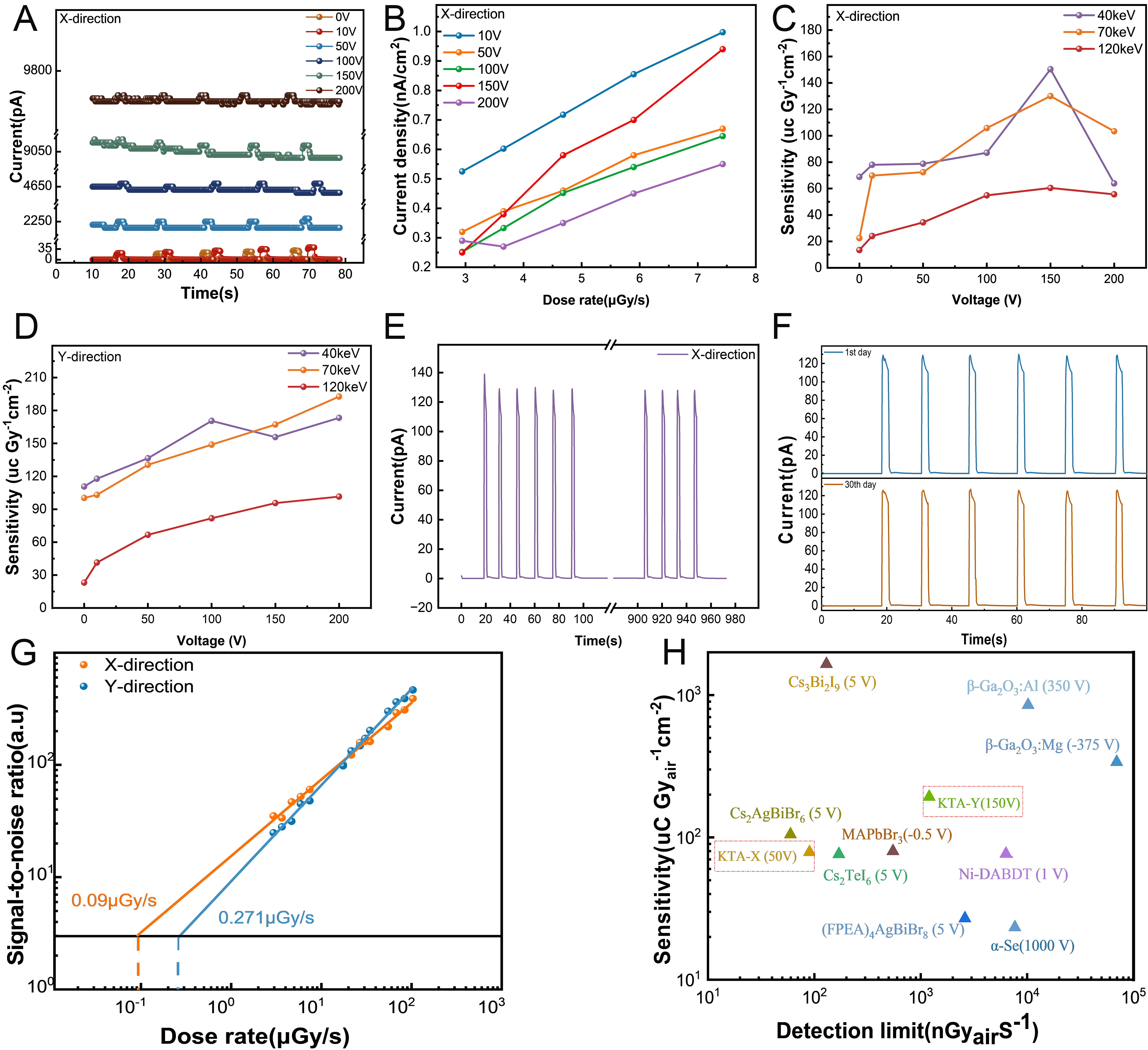
Figure 2. (A) Response of the KTA detector to X-rays at different bias voltages. (B) X-ray photocurrent generated by the KTA detector at different dose rates. (C) Sensitivity of the KTA detector along the X-axis under an applied bias voltage. (D) Sensitivity of the KTA detector along the Y-axis under an applied bias voltage. (E) Stability of the detector after 1,000 s of irradiation. (F) Photocurrent of the detector before and after 30 days of storage at room temperature. (G) Fitted detection limit of the KTA detector at a 50 V bias. (H) Comparison of the performance of KTA and other detector materials under an applied bias[30,32,34-42].
To evaluate the performance stability of the KTA detector under continuous irradiation conditions, we subjected the device to frequent X-ray irradiation at a dose rate of 7.43 μGyair·s-1 and recorded the I-T curves of the switching process over time [Figure 2E]. Notably, the KTA X-ray detector exhibited high photocurrent and stable dark current characteristics. The unencapsulated device retained 95.5% of its initial photocurrent even after being exposed to air for over 1,000 s. The 4.5% loss can be attributed mainly to electrode damage caused by prolonged probe contact, suggesting that operational stability can be further enhanced by optimizing the contact design. Subsequently, after the device was left at room temperature (25 ± 2 °C in ambient air) for 30 days and retested, the photocurrent density showed no significant decrease [Figure 2F], confirming its excellent stability under routine conditions. Admittedly, this stability assessment was limited to intermittent irradiation tests. To verify its practical applicability in clinical and industrial settings, a systematic evaluation of long-term operational stability under continuous X-ray irradiation is still required. However, under standard test conditions described above, the photocurrent values remained highly stable throughout the observation period. Based on this, we can cautiously conclude that the KTA detector has the potential for long-term service under standard operating conditions. Extreme or continuous irradiation scenarios remain within the scope of future optimization and validation and do not undermine the validity of the current conclusion.
The detection limits of KTA detectors are derived from current-time (I-T) curves measured under X-ray exposure. Specifically, the minimum detection limit is defined as the dose rate at which the signal-to-noise ratio (SNR) equals 3[30,31]. This SNR is calculated using the following equation:
Where Iphoto represents the average photocurrent, Idark represents the average dark current without X-ray irradiation, and Ii represents the photocurrent. Figure 2G shows the signal-to-noise ratio of KTA single-crystal detectors. The lowest detection limit of the KTA detector reaches 0.09 μGyair·s-1 (50 V, 40 keV). For comparison, conventional X-ray detectors exhibit detection limits of 5 μGyair·s-1 for α-Se (20 keV)[32],
Comparison of the performance of KTA and other X-ray detectors in the applied voltage mode
| Materials | X-ray energy (keV) | μτ (cm2 V-1) | Sensitivity (μC·Gyair-1 cm-2) | Detection limit (μGyair·s-1) | Ref |
| α-Se | 20 | 4 × 10⁻⁷ | 20 | 5 | [32] |
| β-Ga2O3:Mg | 50 | - | 338.9 | 69.5 | [34] |
| MAPbBr3 | 50 | 1.2 × 10⁻² | 80 | 0.5 | [30] |
| β-Ga2O3:Al | 50 | - | 851.6 | < 9.8 | [35] |
| Ni-DABDT | 20 | 3.26 × 10⁻⁴ | 98.6 | 7.2 | [36] |
| (FPEA)4AgBiBr8 | 40 | - | 27 | 2.6 | [37] |
| Cs2AgBiBr6 | 50 | 1.72 × 10⁻³ | 105 | 0.0597 | [38] |
| Cs3Bi2I9 | 50 | 1.68 × 10⁻³ | 1,652.3 | 0.13 | [39] |
| Cs2TeI6 films | 20 | - | 76.27 | 0.17 | [40] |
| Cs3Cu2I5 | 50 | 1.4 × 10⁻⁴ | 441.45 | 0.335 | [41] |
| TMCM-CdCl3 | 40 | 1.42 × 10⁻⁴ | 128.9 | 1.6 | [42] |
| KTA (Y-axis, 200 V) | 70 | 3.98 × 10⁻⁷ | 192.96 | 1.2 | This work |
| KTA (X-axis, 50 V) | 40 | 3.97 × 10⁻⁷ | 78.8 | 0.09 | This work |
Self-powered X-ray detection
By utilizing the intrinsic electric field inherent in KTA, we developed a self-powered X-ray detection technique. The experimental setup is shown in Supplementary Figure 6. This approach efficiently collects and transports charge carriers generated by X-ray excitation, eliminating the need for an external voltage. As shown in Figure 3A, the dark current is less than 1 pA at 0 V bias, significantly lower than that under an applied bias. Furthermore, at 0 V bias, the detector exhibits both a substantial photocurrent response
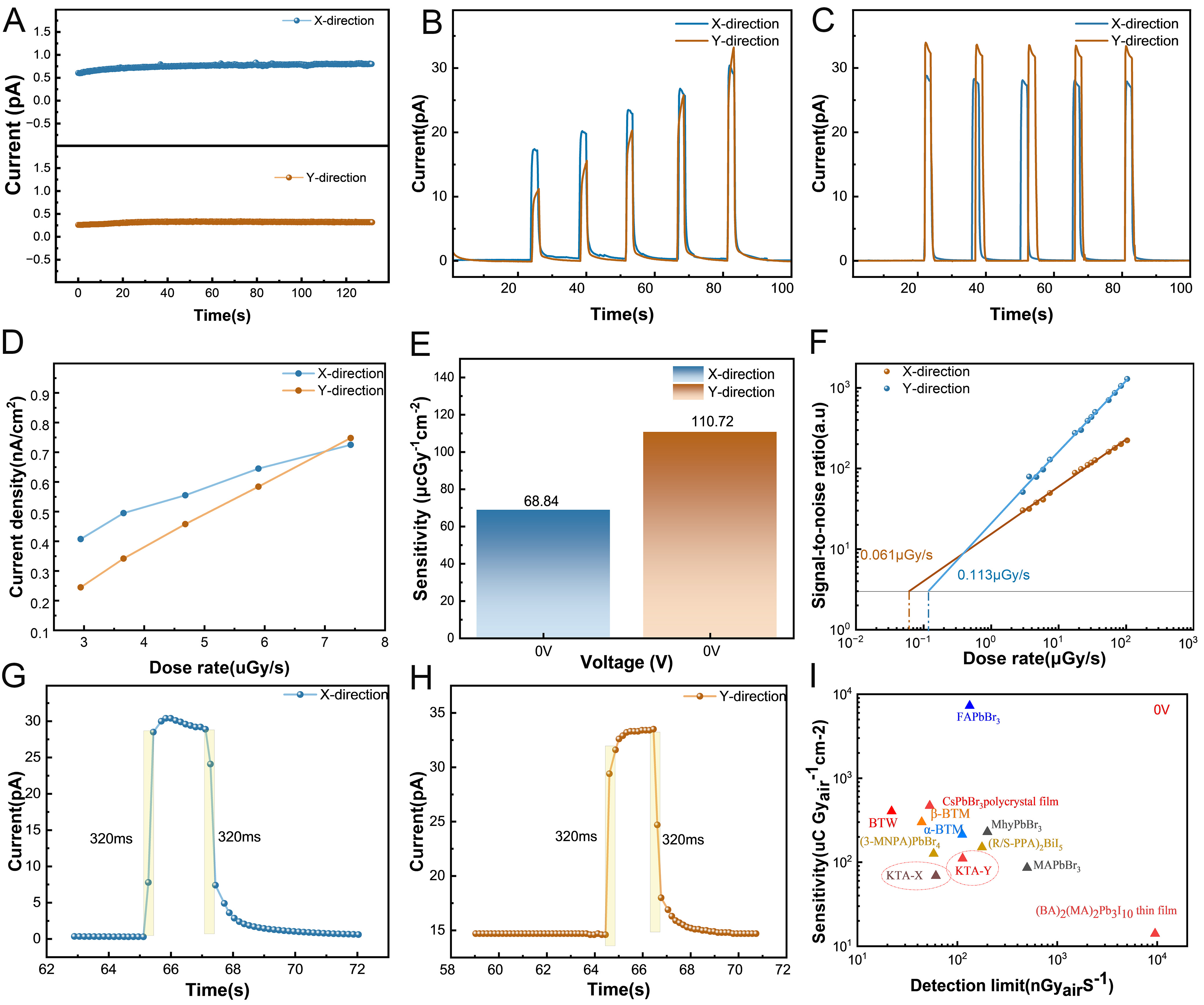
Figure 3. (A) Dark current of the KTA detector at 0 V bias. (B) Photocurrent response of the KTA detector at 0 V bias. (C) Steady-state photocurrent signal of the KTA detector under continuous X-ray irradiation. (D) Photocurrent density without applied bias. (E) Sensitivity without applied bias. (F) Detection limit without an applied bias. (G and H) Response time of the detector without an applied bias. (I) Comparison of detection performance without an applied bias[20,25,26,43-49].
In self-powered detectors, no bias voltage is applied, so the dark current is reduced to the lowest level inherent to the material. This characteristic resolves the significant problem of conventional X-ray detectors where both photocurrent and dark current increase simultaneously, and it also reduces photon noise to some extent, thereby lowering the detection limit. In addition, the performance of the KTA detector in self-powered mode surpasses that of many previously reported self-powered X-ray detectors such as GaN[43], MAPbBr3[44], and MhyPbBr3 (Mhy = methylhydrazine)[45], in the balance of sensitivity and detection limit. It can thus be concluded that polar oxide crystals exhibit good self-powered detection performance. Table 2 compare the detection limits of some previously reported self-powered X-ray detectors with those of the KTA self-powered detector. In practical applications, reducing the response time of X-ray detectors not only significantly lowers radiation exposure but also improves imaging efficiency, which fully meets safety requirements in fields such as radiation protection and clinical practice. To this end, we tested the response time of the KTA crystal X-ray detector. Figures 3G and H show the photocurrent response time of the KTA X-ray detector at an X-ray energy of 40 keV. In self-powered mode, the response time was approximately 320 ms, indicating a rapid signal response. This ability to effectively capture rapidly changing processes and provide precise time-resolved information for clinical diagnosis or real-time imaging further validates its application potential. Meanwhile, we compared its performance with that of self-powered detectors reported in the literature [Figure 3I], finding that the KTA detector achieves an effective balance between sensitivity and detection limit.
Performance comparison of KTA and other X-ray detectors in self-powered mode
| Materials | X-ray energy (keV) | μτ (cm2 V-1) | Sensitivity (μC·Gyair-1 cm -2) | Detection limit (μGyair·s-1) | Ref |
| CsPbBr3 thick film | 35 | - | 470 | 0.053 | [46] |
| MAPbBr3 single crystal | 35 | - | 80 | 0.5 | [44] |
| (BA)2(MA)2Pb3I10 thin film | 10.91 | - | 13 | 10 | [47] |
| GaN | 30 | - | 170 | [43] | |
| (BA)2CsAgBiBr7/Cs2AgBiBr6 heterojunction | 40 | 2.8 × 10⁻³ | 206 | - | [48] |
| (3-MNPA)PbBr4 | 30 | 3.43 × 10⁻³ | 123 | 0.058 | [20] |
| MhyPbBr3 | 50 | 1.1 × 10⁻³ | 220 | 0.2 | [45] |
| α-BTM | 40 | 3.2 × 10⁻³ | 213 | 0.112 | [26] |
| β-BTM | 40 | 5.89 × 10⁻³ | 300.2 | 0.044 | [26] |
| BaTeW2O9 | 120 | 4.03 × 10⁻³ | 404 | 0.022 | [25] |
| FAPbBr3 thin film | 40 | 5.09 × 10⁻⁵ | 7,280 | 0.133 | [49] |
| KTA (Y-axis) | 40 | 4 × 10⁻⁷ | 110.72 | 0.113 | This work |
Dipole moment and electrostatic field
In self-powered mode, the outstanding X-ray detection performance of the KTA detector is attributed to the built-in electric field of the polar crystal, which enables a synergistic combination of high sensitivity, fast response, high stability, and low detection limits. To elucidate the microscopic mechanism underlying the polarity of the KTA crystal, we systematically analyzed the atomic arrangement within the unit cell. The data reveal that the KTA crystal is a polar oxide crystal composed of distorted TiO6 octahedra and AsO4 tetrahedra. Its crystallographic parameters, obtained via computational analysis, are detailed in Supplementary Table 1. The distorted TiO6 octahedra within the unit cell generate intrinsic dipole moments. By performing a vector sum of these dipole moments, we obtained the net dipole moment of a single KTA unit cell. This method directly characterizes the strength of the built-in electric field induced by crystal polarization. The significant dipole moment observed in the KTA crystal [Figure 4A] provides direct experimental evidence for the presence of a strong built-in electric field, with a measured dipole moment of 25.93 × 10⁻⁴ esu·cm·A-3. To quantitatively characterize the built-in electric field strength, we employed a series of positive and negative bias voltage gradients to induce an asymmetric photocurrent response in the device.
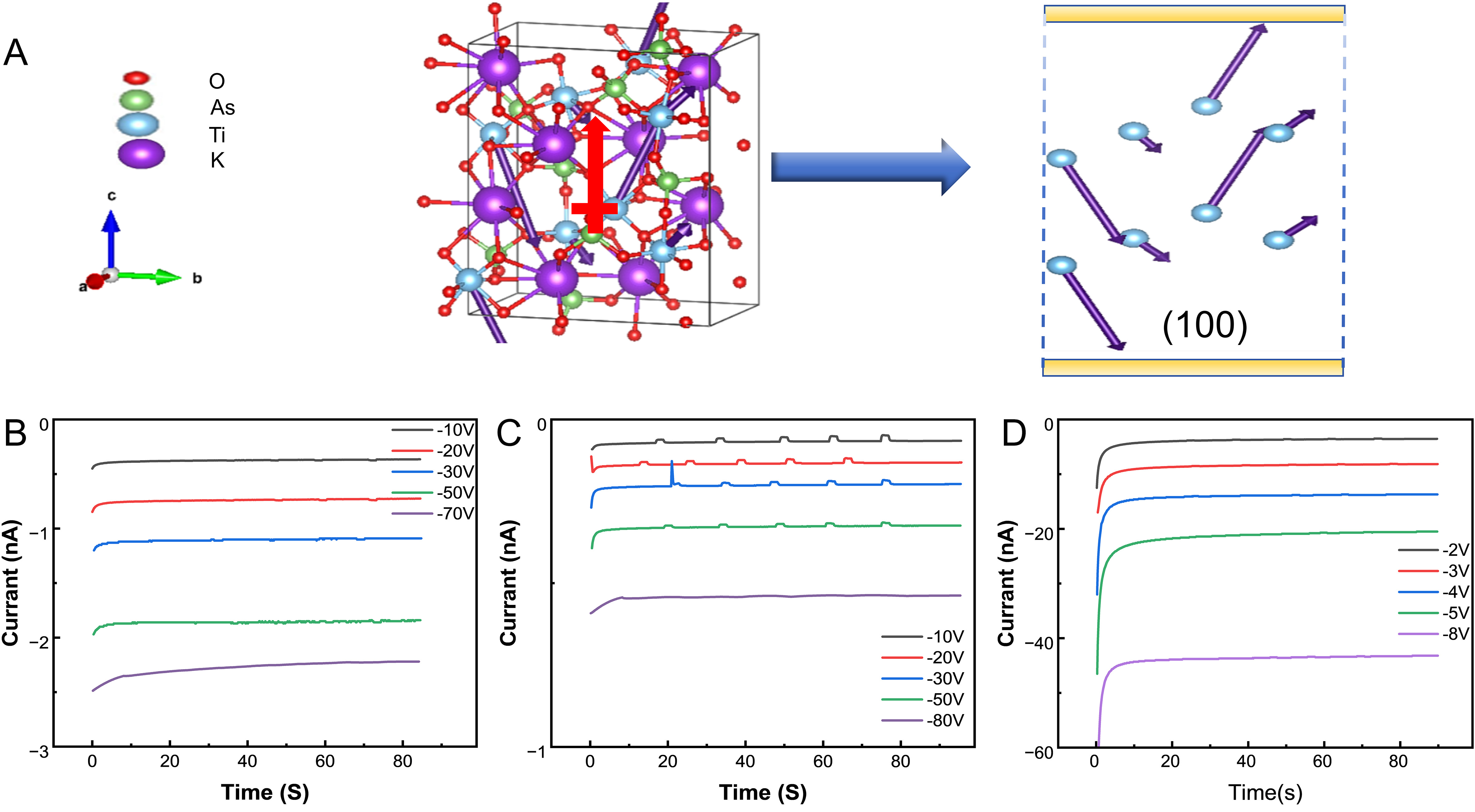
Figure 4. (A) Orientation of the dipole moments in each TiO6 polyhedron of KTA. The direction of the net dipole moment is indicated by the red arrow. (B) Photocurrent response along the X-axis under a negative bias. (C) Photocurrent response along the Y-axis under a negative bias. (D) Photocurrent response along the Z-axis under a negative bias.
Under experimental conditions, the detector is simultaneously exposed to X-ray irradiation and an applied bias voltage. Gradual voltage tuning enables the identification of the null bias condition for zero photocurrent output. At this equilibrium state, the applied voltage completely counteracts the built-in electric field and eliminates the photocurrent response. The strength of the built-in electric field can then be calculated using the following formula:
where Vnull is the voltage at which the photocurrent vanishes, and d is the thickness of the active layer.
As shown in Figure 4B-D, the photocurrent response gradually weakens and eventually disappears as the applied negative voltage increases. Ultimately, the built-in electric field strength of KTA was measured to be 35 V·mm-1 in the X-axis, 40 V·mm-1 in the Y-axis, and 4 V·mm-1 in the Z-axis. This is because KTA crystals exhibit a pronounced multi-domain structure at room temperature; the 180° antiparallel domains along the Z-axis suppress the macroscopic polarization field. Since the crystals were not subjected to deliberate single-domain treatment, the presence of domain walls significantly weakens the built-in electric field along the Z direction[50].
The discrepancy between the experimentally measured spontaneous polarization intensity and the calculated unit cell dipole moment may stem from a variety of factors, including local structural distortions, interfacial interactions, and the limitations of the experimental measurements. Despite discrepancies between measured values and theoretical calculations, the overall trend is consistent with the theoretical predictions. It can be concluded that the built-in electric field primarily originates from the material’s spontaneous polarization, while interfacial effects play a secondary role; together, these two factors enable efficient charge extraction and transport in the absence of an applied bias.
X-ray imaging
The above results demonstrate that the built-in electric field in polar oxide crystals can effectively promote carrier transport, thereby enabling excellent self-powered detection performance. Based on this outstanding detection performance, we further evaluated the imaging capabilities of the self-powered KTA detector. We successfully assembled a complete imaging system, whose structure is shown in Figure 5A. An arrow-shaped metal object (6 mm long × 4.5 mm wide) was selected as the imaging target and placed between the detector and the X-ray source. An X-ray energy of 40 keV was applied [Supplementary Table 2], and imaging tests were conducted in self-powered mode. Figure 5B presents the X-ray imaging of this arrow-shaped metal object. The image clearly reproduces the arrow’s complete outline and edge details, demonstrating that the KTA crystal self-powered detector possesses excellent spatial resolution. This result is consistent with the fast response and high sensitivity described earlier, indicating that the KTA detector is capable of resolving object features on the submillimeter scale in self-powered mode. However, the current spatial resolution is limited by various factors, including the pixel pitch of the electrodes and the crystal thickness. The pixel pitch determines the limiting resolution of the sampling, while the crystal thickness affects lateral diffusion during charge transport and crosstalk between adjacent pixels. By further optimizing the pixel geometry, adjusting the crystal thickness, and employing collimation techniques, it is expected that the resolution of this detector in practical imaging applications will be significantly improved.
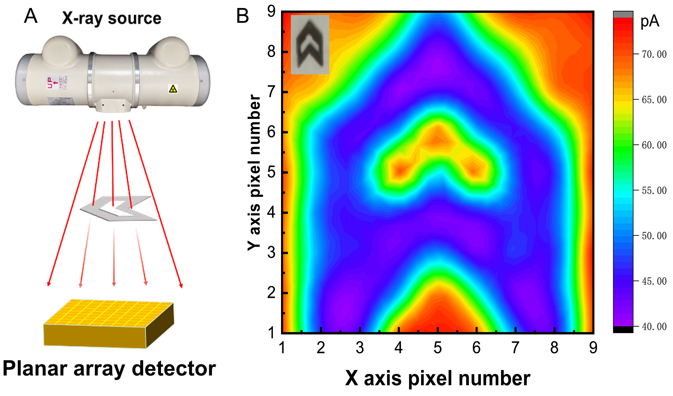
Figure 5. (A) KTA crystal plane array imaging system. (B) X-ray imaging results in self-powered mode.
Anisotropy of structure and properties
As described above, we comprehensively analyzed the performance of the KTA detector under X-ray irradiation at various dose rates. The results indicate that the sensitivity of the KTA detector along the Y-axis is superior to that along the X-axis under all tested dose rates [Figure 6A-C]. This phenomenon can be understood by examining the photocurrent response at low dose rates. At the lowest measured dose rate of 2.94 μGyair·s-1, the photocurrent density along the X-axis (0.40 nA·cm-2) was approximately 66.7% higher than the value along the Y-axis direction (0.24 nA·cm-2). As the dose rate increased to 7.43 μGyair·s-1, the photocurrent along the Y-axis became comparable to that along the X-axis (0.74 and 0.73 nA·cm-2), which is consistent with the higher overall sensitivity slope along the Y-axis (110.72 and 68.84 μC·Gyair-1·cm-2). To explore the underlying causes, we analyzed the crystal structure of the KTA. This anisotropy can be attributed to structural differences along the crystallographic axes: specifically, the parallel, twisted TiO6 chains along the Y-axis form a highly ordered in-plane network of channels, significantly enhancing charge transport. In contrast, the structure along the X-axis exhibits a more staggered arrangement
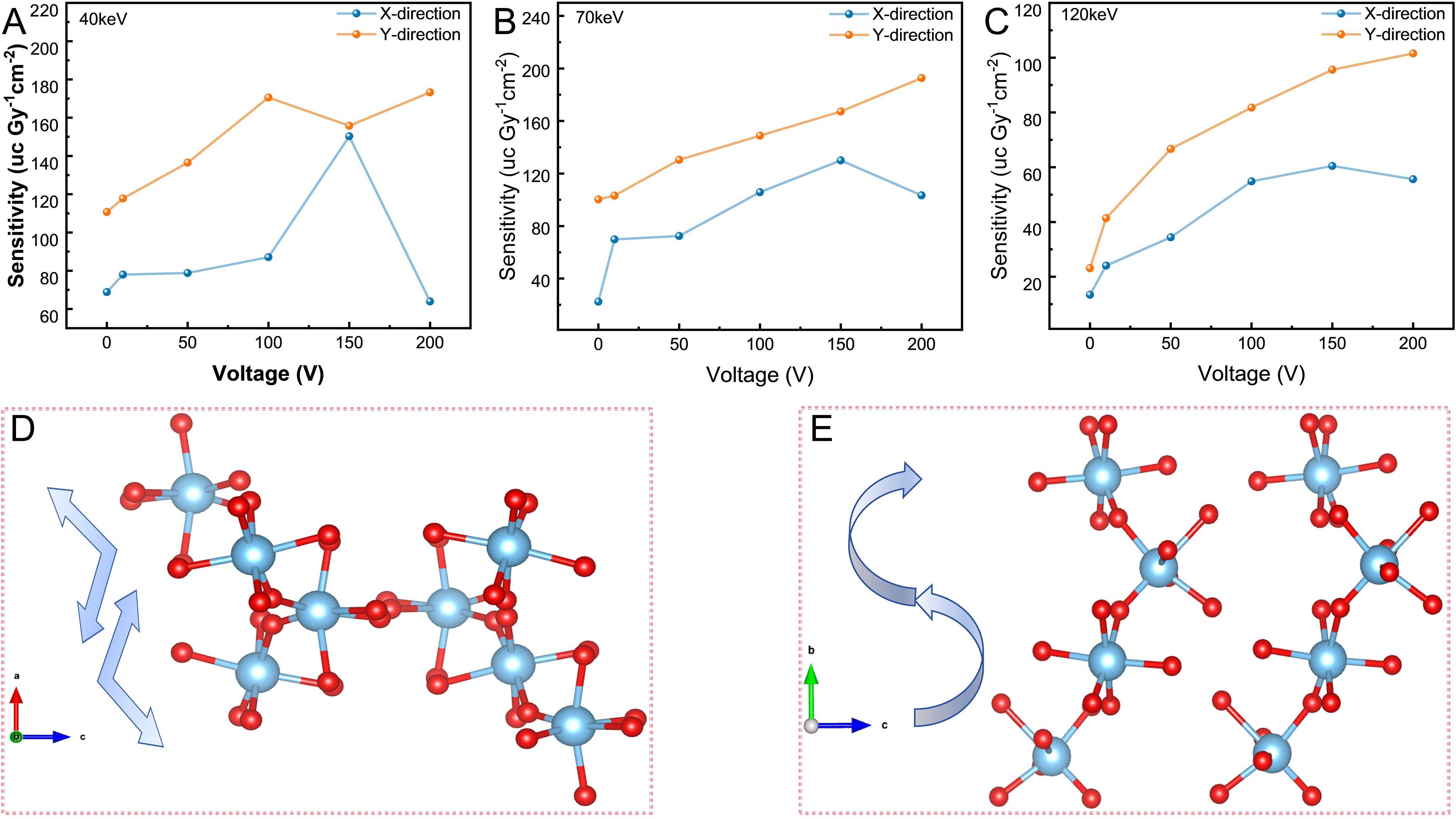
Figure 6. (A) Comparison of X- and Y-axis sensitivity of KTA crystals at X-ray dose rates of 2.94-7.43 μGyair·s-1. (B) at X-ray dose rates of 17.44-34.73 μGyair·s-1. (C) at X-ray dose rates of 50.04-127.322 μGyair·s-1. (D) The distorted TiO6 octahedra in KTA are distributed along the twisted direction of the X-axis. (E) The distorted TiO6 octahedra in KTA exhibit a parallel distribution along the Y-axis direction.
Furthermore, the comprehensive analysis of the experimental data indicates that the lowest detection limit is achieved along the X-axis direction in self-powered mode. This is attributed to a better balance among the built-in electric field strength in the X-axis direction, the charge separation efficiency, and the dark current level. Compared with the stronger built-in electric field along the Y-axis (which may cause slightly higher dark current), the electric field strength along the X-axis ensures that the carrier drift velocity is sufficient to capture weak X-ray signals while effectively suppressing unwanted background current interference. At the same time, although the interlaced crystal structure along the X-axis direction reduces the overall efficiency of charge transport, it also decreases the probability of non-radiative carrier recombination, thereby improving the signal-to-noise ratio for weak signals. The combined effect of these factors enables X-ray detection at lower doses along the X-axis in self-powered mode, thereby achieving the lowest detection limit.
Experimental results show that the structural anisotropy of KTA crystals determines the anisotropic transport of charge carriers and the distribution of the built-in electric field, thereby influencing the detector’s sensitivity and detection limit. This correlation between structure and performance reveals a key physical principle: by controlling the crystal orientation and structure, the balance between sensitivity and detection limit can be systematically adjusted. This design principle applies not only to KTA but can also be extended to other polar oxide detector materials, providing theoretical guidance for the development of next-generation oriented X-ray detectors tailored to specific application scenarios.
CONCLUSION
In summary, this study systematically investigated the X-ray detection performance of the nonlinear optical polar oxide crystal KTA and revealed its significant self-powered anisotropic response. Under external bias conditions, the detector’s sensitivity reaches 192.96 μC·Gyair-1 cm-2, with a detection limit of
DECLARATIONS
Acknowledgement
The authors gratefully acknowledge Guo Feifei for her assistance with experimental instrument usage and data processing methods. We thank Sun Youxuan and Zhai Zhongjun for generously providing the KTA crystals.
Authors' contributions
Investigation, data curation, formal analysis, visualization, writing - original draft: Hu, Y.
Conceptualization, supervision, funding acquisition, review & editing: Sun, W.
Methodology, resources, validation: Feng, X.
Investigation, formal analysis: Hu, F.
Conceptualization, supervision, project administration, funding acquisition, review & editing: Gao, Z.
Availability of data and materials
The data supporting the findings of this study are available within this Article and its Supplementary Materials. Further data are available from the corresponding authors upon reasonable request.
AI and AI-assisted tools statement
During the preparation of this work, the authors used (DeepL, version 25.8.2.17787, 2025-09-01) for language polishing and grammar checking. After using these tools, the authors reviewed and edited the content as needed. The authors take full responsibility for the final content of publication.
Financial support and sponsorship
This work was financially supported by the National Natural Science Foundation of China (grant no. 62175129), the Natural Science Foundation of Shandong Province (No. ZR2025ZD31).
Conflict of interest
All authors declared that there are no conflicts of interest.
Ethical approval and consent to participate
Not applicable.
Consent for publication
Not applicable.
Copyright
© The Author(s) 2026.
Supplementary Materials
REFERENCES
1. Jana, A.; Cho, S.; Patil, S. A.; et al. Perovskite: scintillators, direct detectors, and X-ray imagers. Mater. Today. 2022, 55, 110-36.
2. Aamir, M.; Shahiduzzaman, M.; Taima, T.; Akhtar, J.; Nunzi, J. M. It is an all-rounder! On the development of metal halide perovskite-based fluorescent sensors and radiation detectors. Adv. Opt. Mater. 2021, 9, 2101276.
3. Chen, H.; Li, Y.; Zhao, B.; Ming, J.; Xue, D. Nanocrystals of metal halide perovskites and their analogues as scintillators for x-ray detection. Nano. Futures. 2022, 6, 012001.
4. Cho, S.; Kang, S.; Kwon, C.; et al. Effect of annealing on the X-ray detection properties of nano-sized polycrystalline lead oxide films. Jpn. J. Appl. Phys. 2008, 47, 7393.
5. Zhou, F.; Li, Z.; Lan, W.; Wang, Q.; Ding, L.; Jin, Z. Halide perovskite, a potential scintillator for X-ray detection. Small. Methods. 2020, 4, 2000506.
6. Pauwels, K.; Douissard, P. Indirect X-ray detectors with single-photon sensitivity. J. Synchrotron. Rad. 2022, 29, 1394-406.
7. Geng, X.; Chen, Y. A.; Li, Y. Y.; et al. Lead-free halide perovskites for direct X-ray detectors. Adv. Sci. 2023, 10, 2300256.
8. Wu, H.; Ge, Y.; Niu, G.; Tang, J. Metal halide perovskites for X-ray detection and imaging. Matter 2021, 4, 144-63.
9. Wronski, M. M.; Rowlands, J. A. Direct-conversion flat-panel imager with avalanche gain: feasibility investigation for HARP-AMFPI. Med. Phys. 2008, 35, 5207-18.
10. Kasap, S.; Frey, J. B.; Belev, G.; et al. Amorphous and polycrystalline photoconductors for direct conversion flat panel X-ray image sensors. Sensors 2011, 11, 5112-57.
11. Basiricò, L.; Ciavatti, A.; Fraboni, B. Solution-grown organic and perovskite X-ray detectors: a new paradigm for the direct detection of ionizing radiation. Adv. Mater. Technol. 2020, 6, 2000475.
12. Chaudhari, R.; Ravikant, C. A review on BiI3 perovskites and composites for direct X-ray detection. Sens. Actuator. Phys. 2022, 346, 113863.
13. Sun, X. J.; Xia, M. L.; Xu, Y. S.; Tang, J.; Niu, G. D. Research progress of perovskite direct X-ray imaging. Chin. J. Lumin. 2022, 43, 1014-26.
14. Li, Y.; Adeagbo, E.; Koughia, C.; et al. Direct conversion X-ray detectors with 70 pA cm-2 dark currents coated from an alcohol-based perovskite ink. J. Mater. Chem. C. 2022, 10, 1228-35.
15. Anagnost, K. M.; Lee, E.; Wang, Z.; Liu, J.; Fossum, E. R. Simulating 50 keV X-ray photon detection in silicon with a down-conversion layer. Sensors 2021, 21, 7566.
16. Eisen, Y.; Shor, A.; Mardor, I. CdTe and CdZnTe X-ray and gamma-ray detectors for imaging systems. IEEE. Trans. Nucl. Sci. 2004, 51, 1191-8.
17. Sellin, P. J.; Prekas, G.; Franc, J.; Grill, R. Electric field distributions in CdZnTe due to reduced temperature and X-ray irradiation. Appl. Phys. Lett. 2010, 96, 133509.
18. Zhu, M.; Du, X.; Niu, G.; et al. Template directed perovskite X-ray detectors towards low ionic migration and low interpixel cross talking. Fundam. Res. 2022, 2, 108-13.
19. Huang, Y.; Qiao, L.; Jiang, Y.; et al. A-site cation engineering for highly efficient MAPbI3 single-crystal X-ray detector. Angew. Chem. Int. Ed. 2019, 58, 17834-42.
20. Fan, Y.; Chen, Q.; Li, Z.; et al. Realization of passive X-ray detection with a low detection limit in dion-jacobson halide hybrid perovskite. Small 2023, 19, 2303814.
21. Pan, L.; Liu, Z.; Welton, C.; et al. Ultrahigh-flux X-ray detection by a solution-grown perovskite CsPbBr3 single-crystal semiconductor detector. Adv. Mater. 2023, 35, 2211840.
22. Pisoni, A.; Jaćimović, J.; Barišić, O. S.; et al. Ultra-low thermal conductivity in organic-inorganic hybrid perovskite CH3NH3PbI3. J. Phys. Chem. Lett. 2014, 5, 2488-92.
23. Lee, W.; Li, H.; Wong, A. B.; et al. Ultralow thermal conductivity in all-inorganic halide perovskites. Proc. Natl. Acad. Sci. USA. 2017, 114, 8693-7.
24. Heiderhoff, R.; Haeger, T.; Pourdavoud, N.; et al. Thermal conductivity of methylammonium lead halide perovskite single crystals and thin films: a comparative study. J. Phys. Chem. C. 2017, 121, 28306-11.
25. Guo, F.; Hu, F.; Song, Y.; Wang, L.; Gao, Z.; Tao, X. High-performance oxide crystal BaTeW2O9 X-ray detector with high stability, low detection limit, and ultralow dark current drift. ACS. Appl. Mater. Interfaces. 2025, 17, 9530-8.
26. Hu, F.; Guo, F.; Mu, W.; Cao, T.; Wang, L.; Gao, Z. Effect of internal electrostatic fields on self-powered X-ray detectors with high sensitivity and low detection limits in polar oxide crystals α/β-BaTeMo2O9. Small 2025, 21, 2502928.
27. Jin, P.; Tang, Y.; Xu, X.; et al. Solution-processed perovskite/metal-oxide hybrid X-ray detector and array with decoupled electronic and ionic transport pathways. Small. Methods. 2022, 6, 2200500.
28. Lujan, R. A.; Street, R. A. Flexible X-ray detector array fabricated with oxide thin-film transistors. IEEE. Electron. Device. Lett. 2012, 33, 688-90.
29. Wei, H.; Huang, J. Halide lead perovskites for ionizing radiation detection. Nat. Commun. 2019, 10, 1066.
30. Wei, H.; Fang, Y.; Mulligan, P.; et al. Sensitive X-ray detectors made of methylammonium lead tribromide perovskite single crystals. Nat. Photonics. 2016, 10, 333-9.
31. Zhuang, R.; Wang, X.; Ma, W.; et al. Highly sensitive X-ray detector made of layered perovskite-like (NH4)3Bi2I9 single crystal with anisotropic response. Nat. Photonics. 2019, 13, 602-8.
32. Kasap, S. O.; Rowlands, J. A. Review X-ray photoconductors and stabilized a-Se for direct conversion digital flat-panel X-ray image-detectors. J. Mater. Sci. Mater. Electron. 2000, 11, 179-98.
33. Berger, M. J.; Hubbell, J. H.; Seltzer, S. M.; et al. XCOM: photon cross sections database: NIST standard reference database 8 (XGAM). 2010.
34. Chen, J.; Tang, H.; Liu, B.; et al. High-performance X-ray detector based on single-crystal β-Ga2O3:Mg. ACS. Appl. Mater. Interfaces. 2021, 13, 2879-86.
35. Li, Z.; Chen, J.; Tang, H.; et al. Band gap engineering in β-Ga2O3 for a high-performance X-ray detector. ACS. Appl. Electron. Mater. 2021, 3, 4630-9.
36. Li, Z.; Chang, S.; Zhang, H.; et al. Cu-based metal-organic frameworks for highly sensitive X-ray detectors. Chem. Commun. 2021, 57, 8612-5.
37. Ge, M.; Chen, S.; Fu, X.; Feng, Y.; Wang, D.; Yuan, M. Effects of fluorinated aromatic spacer in Ag-Bi double perovskite for X-ray detector. J. Phys. Chem. 2022, 126, 19417-23.
38. Pan, W.; Wu, H.; Luo, J.; et al. Cs2AgBiBr6 single-crystal X-ray detectors with a low detection limit. Nat. Photonics. 2017, 11, 726-32.
39. Zhang, Y.; Liu, Y.; Xu, Z.; et al. Nucleation-controlled growth of superior lead-free perovskite Cs3Bi2I9 single-crystals for high-performance X-ray detection. Nat. Commun. 2020, 11, 2304.
40. Guo, J.; Xu, Y.; Yang, W.; et al. High-stability flexible X-ray detectors based on lead-free halide perovskite Cs2TeI6 films. ACS. Appl. Mater. Interfaces. 2021, 13, 23928-35.
41. Wei, Q.; Fan, X.; Xiang, P.; et al. Cs3Cu2I5 single crystal for efficient direct X-ray detection. Adv. Opt. Mater. 2023, 11, 2300247.
42. Ma, C.; Chen, F.; Song, X.; et al. Centimeter-sized molecular perovskite crystal for efficient X-ray detection. Adv. Funct. Mater. 2021, 31, 2100691.
43. Zhou, L.; Lu, X.; Wu, J.; et al. Self-powered fast-response X-ray detectors based on vertical GaN p-n diodes. IEEE. Electron. Device. Lett. 2019, 40, 1044-7.
44. Xu, Q.; Zhang, B.; Nie, J.; et al. Self-powered X-ray detector based on methylammonium lead tribromide single crystals. J. Alloys. Compd. 2021, 859, 157826.
45. Guan, Q.; You, S.; Zhu, Z. K.; et al. Three-dimensional polar perovskites for highly sensitive self-driven X-ray detection. Angew. Chem. Int. Ed. 2024, 63, e202320180.
46. Gou, Z.; Huanglong, S.; Ke, W.; et al. Self-powered X-ray detector based on all-inorganic perovskite thick film with high sensitivity under low dose rate. Phys. Status. Solid. RRL. 2019, 13, 1900094.
47. Tsai, H.; Liu, F.; Shrestha, S.; et al. A sensitive and robust thin-film X-ray detector using 2D layered perovskite diodes. Sci. Adv. 2020, 6, eaay0815.
48. Zhang, X.; Zhu, T.; Ji, C.; Yao, Y.; Luo, J. In situ epitaxial growth of centimeter-sized lead-free (BA)2CsAgBiBr7/Cs2AgBiBr6 heterocrystals for self-driven X-ray detection. J. Am. Chem. Soc. 2021, 143, 20802-10.
49. Girolami, M.; Matteocci, F.; Pettinato, S.; et al. Metal-halide perovskite submicrometer-thick films for ultra-stable self-powered direct X-ray detectors. Nano. Micro. Lett. 2024, 16, 182.
Cite This Article
 Open Access
How to Cite
Hu, Y.; Sun, W.; Feng, X.; Hu, F.; Gao, Z. High-performance self-powered X-ray detection materials based on KTiOAsO4 polar crystals. Microstructures 2026, 6, 20260134. https://dx.doi.org/10.20517/microstructures.2026.142
Download Citation
Export Citation File:
Type of Import
Tips on Downloading Citation
Citation Manager File Format
Type of Import
Direct Import: When the Direct Import option is selected (the default state), a dialogue box will give you the option to Save or Open the downloaded citation data. Choosing Open will either launch your citation manager or give you a choice of applications with which to use the metadata. The Save option saves the file locally for later use.
Indirect Import: When the Indirect Import option is selected, the metadata is displayed and may be copied and pasted as needed.
About This Article
Special Topic
Copyright

Data & Comments
Data
0







Comments
Comments must be written in English. Spam, offensive content, impersonation, and private information will not be permitted. If any comment is reported and identified as inappropriate content by OAE staff, the comment will be removed without notice. If you have any queries or need any help, please contact us at support@oaepublish.com.