




fig1
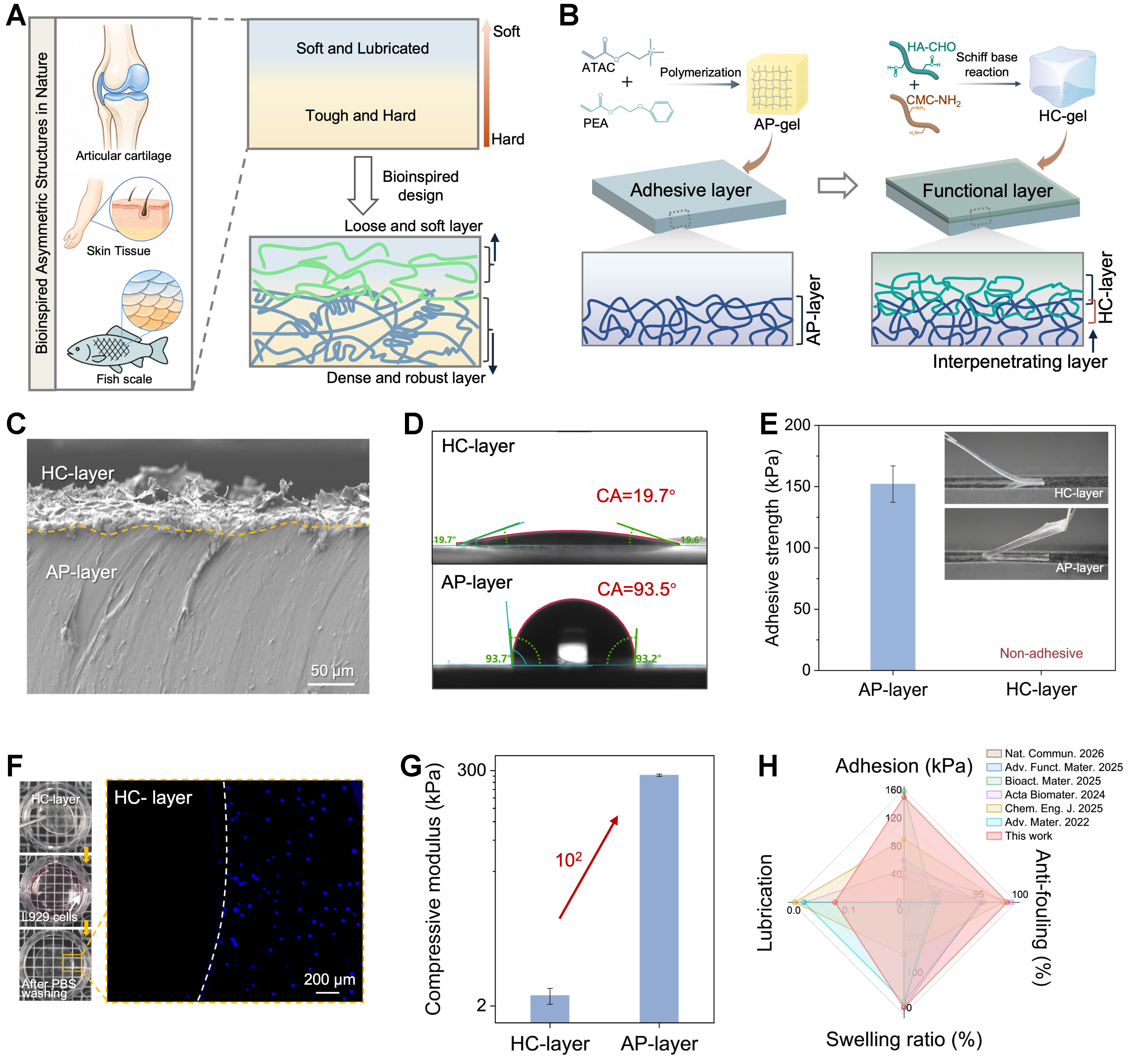
Figure 1. Design and structural characterization of the AP-HC bilayer hydrogel. (A) Schematic illustration of asymmetric biological architectures, including articular cartilage, skin tissue, and fish scales, inspiring the Janus bilayer design; (B) Formation of the HC hydrogel layer on the AP substrate through in situ Schiff base crosslinking between HA-CHO and CMC-NH2, accompanied by interfacial chain interdiffusion and network interpenetration; (C) Cross-sectional SEM image of the AP-HC bilayer hydrogel; (D) Water contact angles of the adhesive AP side and the hydrated lubricating HC side; (E) Comparison of the asymmetric adhesion behavior of the two surfaces of the intact AP-HC hydrogel, showing adhesion on the AP side and negligible adhesion on the HC side; (F) Representative fluorescence images showing reduced L929 cell attachment on the HC surface after 24 h culture and PBS washing; (G) Comparison of the compressive moduli of separately prepared AP-gel and HC-gel; (H) Radar plot comparing the integrated performance of AP-HC with previously reported hydrogel systems[18,33-37]. Data are presented as mean ± SD (n = 3 independent samples). HA-CHO: Aldehyde-functionalized hyaluronic acid; CMC-NH2: amine-functionalized carboxymethyl cellulose; SEM: scanning electron microscope; PBS: phosphate-buffered saline; SD: standard deviation; ATAC: [2-(acryloyloxy) ethyl]trimethylammonium chloride; PEA: 2-phenylethyl acrylate.