




fig2
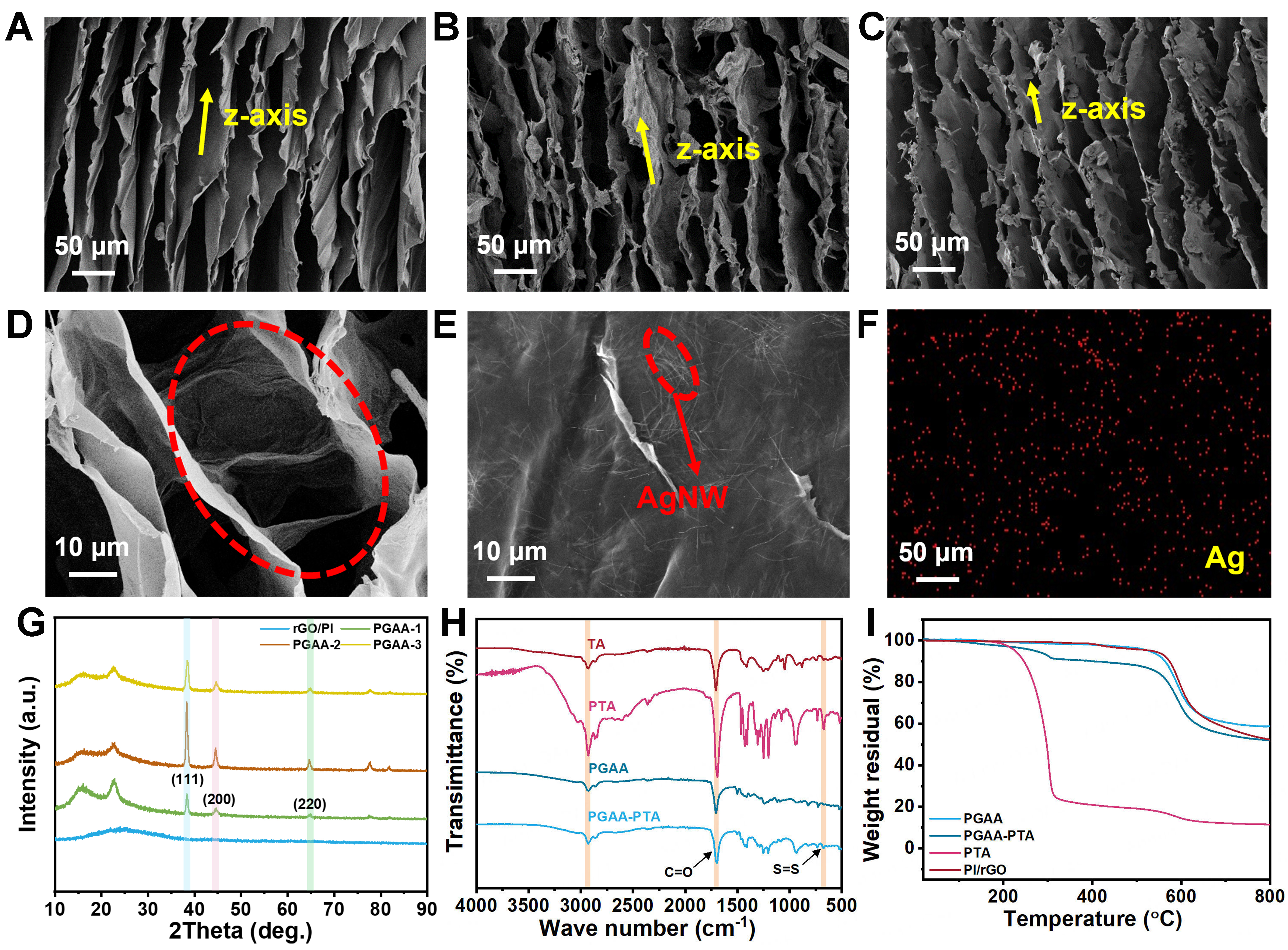
Figure 2. SEM images along the z-axis for (A) PGAA-0, (B) PGAA-2, and (C) PGAA-PTA; (D) Interlayer bridge structure of the PGAA-2 aerogel; (E) Surface morphology of the PGAA-2 lamella; (F) Distribution of Ag elements of PGAA-2; (G) XRD patterns of PGAA samples; (H) FTIR spectra and (I) TGA curves of PGAA and PGAA-PTA. SEM: Scanning electron microscopy; PGAA-PTA: polyimide/reduced graphene oxide/AgNW-poly(thioctic acid) aerogel; XRD: X-ray diffraction; FTIR: Fourier transform infrared spectroscopy; TGA: thermogravimetric analysis; rGO: reduced graphene oxide; PI: polyimide; TA: thioctic acid; AgNW: silver nanowire.





