




fig1
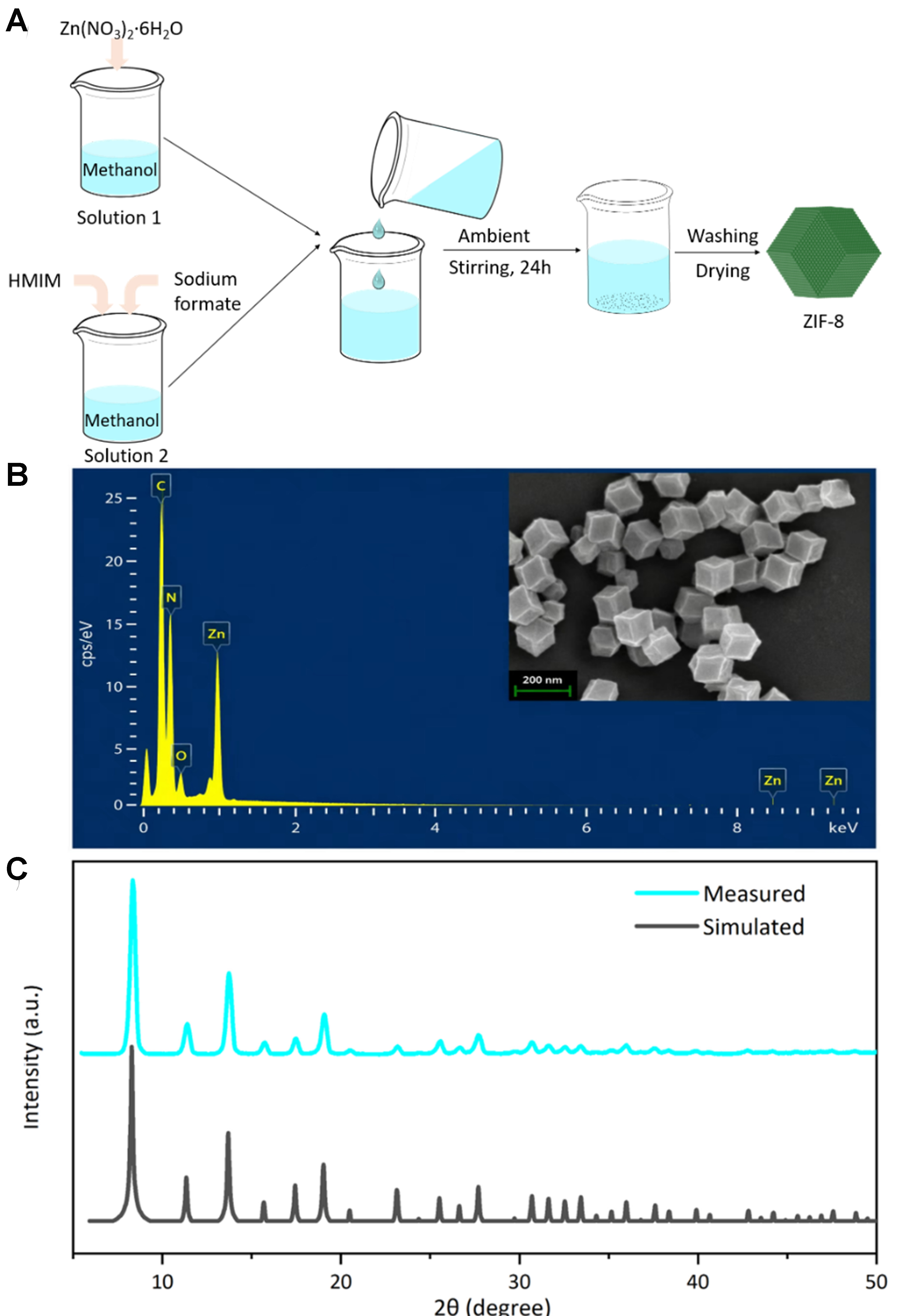
Figure 1. Characterization of the ZIF-8 nanomaterial. (A) Preparation process of ZIF-8; (B) EDS image and the SEM image (inset, with a 200 nm scale bar) of the ZIF-8; (C) PXRD pattern of ZIF-8. ZIF-8: Zeolitic imidazolate framework-8; EDS: energy-dispersive X-ray spectroscopy; SEM: scanning electron microscopy; PXRD: powder X-ray diffraction; HMIM: 2-methylimidazole.





