


fig2
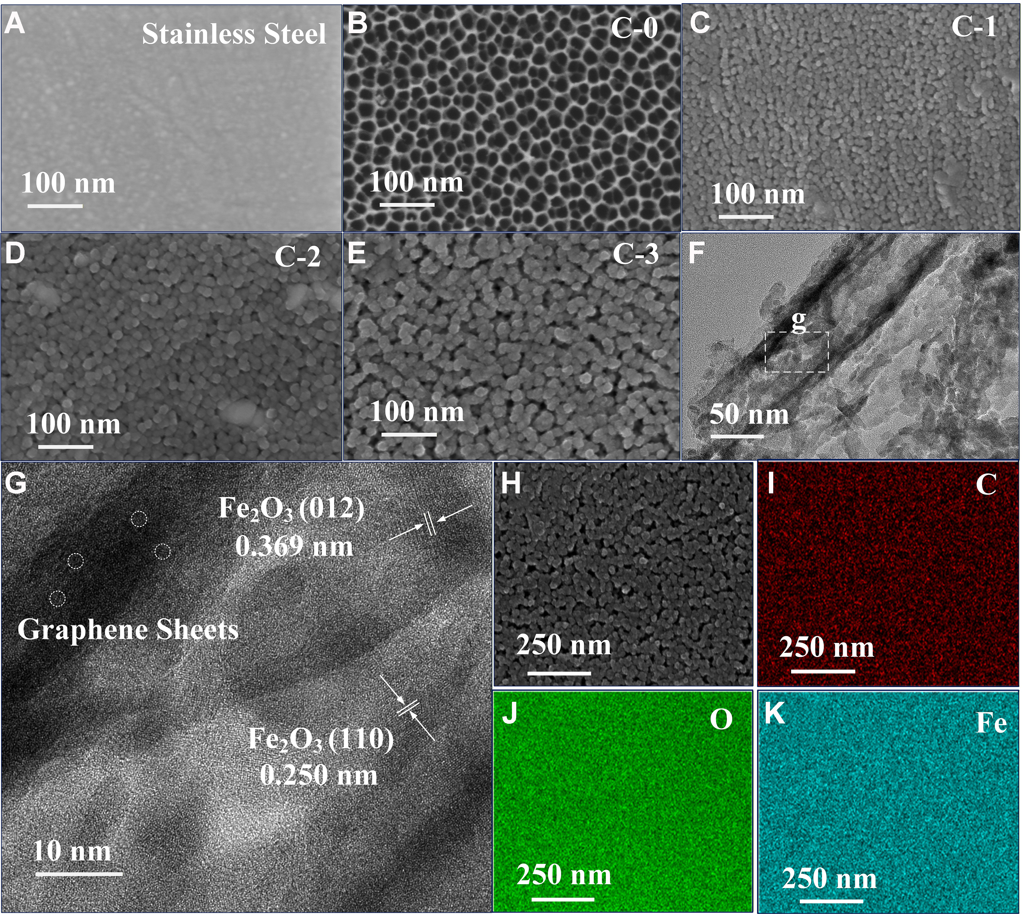
Figure 2. FESEM images of (A) SS substrate, (B) C-0, (C) C-1, (D) C-2, and (E) C-3; (F and G) HRTEM images of C-3; EDS elemental mapping of C-3: (H) HAADF image, (I) C, (J) O, and (K) Fe. FESEM: Field emission scanning electron microscope; SS: stainless steel; HRTEM: high-resolution transmission electron microscopy; EDS: energy dispersive X-ray spectrometer; HAADF: high-angle annular dark-field.


