


fig5
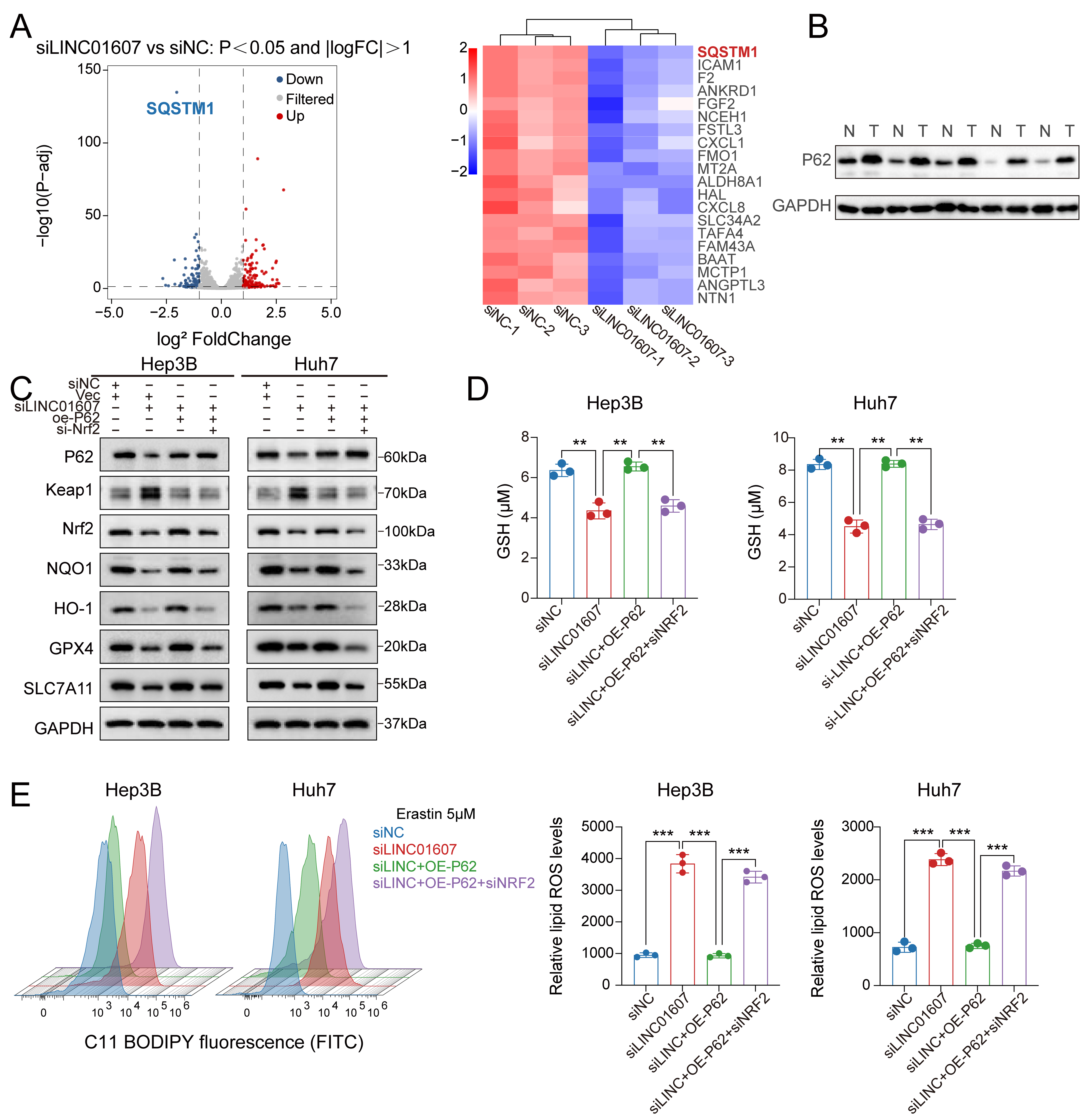
Figure 5. The p62–Keap1–NRF2 pathway is functionally associated with LINC01607-mediated ferroptosis resistance. (A) Volcano plot and heatmap of mRNA expression changes after LINC01607 knockdown in Hep3B cells. SQSTM1/p62 was significantly downregulated. Red, blue, and grey indicate upregulated, downregulated, and unchanged genes, respectively; (B) Western blotting showed higher p62 expression in HCC tumor tissues (T) than in matched normal tissues (N); (C) Western blot analysis in Hep3B and Huh7 cells revealed that LINC01607 knockdown was accompanied by Keap1 accumulation and reduced NRF2, NQO1, HO-1, GPX4, and SLC7A11 levels, whereas p62 overexpression partially reversed these changes. Subsequent Nrf2 silencing abrogated the rescue of the downstream antioxidant and ferroptosis-related proteins, further supporting the functional involvement of the p62–Keap1–NRF2 pathway downstream of LINC01607; (D) The decrease in GSH induced by LINC01607 knockdown was partially rescued by p62 overexpression, whereas subsequent NRF2 silencing abrogated this rescue; (E) Lipid ROS levels, measured using C11-BODIPY fluorescence, indicated that LINC01607 knockdown significantly increased lipid peroxidation. This effect was mitigated by P62 overexpression and further reversed by Nrf2 silencing. Data are presented as mean ± SD from three independent biological replicates unless otherwise indicated. Statistical significance was assessed using two-tailed Student’s t-test for two-group comparisons and one-way ANOVA for multiple-group comparisons, as appropriate. **P < 0.01, ***P < 0.001. HCC: Hepatocellular carcinoma; GSH: glutathione; ROS: reactive oxygen species; SD: standard deviation; ANOVA: analysis of variance; FITC: fluorescein isothiocyanate.