


fig2
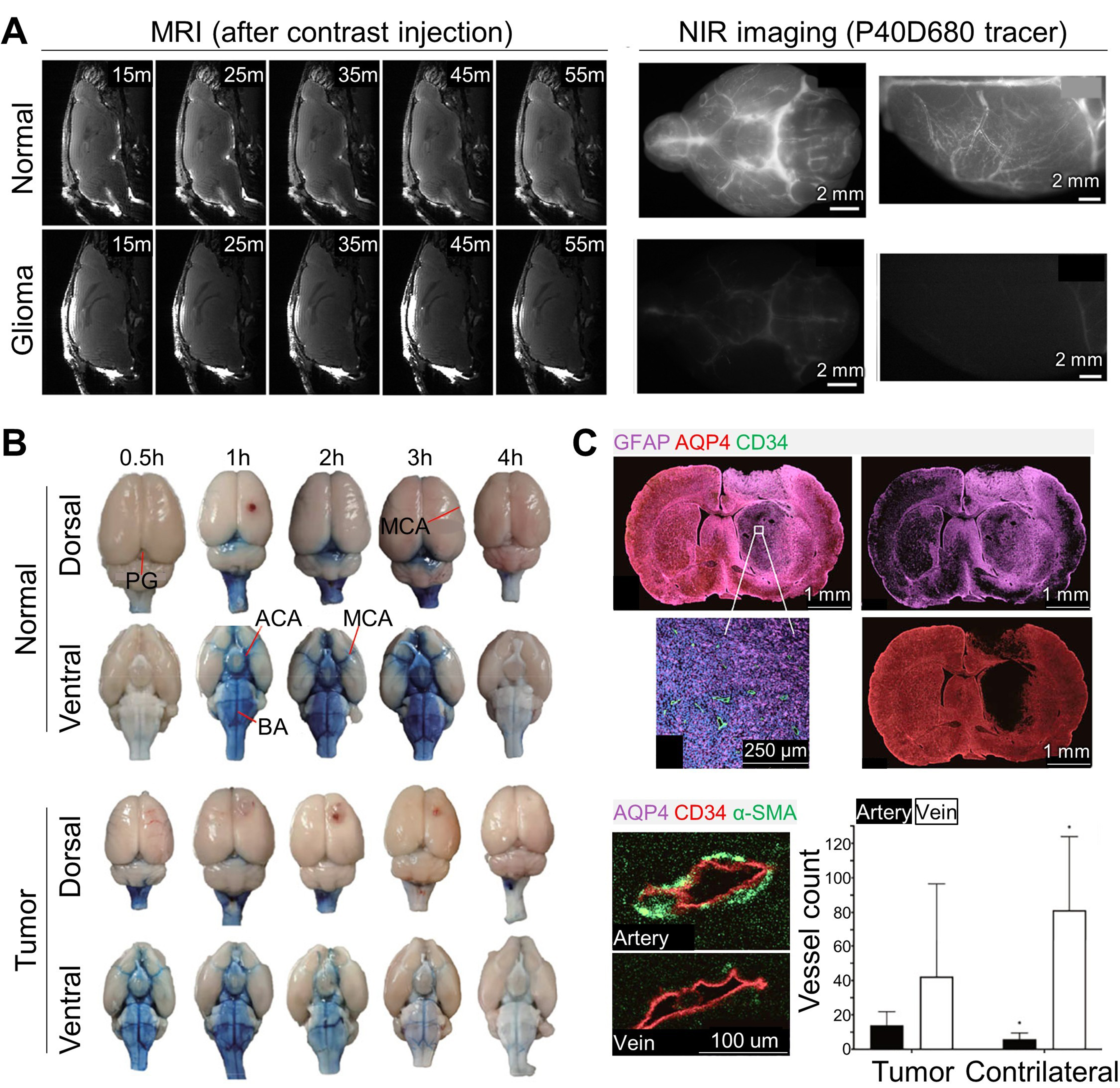
Figure 2. Glymphatic flow alterations in glioma. (A) Contrast-enhanced MRI images (in vivo) acquired 15, 25, 35, 45, and 55 min after cisterna magna injection, and NIR images (ex vivo) of the basal and dorsal regions using the P40D680 tracer, showing suppressed dispersion of contrast agents in glioma models; (B) Evans blue distribution in the dorsal and ventral sides of normal and tumor-bearing brains; (C) Histological images demonstrating the relationship between glymphatic impairment and AQP4 expression and vascular changes. (A) Reproduced from Springer Nature[32] under the CC BY 4.0 license. Copyright © 2019 The Authors. (B and C) Reproduced from Springer Nature[44] under the CC BY 4.0 license. Copyright © 2022 The Authors. MRI: Magnetic resonance imaging; NIR: near-infrared; AQP4: aquaporin-4; PG: pineal gland; MCA: middle cerebral artery; ACA: anterior cerebral artery; BA: basilar artery; GFAP: glial fibrillary acidic protein (astrocytic marker); CD34: cluster of designation 34 (vascular endothelial marker); α-SMA: alpha smooth muscle actin (vascular smooth-muscle/pericyte marker).


