


fig6
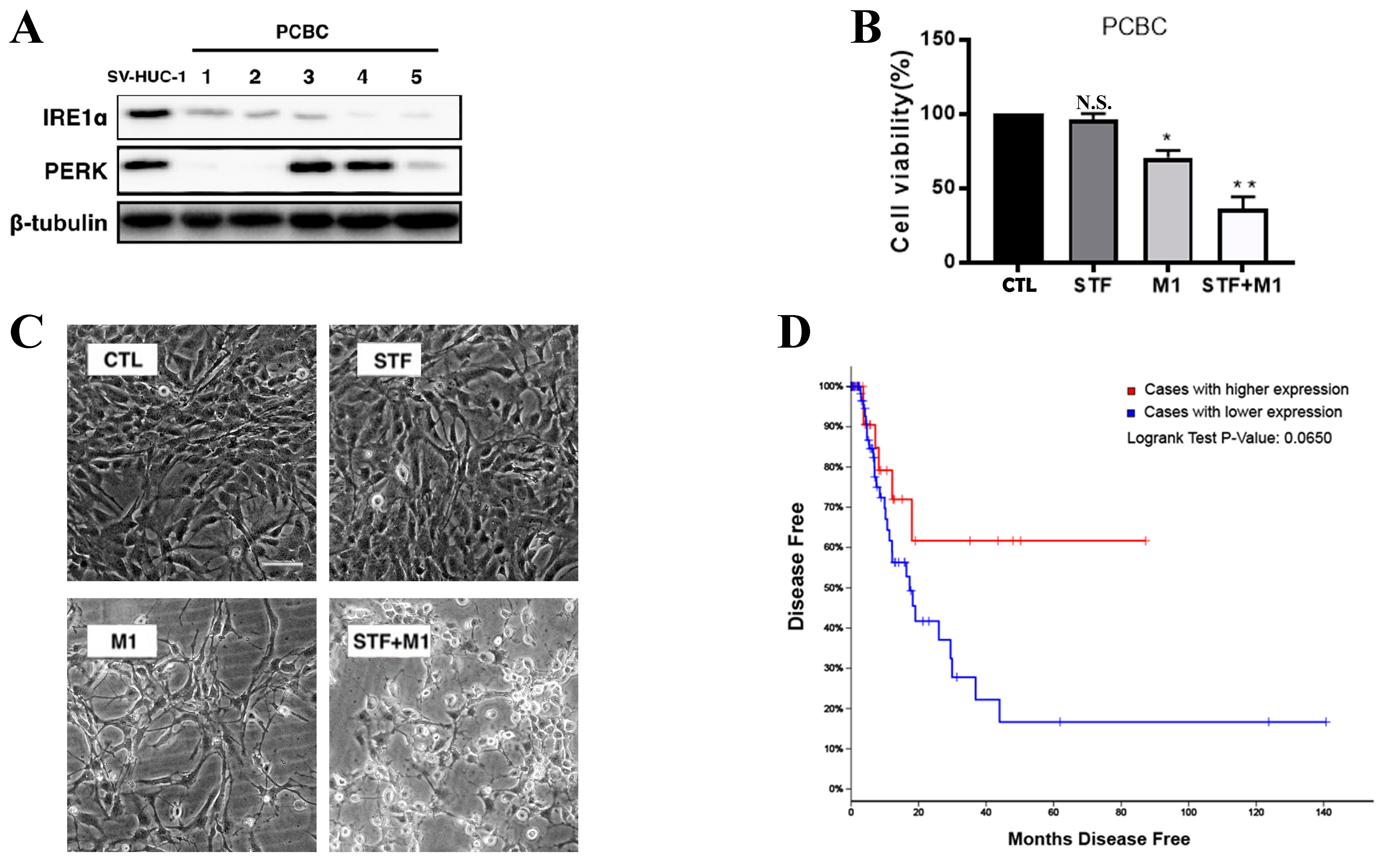
Figure 6. IRE1α is downregulated in bladder cancer and correlates with disease recurrence. (A) Western blot analysis of IRE1α and PERK expression in five PCBC samples compared to the non-tumorigenic bladder epithelial cell line SV-HUC-1. β-tubulin was used as a loading control; (B) Quantification of cell viability in PCBC cells treated with STF (10 μM), M1 (MOI = 0.01), or the combination (STF + M1), relative to the control group (CTL). *P < 0.05, **P < 0.01 vs. CTL group; (C) Morphological changes in PCBC cells treated with control (CTL), STF (10 μM), M1 (MOI = 0.01), or STF + M1 (scale bar = 100 μm); (D) Kaplan–Meier disease-free survival analysis of bladder cancer patients from TCGA cohort, stratified by IRE1α expression levels. Patients with high IRE1α expression (red line) exhibited a trend toward poorer prognosis compared to those with low expression (blue line), with a Log-rank P-value of 0.0650. IRE1α: Inositol-requiring enzyme 1 alpha; PERK: protein kinase RNA-like ER kinase; PCBC: primary cultured bladder cancer; MOI: multiplicity of infection; TCGA: The Cancer Genome Atlas.


