


fig4
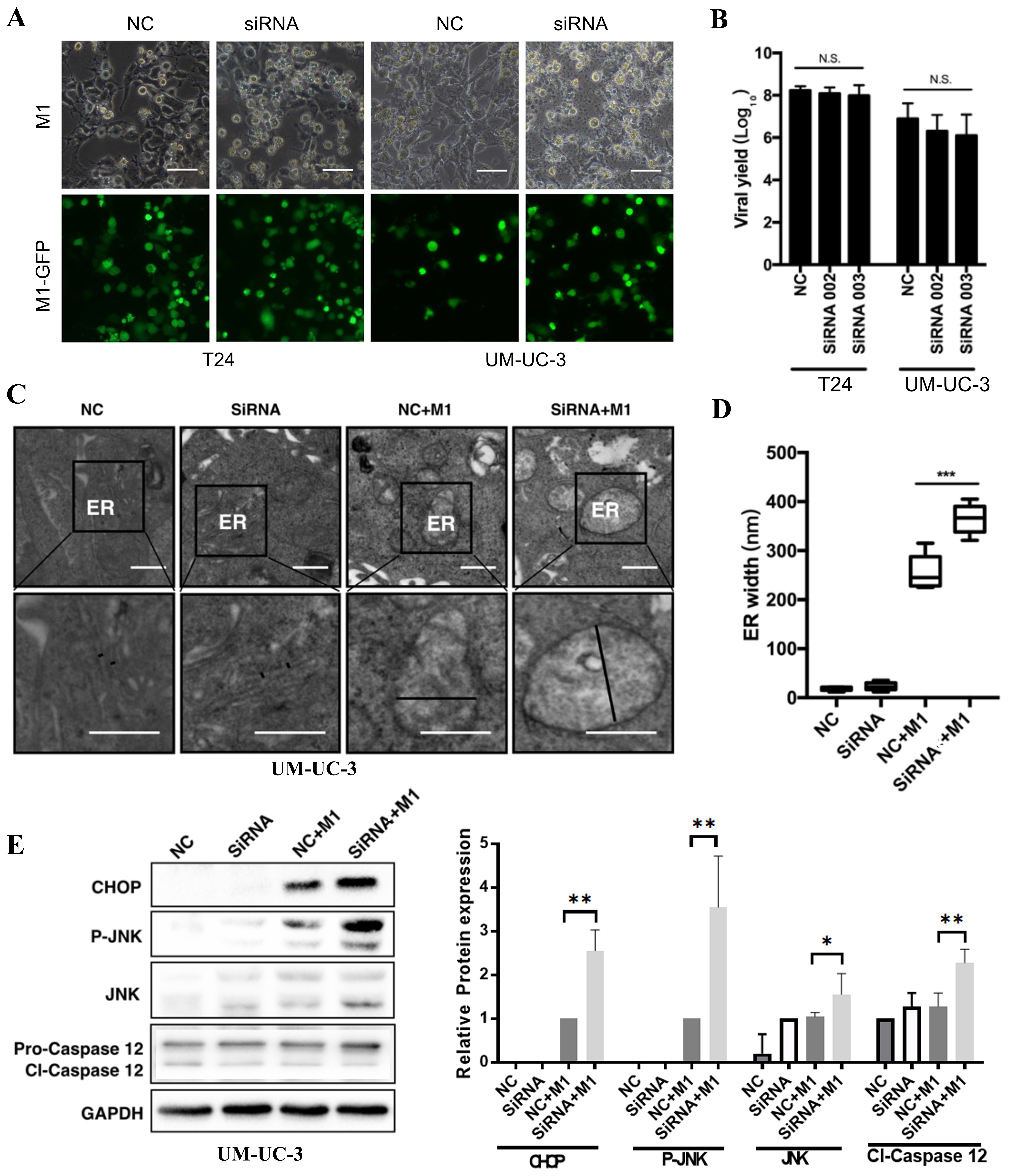
Figure 4. IRE1α knockdown amplifies M1-induced ER stress and activates the CHOP/JNK/caspase-12 pathway. (A and B) Fluorescence imaging and viral yield assay in T24 and UM-UC-3 cells infected with GFP-tagged M1 virus after IRE1α knockdown (siRNA002) or control (NC). Scale bars = 100 μm; (C) TEM of ER morphology in UM-UC-3 cells under the indicated conditions. Marked ER swelling was observed in the siRNA002 + M1 group; (D) Quantification of ER width from TEM images; (E) Western blot and densitometric analysis of ER stress/apoptosis markers CHOP, p-JNK, total JNK, and cleaved caspase-12 (cl-Caspase-12) in UM-UC-3 cells. IRE1α was silenced using siRNA002. GAPDH was used as a loading control. *P < 0.05, **P < 0.01, ***P < 0.001; N.S.: not significant. IRE1α: Inositol-requiring enzyme 1 alpha; ER: endoplasmic reticulum; TEM: transmission electron microscopy; p-JNK: phospho-JNK.


