


fig3
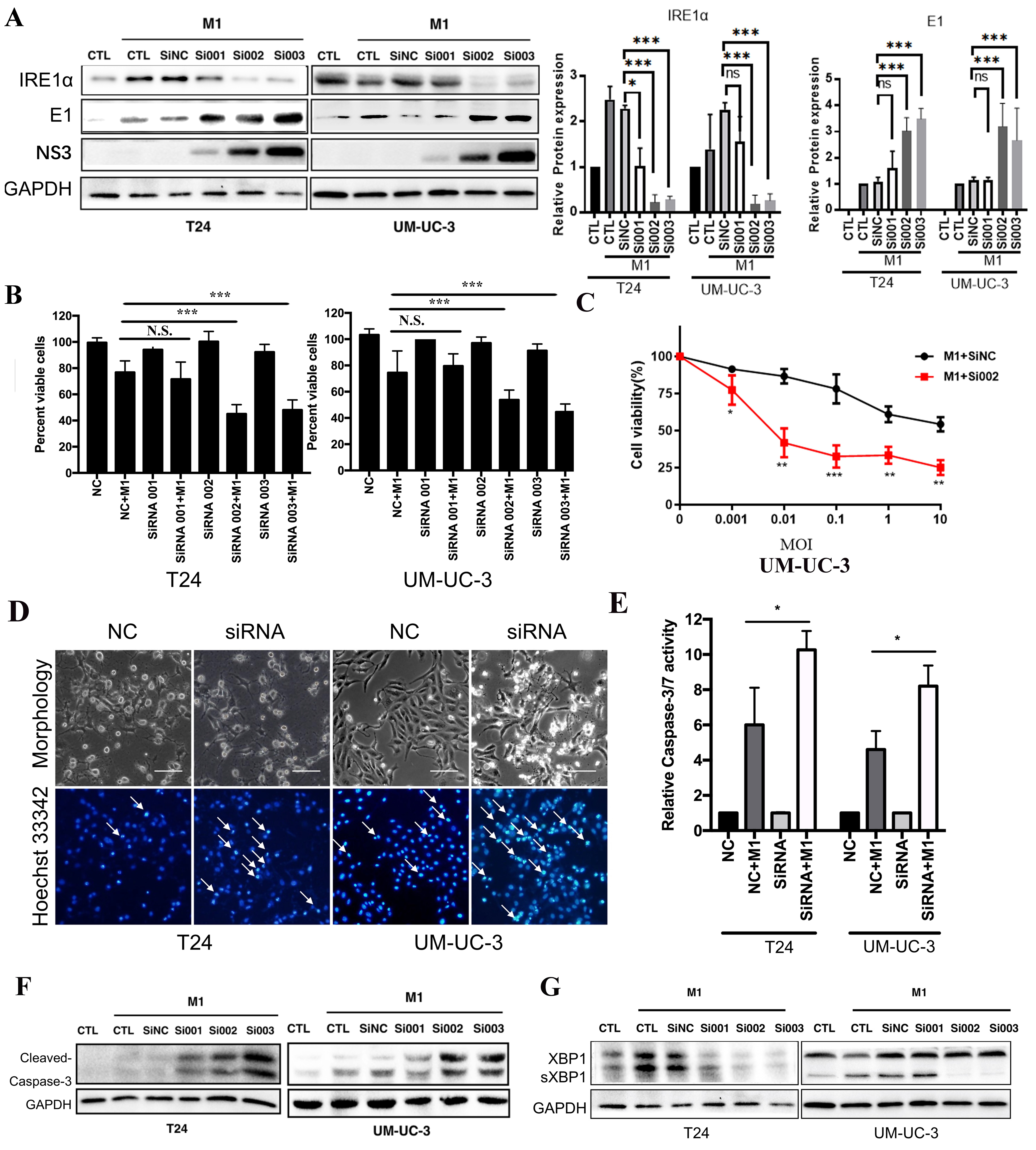
Figure 3. IRE1α silencing enhances M1-mediated apoptosis and viral replication in bladder cancer cells. Western blot analysis and densitometric quantification of IRE1α, E1, and NS3 protein levels in T24 and UM-UC-3 cells transfected with control siRNA (siNC) or IRE1α-specific siRNAs (si001, si002, si003), followed by M1 virus infection. GAPDH was used as a loading control; (B) Quantification of cell viability after M1 treatment in the presence or absence of IRE1α knockdown; (C) Cell viability of UM-UC-3 cells after IRE1α knockdown (si002) or control (siNC) combined with M1 treatment for 48 h; (D) Morphology and Hoechst 33342 nuclear staining of T24 and UM-UC-3 cells showing apoptotic features (arrows) enhanced by IRE1α knockdown (siRNA002); (E) Relative caspase-3/7 activity measurements confirm enhanced apoptotic signaling with IRE1α knockdown (siRNA002); (F) Western blot detection of cleaved caspase-3 in T24 and UM-UC-3 cells, showing increased apoptosis in siRNA + M1 groups; (G) Immunoblot showing elevated M1 viral proteins (E1 and NS3) in IRE1α knockdown groups, indicating increased viral replication. *P < 0.05, **P < 0.01, ***P < 0.001; N.S.: not significant. IRE1α: Inositol-requiring enzyme 1 alpha.


