




Beyond stretchability: preserving quantitative molecular sensing on moving tissues
 0
0 
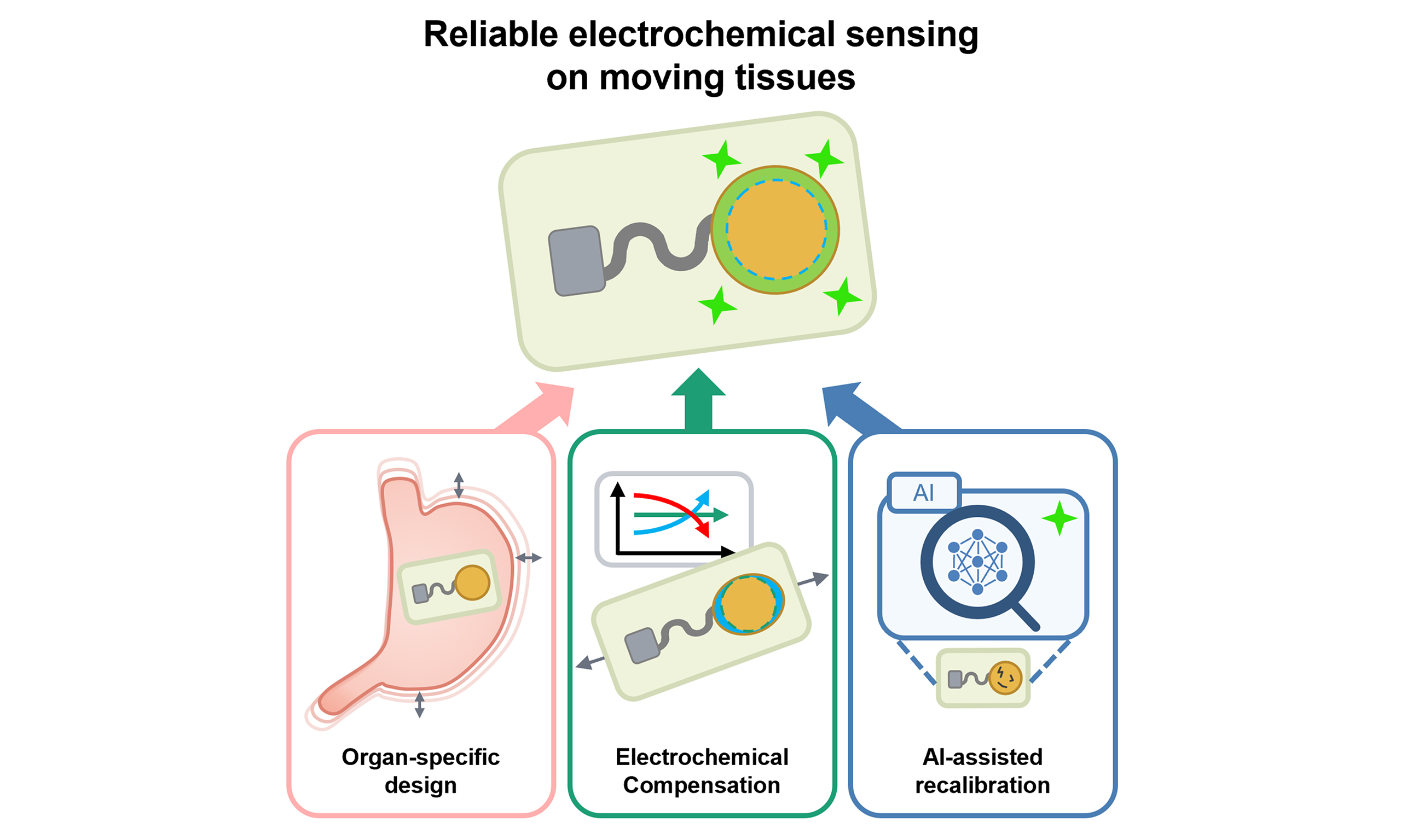
FROM STRAIN TOLERANCE TO ELECTROCHEMICAL COMPENSATION
Stretchable bioelectronics have advanced beyond conforming to soft tissues to sustaining stable signal acquisition during continuous tissue deformation[1,2]. Extending this reliability to molecular sensing is more demanding because electrochemical readouts depend on stable charge transport, controlled electrode–biofluid interactions, and interfacial redox reactions[3,4]. Previous stretchable electrochemical sensors have mitigated strain-induced failure using serpentine or mesh interconnects[5,6], hydrogel electrodes[7,8], composite conductors[5,9,10], liquid-metal conductors[11], soft adhesive interfaces[9,12], and implantable electrodes[13] [Table 1]. However, these strategies stabilize continuity in only one segment of the sensing pathway, rather than the electrochemical interface as a whole, while strain mismatch in multilayer stacks can drive delamination[14-16]. Because deformation alters resistance, electrochemically active area, charge-transfer kinetics, and sensing-layer permeability[17], the central challenge is not stretchability itself but preserving quantitative molecular transduction during deformation[14,18].
Comparison of representative stretchable electrochemical sensing platforms based on independence from geometric strain isolation, intrinsic conductor stretchability, and compensation of strain-induced electrochemical changes
| Platform | Independent of geometric strain isolation | Intrinsic stretchability of conductor | Electrochemical compensation |
| Serpentine Au mesh[6] | × Serpentine mesh | × Au film | × |
| LM island–bridge[11] | × island–bridge | ○ Liquid metal | × |
| 3D micro-patterned PDMS/Au[18] | × Bump–valley microstructure | × Au/Ag–AgCl film | × |
| Printed CNT–PU/Ag–AgCl[5] | △ Free-standing serpentine traces | ○ CNT–PU composite | × |
| Bilayer hydrogel electronics[7] | ○ Intrinsically stretchable | ○ Conductive hydrogel | × |
| Gold nanosheet/CNT nanocomposites[9] | ○ Intrinsically stretchable | ○ Percolation network | × |
| Self-adhesive sweat sensor[12] | ○ Intrinsically stretchable | ○ Composite conductor | × |
| Strain-isolated brittle-film bioelectronics[14] | × Strain-isolating layer | ○ AgNW–PUA composite | △ Circuit-level strain isolation |
| Stretchable PEDOT-based OECT[17] | ○ Intrinsically stretchable | ○ PEDOT conductor | × |
| Implantable NeuroString sensor[13] | ○ Intrinsically stretchable | ○ Graphene nanofiber | × |
| SIRES: SRC–ETI–SFC trilayer[15] | ○ Fully elastomeric trilayer | ○ Liquid-metal SRC | ○ ETI |
To address this coupled problem, Xu and colleagues introduced an intrinsically stretchable interface for resilient electrochemical sensing (SIRES)[15]. Unlike earlier geometric designs that route strain around rigid sensing regions, SIRES makes the entire interface deformable, shifting the design strategy from avoiding strain to compensating for its electrochemical effects. As shown in Figure 1A, SIRES is a fully elastomeric trilayer comprising a liquid-metal strain-resilient conductor (SRC) for charge transport, a carbon nanotube (CNT)–polyurethane electrically tunable interface (ETI) for electromechanical coupling, and a stretchable functional coating (SFC) hosting the sensing chemistry. These layers are covalently integrated within one polyurethane matrix, enabling them to deform together without delamination[15]. Under strain, the SFC exposes a larger electrochemically active area, increasing faradaic current and lowering charge-transfer resistance (Rct)[15,19], while elongation of the conductive pathway increases the series resistance that would otherwise shift or attenuate redox peaks. Because the ETI resistance is tunable through CNT loading[10,15], its strain-dependent increase is matched to the area-driven decrease in Rct. These opposing contributions therefore cancel, and the total resistance remains nearly constant, as captured by a modified Randles model [Figure 1B and C]. Quantitatively, Rct decreases from 253.6 to 173.2 Ω between 0% and 200% strain, while the double-layer capacitance increases by approximately 23%; their product therefore remains within 3% of its initial value at 100% strain. Despite these changes, the voltammetric response is largely retained, with the redox peak current varying by no more than 14% over the same strain range. However, different layers contribute to this stability depending on the analyte. ETI resistance matching operates where faradaic current and electrode area determine the signal, as in voltammetric detection. Amperometric and potentiometric readouts instead depend more strongly on the functional coating and stretchable reference electrode, which preserve the sensing chemistry and potential baseline. Wearable and organ-mounted demonstrations [Figure 1D and E] confirm that this principle extends to complete devices.

Figure 1. Design principle and representative applications of SIRES for wearable and implantable molecular monitoring. (A) Core design concept and cross-sectional schematic of the fully elastomeric trilayer SIRES architecture, comprising an SRC for stable electron transport, an ETI for regulating strain-dependent electrochemical coupling, and an SFC for stabilizing bioactive sensing elements; (B and C) Modified Randles-equivalent-circuit model and relative resistance changes under strain, showing how strain-induced resistance increases are balanced by electrochemically active surface-area modulation; (D) Wireless sweatband implementation for multiplexed sweat monitoring with mobile readout. Scale bar, 5 mm; (E) Implantable SIRES bioelectronics mounted on the rat stomach for gastric glucose monitoring. (A-E) Reprinted in part with permission from[15]. Copyright 2026, AAAS; (F) Viewpoint of this Commentary. Organ deformation (1) alters the electrochemical reaction at the interface (2), and the resulting error is corrected at two levels that operate on different timescales. At the device level, the strain-dependent rise in ETI resistance is matched to the area-driven fall in Rct, so the deviation is canceled as it arises (3.1 and 4.1). At the computational level, an AI-assisted model addresses residual error and slowly accumulating drift, and indicates when recalibration is required (3.2 and 4.2), together sustaining stable quantitative measurement (5). SIRES: Intrinsically stretchable interface for resilient electrochemical sensing; SRC: strain-resilient conductor; ETI: electrically tunable interface; SFC: stretchable functional coating; Rct: charge-transfer resistance; WPU: waterborne polyurethane; CNTs: carbon nanotubes; LM: liquid metal; PU: polyurethane; VIA: vertical interconnect access.
CHALLENGES AND THE ROAD TOWARD RELIABLE ELECTROCHEMICAL SENSING ON MOVING TISSUES
Yet signal stability under strain should not be interpreted as calibration stability during chronic use. The reported robustness over 1,000 cycles at 100% strain was established on a clean interface under repeatable deformation[15], rather than under the fouling-rich, continuously perturbed conditions of on-body or implanted use. More fundamentally, electrochemical compensation assumes that the electrochemically active area varies only with strain. In practice, biofouling, nonspecific adsorption, and biofluid residues progressively block reaction sites, and this area loss distorts the calibrated signal through a pathway that the strain-tuned circuit cannot sense[19,20]. Moreover, a preserved voltammetric readout does not necessarily indicate calibration stability: glucose sensitivity still shifts by approximately 4.7% at 300% strain, and such residual deviations can matter when quantifying subtle biomarker changes, requiring device-specific and potentially strain-state-specific recalibration. Likewise, because glucose, lactate, pH, H2O2, and uric acid rely on distinct enzymes, mediators, and membranes, the optimal electrochemical compensation and calibration cannot be assumed to transfer unchanged among them. Validation is also largely uniaxial, whereas organs deform biaxially and nonuniformly over curved surfaces, often with concurrent shear and torsion[21-23]. Applying biaxial, shear, and torsional loading at organ-relevant amplitudes and frequencies, and reporting calibration stability across directions and repeated cycles rather than signal stability alone, would bring the evaluation closer to actual operating conditions. Reference-electrode drift, mediator instability, and liquid-metal isolation further compound these effects, making calibration stability in complex biofluids during prolonged deformation and implantation challenging[24,25].
Looking forward, the next step is to move from strain-resilient interfaces to organ-specific molecular sensing systems [Figure 1F]. Because each organ deforms differently, calibration should become deformation-aware. Data-driven or artificial intelligence (AI)-assisted models could enable this approach[26-29]. Electrochemical compensation addresses the immediate effect of deformation, whereas the model would address slowly accumulating drift. Using ETI resistance and periodic impedance measurements, it could estimate the mechanical state of the interface, predict drift for a given organ and measurement schedule, and indicate when recalibration is needed, with predictions verified against concurrent reference measurements. Organ-specific design is equally important: acidic gastric distension, intestinal peristalsis, and cyclic bladder filling impose different chemical and mechanical demands, so adhesives, encapsulation, and sensing chemistry cannot be interchangeable[23,30]. Chronic stability must also be built into the sensing layer itself, which should remain selective for the target analyte while resisting fouling that degrades performance. This requires sterilizable encapsulation and long-term tissue compatibility[24,25]. By reframing stretchability as preservation of electrochemical accuracy rather than mechanical survival alone, SIRES marks a conceptual turning point for the field. Building on this foundation, next-generation biointerfaces could enable molecular monitoring that remains reliable across the full range of tissue motion, bringing organ-level biochemical monitoring closer to clinical reality.
DECLARATIONS
Authors’ contributions
Contributed equally to the article, including manuscript writing and figure preparation: Lee, J. Y.; Kim, D.
Supervised the overall study: Yu, K. J.
Availability of data and materials
Not applicable.
AI and AI-assisted tools statement
During the preparation of this manuscript, the authors used the AI tool Claude Opus (version 5, released 2026-07-24; Anthropic) solely for language editing and to improve the clarity and readability of the text. It was not used to develop the scholarly arguments, critical analysis, or conclusions. All authors take full responsibility for the final manuscript.
Financial support and sponsorship
This work was supported by the National Research Foundation of Korea (NRF) grants funded by the Ministry of Science and ICT (MSIT) of the Republic of Korea (RS-2024-00353768, RS-2025-02217919, RS-2025-02215070, RS-2025-18362970, and RS-2024-00400874), the Yonsei Fellowship funded by Lee Youn Jae, and the KIST Institutional Program (Project No. 26E0161-26-050; Yu, K. J.).
Conflicts of interest
All authors declared that there are no conflicts of interest.
Ethical approval and consent to participate
Not applicable.
Consent for publication
Not applicable.
Copyright
© The Author(s) 2026.
REFERENCES
2. Ates, H. C.; Nguyen, P. Q.; Gonzalez-Macia, L.; et al. End-to-end design of wearable sensors. Nat. Rev. Mater. 2022, 7, 887-907.
3. Yang, X.; Cheng, H. Recent developments of flexible and stretchable electrochemical biosensors. Micromachines 2020, 11, 243.
4. Duan, H.; Peng, S.; He, S.; et al. Wearable electrochemical biosensors for advanced healthcare monitoring. Adv. Sci. 2025, 12, e2411433.
5. Bandodkar, A. J.; Jeerapan, I.; You, J. M.; Nuñez-Flores, R.; Wang, J. Highly stretchable fully-printed CNT-based electrochemical sensors and biofuel cells: combining intrinsic and design-induced stretchability. Nano. Lett. 2016, 16, 721-7.
6. Lee, H.; Choi, T. K.; Lee, Y. B.; et al. A graphene-based electrochemical device with thermoresponsive microneedles for diabetes monitoring and therapy. Nat. Nanotechnol. 2016, 11, 566-72.
7. Arwani, R. T.; Tan, S. C. L.; Sundarapandi, A.; et al. Stretchable ionic-electronic bilayer hydrogel electronics enable in situ detection of solid-state epidermal biomarkers. Nat. Mater. 2024, 23, 1115-22.
8. Long, G.; Ge, D.; Yu, H.; Tam, K. C. “Salt washing” adjustment of crosslinking degree to fabricate multifunctional antifreeze hydrogel for self-power and storage integrated flexible sensor. Sci. China. Chem. 2026, 69, 371-81.
9. Oh, S. Y.; Hong, S. Y.; Jeong, Y. R.; et al. Skin-attachable, stretchable electrochemical sweat sensor for glucose and pH detection. ACS. Appl. Mater. Interfaces. 2018, 10, 13729-40.
10. Xu, Y.; Ye, Z.; Zhao, G.; et al. Phase-separated porous nanocomposite with ultralow percolation threshold for wireless bioelectronics. Nat. Nanotechnol. 2024, 19, 1158-67.
11. Silva, C. A.; lv, J.; Yin, L.; et al. Liquid metal based island‐bridge architectures for all printed stretchable electrochemical devices. Adv. Funct. Mater. 2020, 30, 2002041.
12. Wang, X.; Zeng, M.; Torrens, M.; et al. Eco-friendly conformal and self-adhesive electrochemical sensors for sweat monitoring. ACS. Appl. Mater. Interfaces. 2025, 17, 54411-22.
13. Li, J.; Liu, Y.; Yuan, L.; et al. A tissue-like neurotransmitter sensor for the brain and gut. Nature 2022, 606, 94-101.
14. Zhao, Y.; Wang, B.; Tan, J.; et al. Soft strain-insensitive bioelectronics featuring brittle materials. Science 2022, 378, 1222-7.
15. Xu, Y.; Ma, X.; Fan, K.; et al. Strain-resilient intrinsically stretchable electrochemical biointerfaces. Science 2026, 392, eaed1630.
16. Shao, Y.; Yan, J.; Zhi, Y.; et al. A universal packaging substrate for mechanically stable assembly of stretchable electronics. Nat. Commun. 2024, 15, 6106.
17. Chen, J.; Huang, W.; Zheng, D.; et al. Highly stretchable organic electrochemical transistors with strain-resistant performance. Nat. Mater. 2022, 21, 564-71.
18. Kim, B.; Lee, H.; Lee, N. A durable, stretchable, and disposable electrochemical biosensor on three-dimensional micro-patterned stretchable substrate. Sens. Actuators. B. Chem. 2019, 283, 312-20.
19. Veselinovic, J.; AlMashtoub, S.; Nagella, S.; Seker, E. Interplay of effective surface area, mass transport, and electrochemical features in nanoporous nucleic acid sensors. Anal. Chem. 2020, 92, 10751-8.
20. Russo, M. J.; Han, M.; Desroches, P. E.; et al. Antifouling strategies for electrochemical biosensing: mechanisms and performance toward point of care based diagnostic applications. ACS. Sens. 2021, 6, 1482-507.
21. Wang, S.; Nie, Y.; Zhu, H.; et al. Intrinsically stretchable electronics with ultrahigh deformability to monitor dynamically moving organs. Sci. Adv. 2022, 8, eabl5511.
22. Kim, J.; Kim, K. S.; Kim, S.; Lee, Y.; Koo, J. Omnidirectionally stretchable, biodegradable mesh electrode with re-entrant structure for spatial-stable functional position on dynamic organs. Front. Nanotechnol. 2025, 7, 1634033.
23. Han, W. B.; Jang, T. M.; Shin, B.; Naganaboina, V. R.; Yeo, W. H.; Hwang, S. W. Recent advances in soft, implantable electronics for dynamic organs. Biosens. Bioelectron. 2024, 261, 116472.
24. Xu, J.; Lee, H. Anti-biofouling strategies for long-term continuous use of implantable biosensors. Chemosensors 2020, 8, 66.
25. Ben-Aissa, S.; Tripathy, S.; Cass, A. E. G. Implantable electrochemical biosensors: challenges, strategies, and applications. Curr. Opin. Electrochem. 2025, 54, 101745.
26. Qiao, Y.; Luo, J.; Cui, T.; et al. Soft electronics for health monitoring assisted by machine learning. Nanomicro. Lett. 2023, 15, 66.
27. Nashruddin, S. N. A. B. M.; Salleh, F. H. M.; Yunus, R. M.; Zaman, H. B. Artificial intelligence-powered electrochemical sensor: recent advances, challenges, and prospects. Heliyon 2024, 10, e37964.
28. Bocan, A.; Siavash Moakhar, R.; Del Real Mata, C.; et al. Machine-learning-aided advanced electrochemical biosensors. Adv. Mater. 2025, 37, e2417520.
29. Ruan, S.; Zhu, S.; Dong, Y.; Gong, R.; Yu, H. Y. Ion coordination–reduction synergy constructs CNFene/Cu nanoparticles to regenerate silk fibroin fibers for reliable and precise smart pulse diagnosis. Adv. Fiber. Mater. 2026, 1-17.
Cite This Article
 Open Access
How to Cite
Lee, J. Y.; Kim, D.; Yu, K. J. Beyond stretchability: preserving quantitative molecular sensing on moving tissues. Soft Sci. 2026, 6, 91. https://dx.doi.org/10.20517/ss.2026.184
Download Citation
Export Citation File:
Type of Import
Tips on Downloading Citation
Citation Manager File Format
Type of Import
Direct Import: When the Direct Import option is selected (the default state), a dialogue box will give you the option to Save or Open the downloaded citation data. Choosing Open will either launch your citation manager or give you a choice of applications with which to use the metadata. The Save option saves the file locally for later use.
Indirect Import: When the Indirect Import option is selected, the metadata is displayed and may be copied and pasted as needed.
About This Article
Copyright

Data & Comments
Data
0








Comments
Comments must be written in English. Spam, offensive content, impersonation, and private information will not be permitted. If any comment is reported and identified as inappropriate content by OAE staff, the comment will be removed without notice. If you have any queries or need any help, please contact us at support@oaepublish.com.