




fig3
From: Role of Mn in partitioning behavior and precipitation evolution in multicomponent Fe-Cu-based steels
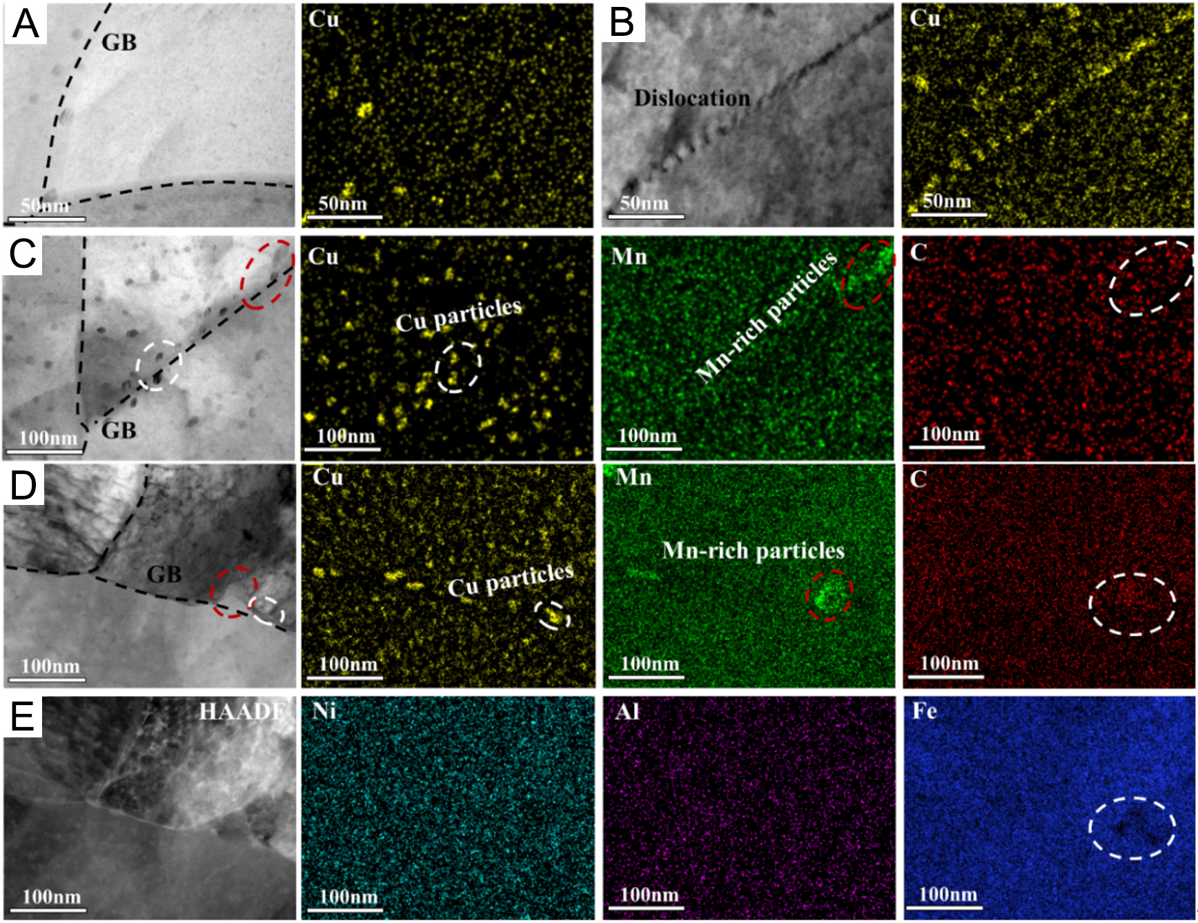
Figure 3. STEM bright-field images and energy-dispersive X-ray spectroscopy maps of Cu, Mn, C, Ni, Al and Fe in samples aged for 60 min: (A and B) Cu-0Mn steel, (C) Cu-4Mn steel, (D) CuNiAl-4Mn steel, and (E) the corresponding high-angle annular dark-field (HAADF) image of (D).