


fig3
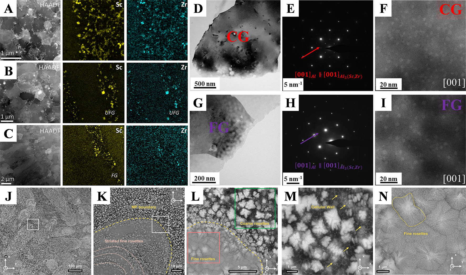
Figure 3. Heterogeneous microstructures induced by phase distribution. (A-C) Scanning transmission electron microscopy (STEM) and corresponding energy dispersive spectroscopy (EDS) maps of Sc and Zr elements in different regions of L-PBFed Al-Mg-Mn-Sc-Zr alloy, showing the uneven distribution of the L12-ordered Al3(Sc,Zr) nanoparticles[51]. (D) Bright-field (BF)-TEM image from the CG region of the L-PBFed Al-Mg-Sc-Zr alloy during direct aging; (E) The corresponding selected area electron diffraction (SAED) pattern; (F) High-angle annular dark field (HADDF)-STEM image; (G) BF-TEM image in FG region; (H) The corresponding SAED pattern; (I) HADDF-STEM image[71]. (J) Microstructure on the horizontal view of Al92Ti2Fe2Co2Ni2 alloy produced via L-PBF; (K) Enlarged region outlined in the white box of (J) showing a representative melt pool micrograph; (L) Magnified region outlined in the white box of (K) showing the microstructure across the melt pool boundaries; (M) High-magnification SEM micrograph of the coarse rosettes region taken from the green box of (L); (N) High-magnification SEM micrograph of the fine rosettes region taken from the pink box of (L)[72].


