


fig6
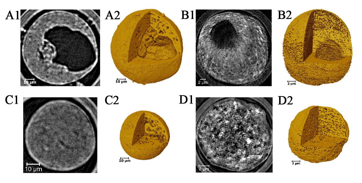
Figure 6. Multiscale CT images of four single catalyst particles: (A) and (B) acquired by micro-CT with a pixel size of 325 nm, and (C) and (D) acquired by nano-CT with a pixel size of 45 nm. (A1-D1) Central x-y plane slices of the four catalyst particles, revealing a significant amount of internal porosity; (A2-D2) 3D images of the four catalyst particles, demonstrating the morphology and internal structure, clearly distinguishing between solid and hollow particles. CT: Computed tomography.


