


fig5
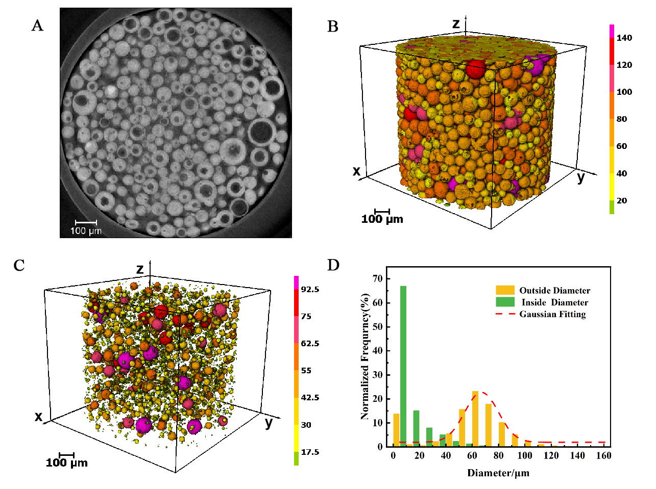
Figure 5. Micro-CT (650 nm/pixel) analysis of multiparticle catalyst structure. (A) Slice from the transverse x-y plane of the original data; (B) 3D reconstruction image of the multiparticle catalyst, showing the spheroid shape of the particles; (C) A detailed view of the central large pore within a representative hollow catalyst particle, highlighting the complex pore structure. The color of the particles and pore structure corresponds to the equivalent diameter on the scale to distinguish the particles of different sizes; (D) Normalized frequency distribution diagram of catalyst particle outer diameter and hollow particle inner diameter, showing the particle size and hollow degree of catalyst. CT: Computed tomography.


