



fig2
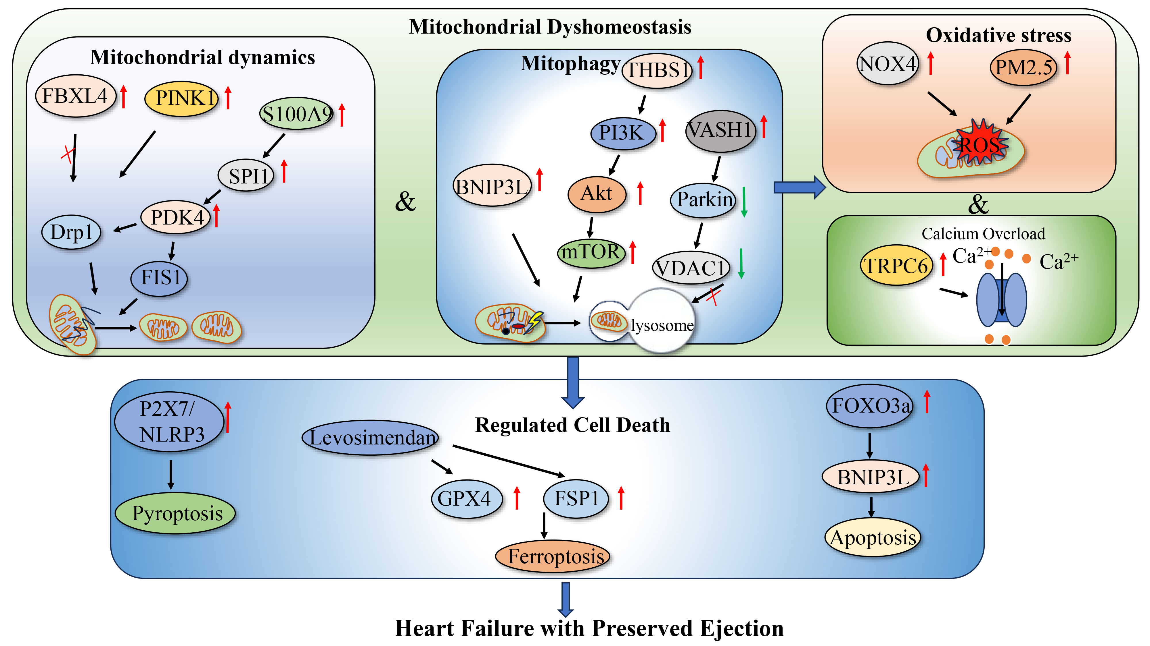
Figure 2. Mitochondrial Dyshomeostasis in HFpEF: Mechanisms and Pathogenesis. Schematic illustrating the major mechanisms linking mitochondrial dyshomeostasis to myocardial injury in HFpEF. Disrupted mitochondrial dynamics and defective mitophagy promote the accumulation of damaged mitochondria, while oxidative stress and Ca2+ overload further impair mitochondrial function and activate regulated cell death, including pyroptosis, ferroptosis, and apoptosis. These interconnected processes contribute to myocardial remodeling and diastolic dysfunction. In the schematic, red upward and green downward arrows indicate increased and decreased expression or activity, respectively. Solid arrows indicate regulatory relationships or signaling direction. Drp1: Dynamin-related protein 1; FBXL4: F-box and leucine-rich repeat protein 4; PINK1: PTEN-induced kinase 1; BNIP3L: BCL2/adenovirus E1B 19-kDa interacting protein 3-like; VASH1: vasohibin 1; VDAC1: voltage-dependent anion channel 1; NOX4: NADPH oxidase 4; ROS: reactive oxygen species; TRPC6: transient receptor potential canonical 6; mPTP: mitochondrial permeability transition pore; NLRP3: NOD-like receptor family pyrin domain-containing 3; GPX4: glutathione peroxidase 4; FSP1: ferroptosis suppressor protein 1; FOXO3a: forkhead box O3a; BNIP3: BCL2/adenovirus E1B 19-kDa interacting protein 3; HFpEF: heart failure with preserved ejection fraction; PDK4: pyruvate dehydrogenase kinase 4; PI3K: phosphoinositide 3-kinase; mTOR: mammalian target of rapamycin; NOX4: NADPH oxidase 4; SPI1: spi-1 proto-oncogene; FIS1: mitochondrial fission 1 protein; THBS1: thrombospondin 1; Akt: protein kinase B.




