



fig2
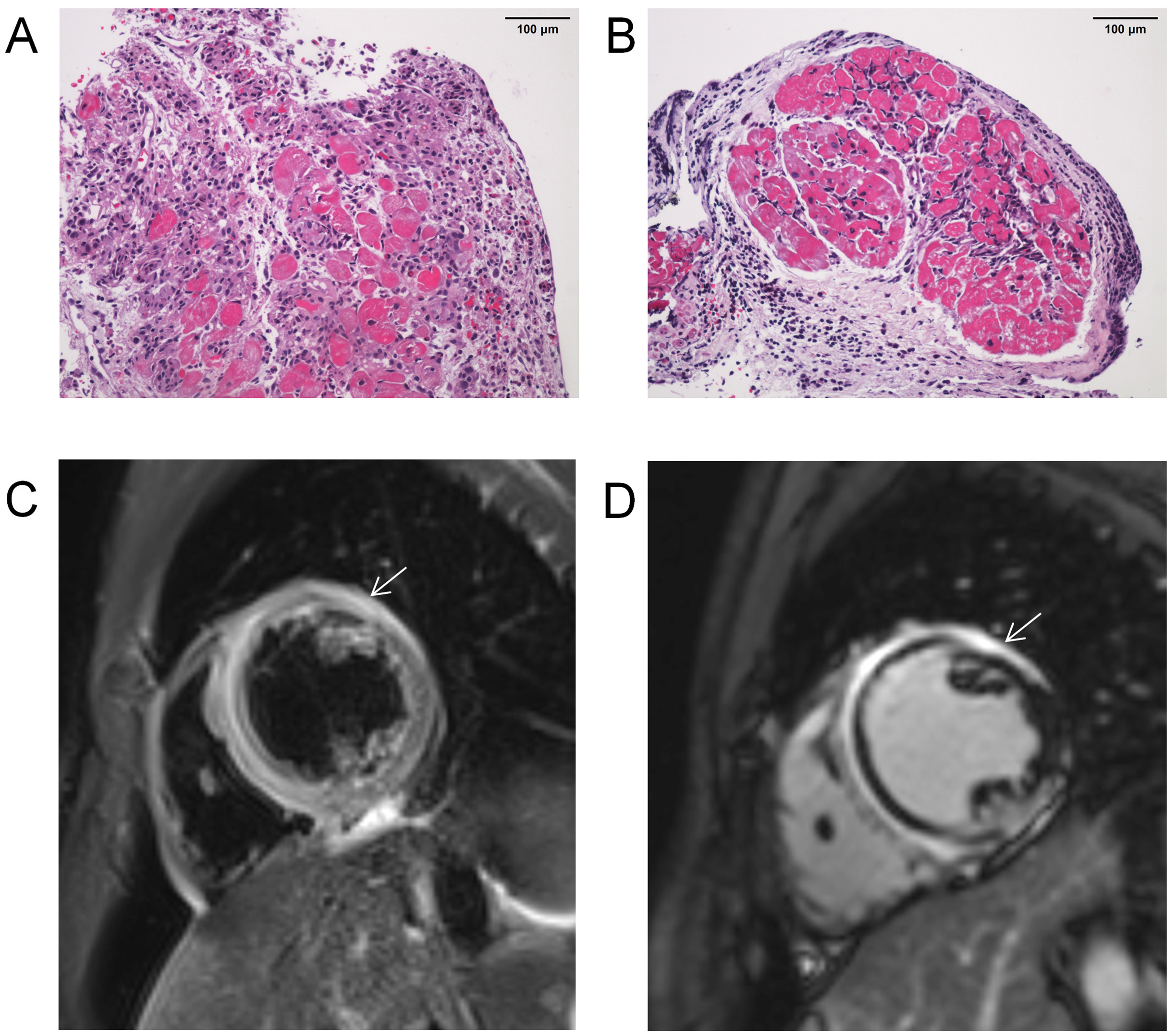
Figure 2. Representative pathological and CMR images from cases with FM. (A and B) Hematoxylin and eosin (H&E) staining of EMB specimens showing cardiac inflammatory infiltration (200×), scale bar = 100 μm; (C) T2-weighted imaging demonstrating marked myocardial edema (white arrow); (D) LGE imaging revealing myocardial injury (white arrow). CMR: Cardiac magnetic resonance imaging; LGE: late gadolinium enhancement; FM: fulminant myocarditis.