


fig1
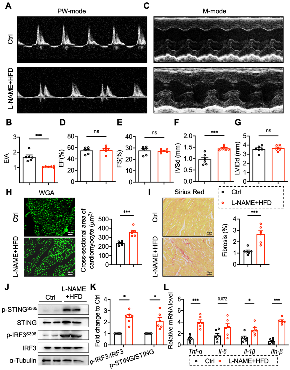
Figure 1. STING was activated in the heart tissues of HFpEF mice. (A and B) PW mode analysis of early to late diastolic flow velocity ratio (E/A); (C-G) M-mode analysis of left ventricular ejection fraction (EF) and fractional shortening (FS); (H) Representative image and analysis of WGA staining; (I) Representative image and analysis of Sirius Red staining; (J and K) Protein and phosphorylated levels of STING (Ser365) and IRF3 (Ser396) in cardiac tissues; (L) mRNA levels of TNF-α, IL-6, IL-1β and IFN-β in cardiac tissues. Mean ± SEM; n = 6 in each group, *P < 0.05, ***P < 0.001, ns, no significance. Student’s t-test for 1B-I, 1K-L. STING: Stimulator of interferon genes; HFpEF: heart failure with preserved ejection fraction; WGA: wheat germ agglutinin; mRNA: messenger RNA; SEM: standard error of the mean; TNF-α: tumour necrosis factor; IL-1β: interleukin-1β; IFN-β: interferon-β; IL-6: interleukin-6; PW: pulsed wave; IRF3: interferon regulatory factor 3; L-NAME: Nω-nitro-L-arginine methyl ester; HFD: high-fat diet.


