




fig3
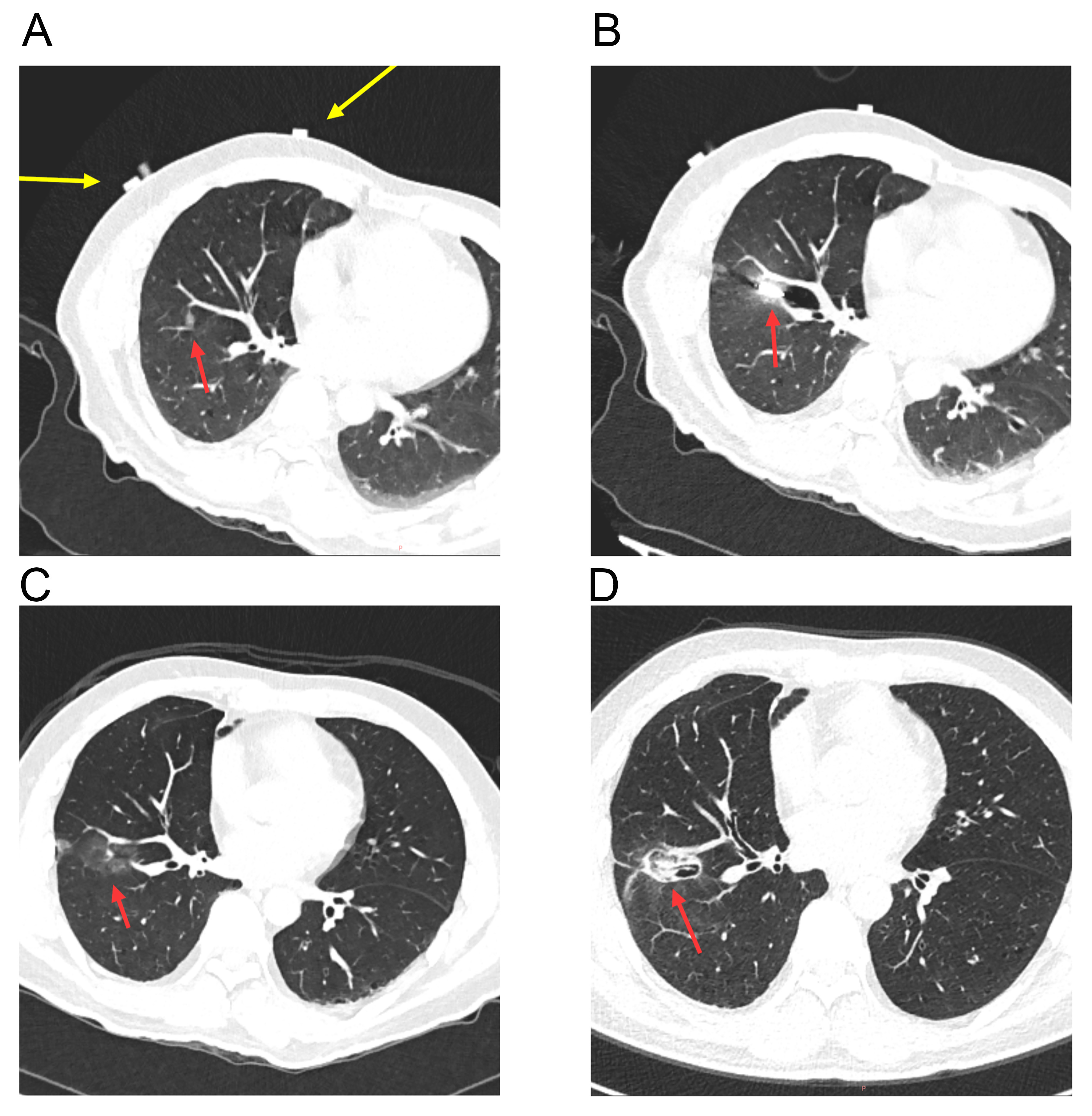
Figure 3. A robotic-assisted navigation system-guided MWA case. (A) The nodule is located in the right lower lobe. The yellow arrow indicates the CT visualization of the body surface positioning belt of the robotic-assisted navigation system, and the red arrow indicates the target nodule; (B) The puncture needle reaches the target in one pass, and MWA is initiated; (C) Immediate postoperative CT scan shows a clear ablation zone, with no pneumothorax or hemorrhage observed; (D) One-month follow-up CT after ablation reveals shrinkage and fibrosis of the ablation zone. MWA: Microwave ablation; CT: computed tomography.