

Synergistic regenerative rehabilitation for osteoarthritis: from biochemical microenvironment remodeling to multimodal physio-biological coupling
 0
0 Abstract
Osteoarthritis (OA) is a prevalent degenerative joint disease characterized by a complex dual imbalance of both the biochemical and biomechanical microenvironment. Conventional monotherapies targeting a single pathological aspect are often insufficient to achieve long-term functional joint restoration. In recent years, the integrated regenerative rehabilitation strategy, combining biological repair with physical rehabilitation, has emerged as a crucial direction in OA research. This review systematically evaluates recent advances in integrating biological repair with physical rehabilitation for OA management. First, we examine bioactive materials and cell/exosome-derived therapies as foundational tools to reconstruct the pathological biochemical niche, focusing on immune-metabolic regulation and ROS scavenging. Subsequently, we highlight the pivotal role of exogenous physical modalities - including optical, acoustic, and electromagnetic stimulations - as dynamic triggers. These modalities not only enable the spatiotemporally controlled release of therapeutics from smart hydrogels but also guide stem cell differentiation and matrix deposition via mechanotransduction. Furthermore, by evaluating recent animal studies and clinical trials, we objectively analyze the practical application outcomes of this combinatorial strategy. Finally, we critically discuss the current translational bottlenecks, aiming to provide a valuable reference for clinical treatment and subsequent research of osteoarthritis. This synergistic strategy represents a highly promising approach to shift OA treatment from passive symptom relief to active functional reconstruction.
Keywords
INTRODUCTION
Osteoarthritis (OA) is currently one of the most common degenerative joint diseases, pathologically characterized by articular cartilage degeneration, chronic synovial inflammation, and subchondral bone sclerosis[1-3]. Driven by the accelerating global aging process, rising obesity rates, and the frequent occurrence of sports injuries, the incidence of OA continues to climb, making it the leading cause of chronic pain and motor dysfunction among middle-aged and elderly populations[3,4]. According to the Global Burden of Disease study, there are over 500 million OA patients worldwide, with knee osteoarthritis being particularly prevalent[4]. OA not only severely compromises the long-term quality of life of patients but also imposes a profound economic burden on global healthcare systems. Currently, traditional clinical interventions for OA primarily focus merely on pain relief and short-term symptomatic management, with conventional approaches including oral nonsteroidal anti-inflammatory drugs (NSAIDs), intra-articular injections of glucocorticoids, and end-stage joint replacement surgery[5,6]. However, these conventional conservative treatments often fail to fundamentally reverse the cartilage degeneration process and cannot achieve the long-term restoration of joint biological functions. Therefore, exploring novel interventional strategies capable of realizing in situ regeneration and functional reconstruction of damaged cartilage has become a core objective in current OA research.
The intractable nature of OA stems from its highly complex pathological microenvironment, which is fundamentally driven by a vicious cycle of biochemical and biomechanical imbalances. Biochemically, OA is widely recognized as an immune-mediated disease accompanied by chronic inflammation. The polarization imbalance of synovial macrophages triggers an excessive accumulation of reactive oxygen species (ROS), which induces mitochondrial DNA damage, lipid peroxidation, and subsequent chondrocyte senescence via the p53/p21 and nuclear factor - κB (NF-κB) signaling axes[7-14]. This inflammatory cascade is further exacerbated by a severe hypoxic microenvironment, which synergistically disrupts the metabolic homeostasis of intra-articular cells[15,16]. Crucially, from a biomechanical perspective, articular cartilage is a specialized load-bearing tissue highly dependent on mechanical stimulation for matrix synthesis. In advanced OA, pathological mechanical stress and the loss of physiological hydrostatic pressure directly impair chondrocyte mechanotransduction. This dual deterioration - where biochemical toxicity couples with biomechanical collapse - renders conventional single-target biological therapies largely ineffective, as they fail to reconstruct a holistic pro-regenerative niche.
Before examining specific technical strategies, it is imperative to clearly define the conceptual framework of “Regenerative Rehabilitation” and its underlying “physio-biological coupling” mechanisms in the context of OA. Historically, biomaterial-assisted OA therapies have predominantly focused on remodeling the biochemical niche statically, whereas mechanobiology explores the fundamental principles of cellular sensing of physical forces. The Regenerative Rehabilitation paradigm transcends both by providing an integrated, bidirectional framework. Within this paradigm, physio-biological coupling is defined not merely as a mere arithmetic sum of physical therapy and biological materials (1 + 1 = 2), but rather as a non-linear, synergistic integration (1 + 1 > 2). In true coupling, physical cues do not merely function as passive triggers for stimulus-responsive drug delivery; instead, they operate as active, dynamic modulators that fundamentally alter the cellular response to the biomaterial. For example, mechanical loading or ultrasound can activate specific mechanosensors (e.g., Piezo1, integrins), thereby sensitizing chondrocytes or stem cells to the biochemical cues presented by the 3D hydrogel matrix. Therefore, a truly “optimized” biomechanical environment in OA entails not only the macroscopic restoration of joint loading distribution but also the microscopic normalization of mechanosensitive ion channel activity to favor matrix anabolism over catabolism.
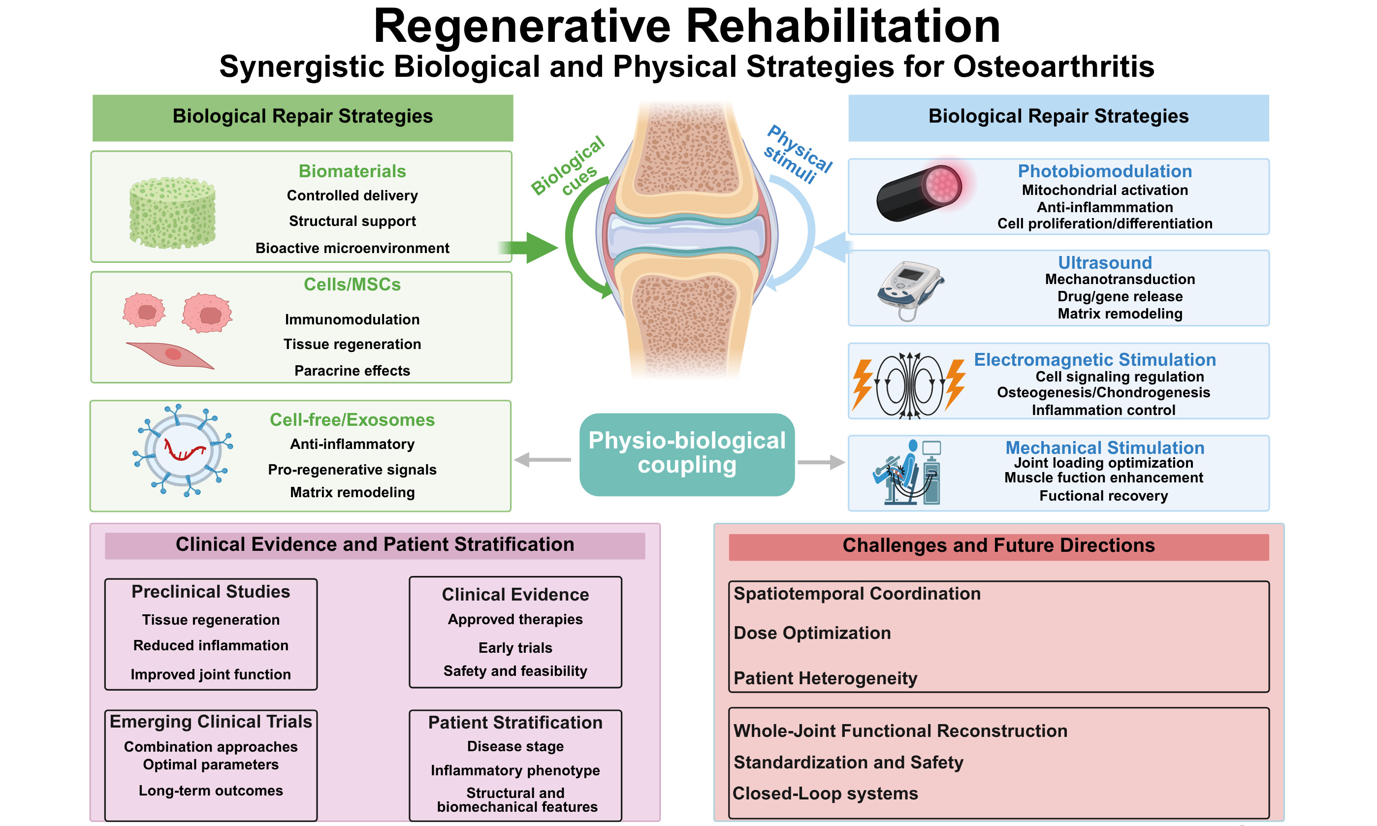
To address these multifaceted pathological challenges, the profound intersection of regenerative medicine and physical medicine has catalyzed the emergence of the “Regenerative Rehabilitation” paradigm[17,18]. This synergistic strategy advocates the deployment of advanced biological approaches (e.g., smart hydrogels, exosomes, and mesenchymal stem cells) to remodel the biochemical niche[19-22], intricately coupled with physical rehabilitation modalities to optimize the local biomechanical environment [Figure 1]. In this review, we systematically summarize the recent advances of this integrated strategy in OA management. Specifically, we first outline the foundational role of bioactive materials in alleviating oxidative stress and immune inflammation. Subsequently, we deeply elucidate the mechanisms of multimodal physical interventions - comprising optical, mechanical, electromagnetic, and ultrasound stimulations - act as dynamic triggers for the spatiotemporal controlled release of therapeutics and regulators of mechanotransduction[23-25]. Finally, by evaluating recent preclinical and clinical studies, we highlight the translational potential of multimodal physio-biological coupling, aiming to provide a comprehensive reference for shifting OA treatment toward active, functional joint reconstruction.

Figure 1. Overview of the synergistic regenerative rehabilitation treatment for osteoarthritis. Created in BioRender. Zhao, W. (2026) https://BioRender.com/oiagtll
APPLICATION OF BIOLOGICAL REPAIR STRATEGIES IN OSTEOARTHRITIS TREATMENT
Biomaterial-based cartilage repair strategies
In recent years, biomaterials-particularly natural and synthetic polymer hydrogels have demonstrated significant application value in the minimally invasive intervention of OA, owing to their excellent biocompatibility, high water content, and tunable physicochemical properties[16]. Hydrogels can closely mimic the native cartilaginous extracellular matrix (ECM) microenvironment, providing physical support for cell adhesion, proliferation, and differentiation, while serving as excellent delivery vehicles for drugs, cells, and bioactive factors, and importantly, acting as an ideal responsive matrix to anchor external physical stimuli[26].
Among them, gelatin methacryloyl (GelMA) remains one of the most widely investigated naturally derived hydrogels; it not only retains favorable biocompatibility but also forms a stable three-dimensional network via photocrosslinking[27]. For instance, Li et al. developed analgesic microspheres using a composite GelMA and hyaluronic acid methacryloyl (HAMA) hydrogel as a carrier. Demonstrating an elegant biological-physical synergy, this system is triggered by ultrasound to generate a piezoelectric effect, which effectively mitigated the pathological degeneration of articular cartilage, upregulated type II collagen expression, and suppressed MMP13 activity, thereby decelerating ECM degradation and loss[28]. Furthermore, hyaluronic acid (HA), a core component of the native cartilage matrix, is widely utilized in the development of biomimetic hydrogels[29]. Wu et al. designed a self-healing, injectable, and adhesive HA-based hydrogel capable of achieving stable in situ adhesion to the cartilage surface[30]. This material not only reduces joint friction and prolongs intra-articular retention time but also synergistically promotes chondrocyte proliferation and anabolism, demonstrating the potential to delay early OA progression through the dual dimensions of biolubrication and matrix repair[30].
With the advancement of materials science, the introduction of double-network and nanocomposite hydrogels has further addressed the mechanical shortcomings of traditional hydrogels[31]. Chen et al. developed an OGPGM nanocomposite hydrogel by incorporating poly(gallic acid)-manganese (PGA-Mn) nanoparticles into an oxidized sodium alginate-gelatin network crosslinked by transglutaminase (TG)[32]. This system not only enhanced overall mechanical strength, but the incorporation of PGA-Mn also endowed the material with the capability to scavenge local ROS, exhibiting therapeutic efficacy in reducing osteophyte formation and protecting cartilage structures in vivo[32].
Concurrently, smart-responsive hydrogels (e.g., ROS-, pH-, and near-infrared-responsive systems) have emerged as a crucial bridge connecting material science with physical rehabilitation[33]. These materials can achieve precise, on-demand drug release in response to changes in the pathological OA microenvironment or external physical triggers such as near-infrared (NIR) light, laying the structural foundation for the spatiotemporal controlled synergistic therapies discussed later in this review.
Cell-based and cell-free exosome regenerative strategies
Mesenchymal stem cells (MSCs) are considered one of the most promising cell sources for OA regenerative interventions due to their capacity for self-renewal, multipotent differentiation, and remarkable immunomodulatory properties[34-36]. Currently, bone marrow-derived MSCs (BMSCs), adipose-derived MSCs (ADSCs), and synovium-derived MSCs (SMSCs) have been extensively investigated in preclinical and clinical studies[36-38]. Mounting evidence suggests that MSCs primarily exert their therapeutic effects through paracrine mechanisms, releasing a plethora of cytokines and growth factors that promote chondrocyte proliferation and effectively suppress local inflammatory responses[39-41]. For instance, Platas et al. systematically evaluated the modulatory effects of human ADSC (hADSC)-conditioned medium on IL-1β-induced senescent characteristics in OA chondrocytes[42]. The results demonstrated that the conditioned medium could effectively reverse the senescent phenotype of chondrocytes by upregulating Sirt1 expression and inhibiting both the p53/p21 signaling axis and oxidative stress, thereby exerting a definitive chondroprotective effect[42].
To circumvent the rapid clearance and compromised engraftment of free stem cells within the joint cavity, biomaterial-mediated microenvironmental remodeling has been extensively employed; nevertheless, orchestrating the terminal functional maturation of these cells inherently necessitates the synergistic integration of dynamic biomechanical cues. Huang et al. developed a modular hydrogel niche (PPT hydrogel) integrating microenvironment detoxification and cell fate redirection functions[43]. By scavenging ROS under pathological conditions and releasing anti-inflammatory factors, this system significantly prolonged the intra-articular retention time of BMSCs and maintained their stemness. Concurrently, the sustained release of kartogenin (KGN) precisely directed the differentiation of BMSCs toward hyaline cartilage, preventing the formation of fibrocartilage or hypertrophic cartilage typically induced by the inflammatory microenvironment[43]. Furthermore, efficient delivery based on microcarriers has demonstrated unique advantages. Peng et al. utilized gelatin microspheres as 3D culture matrices, which not only achieved efficient enzyme-free in vitro expansion of ADSCs but also significantly enhanced their paracrine secretion of key repair factors[44]. This microsphere delivery system synergistically exerted the effects of suppressing inflammation, attenuating matrix degradation, and reducing joint friction, realizing highly efficient therapy with a low stem cell dosage[44].
Recently, extracellular vesicles (e.g., exosomes) have emerged as a research hotspot in “cell-free” therapeutics. MSC-derived exosomes can directly deliver anti-inflammatory cytokines, growth factors, and regulatory non-coding RNAs to damaged cells, exhibiting comparable or even superior potential to their parent cells in reducing joint inflammation and enhancing in situ cartilage repair[45-47]. However, direct intra-articular injection of free exosomes remains limited by the bottleneck of rapid clearance. Integrating exosomes with advanced biomaterials has become an essential approach to achieve their long-term controlled release[48]. Zhang et al. verified the functional complementarity in the miRNA transcriptomes of BMSC-derived exosomes and cartilage progenitor cell-derived exosomes (the former prioritizing anti-inflammation, the latter favoring pro-chondrogenesis and anti-apoptosis)[49]. Based on this, they utilized microfluidic technology to precisely fabricate HAMA hydrogel microspheres loaded with a dual-source exosome combination. This microfluidic microsphere system achieved the long-term sustained release of active components and multi-target synergistic interventions within the joint cavity, significantly improving cartilage structure and alleviating OA symptoms[49]. Similarly, Deng et al. loaded self-assembled bifunctional targeted exosomes into decellularized cartilage extracellular matrix (HECM) microgels via a single-step method[50]. This delivery platform can precisely target and accumulate in damaged cartilage regions, regulating cellular metabolism via the PI3K/AKT pathway to safely and efficiently decelerate OA progression[50].
In summary, whether through material-based interventions utilizing functionalized polymer networks or biological interventions relying on cells or derived vesicles, these strategies demonstrate an irreplaceable role in remodeling the biochemical OA microenvironment. The core mechanistic features of various biological repair vehicles, along with the specific technical bottlenecks encountered toward clinical translation (e.g., standardized preparation and long-term spatiotemporal controlled release), are systematically compared and summarized in Table 1. However, these purely biological or material-based approaches remain inherently static. To overcome the limitations of passive release and to address the pathological biomechanical deterioration of the joint, integrating these bioactive carriers with active physical rehabilitation modalities has become an inevitable evolutionary step. The profound synergy of this Regenerative Rehabilitation paradigm will be detailed in the following section.
Quantitative and mechanistic evaluation of biological repair strategies in osteoarthritis
| Treatment strategy | Classification | Specific interventions & carriers | Quantitative dosing / parameters | Evaluation model & duration | Efficacy readouts / outcomes | Refs. |
| Biomaterials | Piezoelectric Hydrogel | Ultrasound-responsive BaTiO3-RVG@GA/HA microspheres + LIFU | 2 mg/mL BaTiO3; US: 1 MHz, 0.5 W/cm2, 5 min | ACLT rat model, 8 weeks | Significant Nav1.7 blockade; 60% reduction in MMP13; Sustained analgesia | [28] |
| Adhesive Hydrogel | AHA/CHA self-healing hydrogel via dihydrazide/catechol modification | 50 μL injection volume; Friction coefficient reduced to ~ 0.02 | DMM murine model, 8 weeks | Downregulation of THBD pathway; Enhanced ECM boundary lubrication; OARSI score decreased by 40% | [30] | |
| Nanocomposite Hydrogel | PGA-Mn nanoparticles + oxidized sodium alginate-gelatin | PGA-Mn (0.5-2.0 wt.%); Gelation time < 3 min | MIA-induced OA rat model, 6 weeks | 80% local ROS scavenging efficiency; Significant reduction in osteophyte formation | [32] | |
| Cell therapy | MSC Secretome | Human AMSC-conditioned medium for chondrocyte senescence | 50% conditioned medium concentration in vitro | IL-1β-induced OA chondrocytes, 48 h | Upregulation of SIRT1 by 2.5-fold; Significant decrease in p53/p21 senescence markers | [42] |
| BMSC niche | Thermosensitive hydrogel co-delivering BMSCs and Kartogenin (KGN) | 1 × 10⁶ BMSCs/mL; KGN sustained release over 28 days | DMM rat model, 12 weeks | Restoration of Hedgehog signaling; robust hyaline cartilage regeneration (Col II/I ratio > 3) | [43] | |
| ADSC microcarriers | ADSCs cultured on 180 μm gelatin microcarriers | 1 × 10⁵ cells (10-fold lower dose than free cells) | ACLT rat model, 8 weeks | Enhanced paracrine secretion; physical ball-bearing lubrication reducing joint friction | [44] | |
| Exosomes | Dual-Exosome | BMSC-Exo + CPC-Exo loaded in HAMA hydrogel microspheres | 100 μg total exosome protein/mL hydrogel | DMM murine model, 8 weeks | Synergistic macrophage polarization (M2) and chondrocyte apoptosis inhibition | [49] |
| Biomimetic microgels | Bifunctional targeted exosomes in decellularized cartilage matrix (HECM) | 50 μg exosomes in 2% HECM microgels | DMM rat model, 8 weeks | Specific targeting via PI3K/AKT pathway; highly localized matrix production. | [50] |
MULTIMODAL PHYSIO-BIOLOGICAL COUPLING: SYNERGISTIC MECHANISMS AND FUNCTIONAL INTEGRATION
While the bioactive materials and derived vesicles discussed in Section APPLICATION OF BIOLOGICAL REPAIR STRATEGIES IN OSTEOARTHRITIS TREATMENT establish a robust biochemical foundation for OA treatment, their static nature limits their adaptability to the progressively deteriorating biomechanical niche of the joint. To achieve effective Regenerative Rehabilitation, exogenous physical modalities must be integrated as active drivers rather than passive adjunctive therapies. Rather than categorizing these interventions merely by their physical origins (e.g., optical, mechanical, acoustic, or electromagnetic), it is more mechanistically rigorous to classify them based on their mode of physio-biological coupling. In this section, we systematically explore how multimodal physical interventions synergize with smart biomaterials through four distinct functional paradigms: (1) stimulus-triggered spatiotemporal release, (2) mechanotransduction and biomechanical niche remodeling, (3) remote deep-tissue regulation, and (4) self-powered mechanoelectrical systems. To provide a comprehensive overview of this framework, Table 2 systematically compares and summarizes the key physical parameters, core biological mechanisms, primary therapeutic targets, and safety limitations of these diverse physical modalities.
Comparative analysis of physical modalities in regenerative rehabilitation
| Physical modality | Typical parameters | Core biological mechanisms | Primary therapeutic targets | Safety considerations & limitations | Refs. |
| Photobiomodulation (PBM) | NIR (650-1,064 nm); Low power density (< 500 mW/cm2) | Photon absorption by mitochondrial cytochrome c oxidase; ATP synthesis; Intracellular NO release | Mitochondrial metabolism; inflammatory regulation; chondrocyte and immune-cell responses | Generally favorable safety profile; limited penetration into deep tissues may restrict efficacy in large joints | [52] |
| Photothermal therapy (PTT) | NIR irradiation, typically 808 nm; photothermal nanomaterials; controlled local temperature elevation | Hotothermal conversion generates moderate localized hyperthermia; temperature-responsive drug release; regulation of cellular stress responses and ROS metabolism | Spatiotemporal drug release; ROS regulation; chondrocyte protection and regeneration | Excessive heating may cause thermal injury or protein denaturation; precise control of irradiation dose and temperature is required | [58] |
| Focused ultrasound (Cavitation) | Focused high-intensity acoustic waves; sonosensitive carriers or responsive chemical bonds | Acoustic cavitation and mechanical forces induce bond cleavage, carrier disruption, or transient sonoporation, enabling on-demand therapeutic release | Stimulus-responsive drug release; enhanced local therapeutic delivery; increased cellular uptake | Requires strict control of acoustic pressure and exposure parameters to avoid excessive cavitation and tissue damage | [60] |
| Pulsed electromagnetic fields (PEMF) | Low frequency (1-100 Hz); Low intensity (0.1-5 mT) | Modulates cell membrane receptors (e.g., Adenosine A2A/A3); Regulates Wnt/β-catenin and MAPK pathways | Inflammation control; pain relief; extracellular matrix preservation; chondrogenic regulation | Non-invasive with good tissue penetration; clinical efficacy may vary according to disease phenotype, treatment parameters, and patient characteristics | [71] |
| Low-intensity pulsed ultrasound (LIPUS) | Frequency approximately 1-3 MHz; low acoustic intensity; pulsed exposure | Acoustic micro-mechanical stimulation and mechanotransduction; activation of mechanosensitive signaling; regulation of YAP/RIPK1/NF-κB and autophagy | Chondrocyte homeostasis; cartilage matrix metabolism; suppression of OA progression | Generally non-destructive and suitable for deep-tissue stimulation; therapeutic effects remain dependent on acoustic parameters and treatment duration | [83] |
| Mechanoelectrical Conversion (Piezoelectric) | Driven by physiological gait frequency (approx. 1 Hz) and compressive load | Conversion of mechanical deformation into local electrical/streaming potentials; modulation of cellular electrophysiological and mechanotransduction signaling | Chondrocyte matrix anabolism; osteochondral regeneration; restoration of the local electromechanical niche | Long-term mechanical fatigue, material degradation, and potential wear debris require evaluation; some systems may require implantation | [87] |
Stimulus-triggered spatiotemporal release: optical and acoustic triggers
Among various physical rehabilitation interventions, optical stimulation has garnered substantial attention in the adjunctive treatment of OA due to its non-invasive nature and favorable tissue penetration depth. Particularly, NIR light is extensively utilized in cartilage tissue regeneration and local inflammation regulation studies, owing to its low phototoxicity and excellent spatiotemporal controllability[51]. When strategically coupled with photoresponsive biomaterials, NIR light transitions from a simple physical therapy to a highly precise spatiotemporal switch. Accumulating evidence indicates that NIR irradiation at specific wavelengths (typically 650-1,064 nm) can modulate various cellular processes via photobiomodulation. In the integration of these stimuli with smart-responsive biomaterials, it is mechanistically crucial to distinguish between two optical interventions: photobiomodulation (PBM) and photothermal therapy (PTT).
PBM relies on direct photon absorption by mitochondrial chromophores (e.g., cytochrome c oxidase) without significant temperature changes. This process promotes mitochondrial ATP synthesis, suppresses the secretion of pro-inflammatory cytokines (e.g., TNF-α, IL-6, and IL-1β) by immune cells[52,53], and triggers intracellular nitric oxide (NO) release via photodissociation. As a pivotal vasoactive molecule, free NO diffuses into vascular smooth muscle cells, inducing microvascular dilation that improves local blood circulation, alleviates joint pain, and accelerates tissue repair[54-56].
In contrast, PTT utilizes photothermal agents integrated within biomacromolecular systems to convert NIR light into localized heat. For instance, Hong et al. developed a photoresponsive system utilizing gold-core platinum-shell (Au@Pt) nanospheres; under 808 nm NIR irradiation, this material achieves efficient photothermal conversion to promote chondrocyte proliferation and catalytically degrade abnormally accumulated ROS[57]. Furthermore, Chen et al. engineered a cartilage-adhesive microsphere system incorporating MXene nanosheets for the photothermally controlled release of KGN[58]. The induced moderate thermal stress (approximately 42 °C) drives a precise phase transition to accelerate KGN release while concurrently upregulating cellular heat shock proteins (e.g., HSP70) to maintain protein homeostasis. Following this NIR-triggered KGN release, BMSCs demonstrated chondrogenic differentiation in vitro [Figure 2A], accompanied by increased expression of cartilage-specific markers such as COL-2a and SOX-9 [Figure 2B]. Furthermore, in vivo radiographic assessments in an osteoarthritis rat model indicated that this combined approach helped mitigate osteophyte formation and maintain joint space width [Figure 2C]. Similarly, Shi et al. developed a NIR-responsive system utilizing polyoxometalate (POM) nanoclusters[59]. Based on their mechanistic design and in vitro fluorescence evaluations, the moderate photothermal effect induced by NIR irradiation improved the ROS-scavenging capability of the POM clusters. This temperature-enhanced antioxidant activity consequently aided in neutralizing excessive intracellular ROS in chondrocytes under inflammatory conditions[59].

Figure 2. Optical and ultrasound-responsive delivery systems for targeted osteoarthritis therapy. (A) Schematic illustration of NIR-induced chondrogenic differentiation; (B) Western blot analysis of COL-2a and SOX9 protein levels; (C) X-ray analysis of joint space width and osteophyte formation. (A-C) are reprinted from Ref.[58] with permission; (D) P(AzoAA24-stat-MAA10) polymer was self-assembled into nanobowls. Afterward, MnO2 nanoparticles were grown in situ onto nanobowls; (E) Mn-nanobowls motion images captured at different ultrasound amplitudes (5,10,15, and 20 Vp-p) in 5 s; (F) The average velocity of Mn-nanobowls was analyzed from the tracking trajectory at different ultrasound amplitudes (n = 3); (G) Penetration assay of ultrasound-propelled Mn-nanobowls in rat knee joints. (D-G) are reprinted from Ref.[61] with permission. BMSC: Bone marrow-derived mesenchymal stem cell; NCA: nano cationic amylose; KGN: kartogenin; COL-2a: Collagen type II alpha-1; SOX9: SRY-box transcription factor 9; NIR: near-infrared.
Beyond optical triggers, which can be limited by tissue penetration depth, ultrasound provides a robust acoustic trigger for deep weight-bearing joints. Ultrasound-induced stable cavitation can mechanically disrupt sonosensitive chemical bonds within the carrier, enabling the precise spatiotemporal release of pre-loaded therapeutics. Simultaneously, transient sonoporation safely and reversibly increases the permeability of the cellular membrane, substantially enhancing the intracellular uptake of exogenous therapeutic macromolecules. For instance, Zhou et al. utilized ultrasound to precisely trigger the cleavage of Se-Se bonds in a hyaluronic acid-diselenide (HA-Se) hydrogel, enabling the on-demand release of active selenium[60]. Similarly, Gong et al. developed MnO2-decorated asymmetrical nanobowls that utilize ultrasound for active propulsion [Figure 2D][61]. According to their mechanistic design and in vitro trajectory evaluations, acoustic stimulation enables these nanocarriers to achieve directional movement with controllable velocity [Figure 2E and F]. Furthermore, in vivo penetration assays indicated that this ultrasound-driven motion assists the nanobowls in overcoming the dense extracellular matrix barrier, allowing them to reach deeper cartilage regions to scavenge ROS [Figure 2G][61]. These mechanisms transform acoustic mechanical energy into a precise biochemical delivery trigger.
Mechanotransduction and biomechanical niche remodeling
As a highly specialized weight-bearing tissue, articular cartilage exhibits a profound dependence on external mechanical stimulation for phenotype maintenance. Therefore, mechanical rehabilitation is not a macroscopic adjunct, but a microscopic necessity for cell fate determination. Moderate compressive stress, shear stress, or hydrostatic pressure have been well documented as essential physical prerequisites for promoting the chondrogenic differentiation of stem cells and upregulating ECM secretion[24]. Under physiological conditions, appropriate mechanical loading triggers biochemical signaling cascades that enhance the anabolic activity of chondrocytes, thereby preserving the structural integrity of articular cartilage[62]. Conversely, abnormal or excessive biomechanical stress induces cartilage degeneration and local inflammation[63]. Therefore, guided by the integrated regenerative rehabilitation paradigm, mechanical stimulation-based rehabilitation strategies such as in vivo rehabilitative exercise training and in vitro bioreactor loading are extensively employed to simulate and remodel a healthy mechanical microenvironment within the joint cavity. This approach accelerates cartilage regeneration and enhances the mechanical adaptability of the nascent tissue[64,65]. At the microscopic level, mechanical rehabilitation primarily relies on mechanotransduction pathways to translate physical cues into biochemical signals. When chondrocytes or MSCs are exposed to dynamic compression, cell surface integrin receptors and mechanosensitive ion channels are activated, initiating robust intracellular signaling cascades.
Specifically, the activation of integrins by matrix stiffness and mechanical loading induces the phosphorylation of focal adhesion kinase (FAK), which subsequently triggers the mitogen-activated protein kinase / Extracellular-Signal-Regulated Kinase (MAPK/ERK) pathways to promote cell proliferation and matrix deposition. Simultaneously, the mechanosensitive transcriptional co-activators YAP/TAZ play an indispensable role; mechanical stress induces Yes-Associated Protein / Transcriptional Co-Activator with PDZ-binding Motif (YAP/TAZ) nuclear translocation, directing the chondrogenic differentiation trajectory of MSCs. However, abnormal pathological stress leads to deleterious mechanotransduction. Zhou et al. revealed that pathological mechanical stress aberrantly activates the Piezo1 channel on the surface of MSCs[66]. This pathological calcium influx upregulates HK2-mediated glycolysis, subsequently promoting the secretion of macrophage migration inhibitory factor (MIF) and driving Th17 cell polarization, thereby exacerbating OA progression.
To counteract this, integrating mechanical rehabilitation with advanced biomaterials offers a pathway to remodel the biomechanical niche. For example, Sun et al. developed a mechanoresponsive drug delivery system utilizing the dynamic compressive pressure generated during daily joint weight-bearing as a trigger[67]. Through in vitro dynamic compression simulations, they demonstrated that the release of the encapsulated KGN increased proportionally with the magnitude of the applied compressive strain. Furthermore, in vivo histological evaluations revealed that this mechanically triggered drug release significantly promoted functional cartilage regeneration and effectively repaired osteochondral defects in an osteoarthritis model[67]. The mechanical deformation of the F127DA micelles enables the on-demand release of KGN, converting the mechanical energy of routine activities into a biochemical driver for cartilage regeneration. Furthermore, Wang et al. demonstrated that moderate-intensity mechanical stimulation effectively activates the Transforming growth factor-β1 (TGF-β1)/Smad2/3 signaling pathway while synchronously inhibiting the NF-κB inflammatory axis, synergistically attenuating chondrocyte pyroptosis[68]. Beyond these anti-inflammatory and molecular regulatory effects, it is imperative to precisely define how the specific magnitude of such physical stimuli ultimately governs the macroscopic quality of the regenerated tissue, particularly in distinguishing between functional hyaline cartilage and inferior fibrocartilage within biomaterial scaffolds. To systematically elucidate this dose-dependent mechanotransduction, recent studies have employed three-dimensional (3D)-bioprinted alginate hydrogel models impregnated with chondrogenic cells. In efforts to mimic the physiological mechanical environment of joints during distinct ambulatory activities (e.g., walking, running, and jumping), these cell-laden constructs were subjected to dynamic cyclic compressive strains of varying magnitudes (6%, 12%, and 24%) utilizing a customized bioreactor system [Figure 3A]. This macroscopic, pure physical compressive stimulus profoundly modulated cellular phenotypes and ECM secretion within the microenvironment. Immunofluorescence analyses revealed the differential spatial deposition of Type II collagen (Col2, indicative of hyaline cartilage) versus Type I collagen (Col1, indicative of fibrocartilage) across distinct strain conditions [Figure 3B]. Crucially, subsequent quantitative assessments confirmed that moderate physical compression (12% strain) maximized the deposition of hyaline-like cartilage ECM. In sharp contrast, elevated mechanical strain (24%) significantly augmented the formation of the biologically and mechanically inferior fibrocartilage-like ECM [Figure 3C and D]. Collectively, this experimental paradigm substantiates that the precise tuning of the applied physical compressive strain magnitude on biomaterials serves as a decisive factor orchestrating the quality of cartilage regeneration, thereby transforming a passive biomechanical niche into a highly tailored, pro-regenerative microenvironment[69].

Figure 3. Synergistic interventions based on mechanical rehabilitation coupled with biomaterial/biological therapies for joint repair. (A) Representation of the macroscopic mechanical loading regimes, applying cyclic compressive strains (e.g., 12% and 24% at 1 Hz) to mimic varying intensities of joint activities; (B) (A1-D1) Staining with DAPI of different groups showed that cell populations seemed to be lower in the 12% and 24% loaded groups. (E1-H1) Col2 immunofluorescence staining confirmed chondrogenic differentiation of most of the cells for all groups. (I1-L1) Col1 immunofluorescence staining showed its deposition from most of the cells in all groups suggesting that cells were differentiated into fibrochondrocytes (scale bars = 200 µm); (C) %hyaline-like cartilage and (D) %fibrocartilage-like ECM tended to be lowest and highest, respectively, in 12% loaded group. The bar graphs show median and interquartile range. n = 4 for each group, n.s. = not significant, *P < 0.05, and **P < 0.01. The figure is reprinted from Ref.[69] with permission. ECM: Extracellular matrix; DAPI: 4',6-diamidino-2-phenylindole.
Remote regulation for deep-tissue interventions
Compared to optical interventions limited by poor tissue penetration, electromagnetic and acoustic fields offer unique advantages for deep-joint targeting. Low-frequency pulsed electromagnetic fields and low-intensity pulsed ultrasound are well-established physical modalities that exert robust non-invasive regulatory effects on deep tissues without relying on thermal damage or cavitation.
Pulsed electromagnetic field (PEMF) has been validated as an effective non-invasive intervention for OA. Its clinical benefits primarily manifest in rapid analgesia, the attenuation of synovial edema, and the long-term protection of the compromised extracellular matrix[70,71]. Extensive studies have elucidated the profound biological mechanisms of PEMF: by acting on cell membrane receptors, modulating calcium channels, and activating adenosine A2A/A3 receptors, PEMF triggers a cascade of crucial signaling pathways including Wnt/β-catenin, MAPK/ERK, TGF-β/Smad, PI3K/AKT, and NF-κB, thereby exerting comprehensive regulatory effects on cell proliferation, chondrogenic differentiation, and anti-inflammatory repair[70-74].
However, while stand-alone electromagnetic rehabilitation effectively ameliorates the intra-articular inflammatory microenvironment, it is insufficient to independently induce the in situ regeneration of cartilage for established substantial defects lacking a 3D physical anchor. To address this, integrating advanced biomacromolecular hydrogels with PEMF creates a synergistic paradigm. The hydrogel provides a stable 3D biomimetic microenvironment that seamlessly adapts to complex tissue defects and robustly supports cell colonization and matrix deposition. Concurrently, PEMF significantly enhances the chondrogenic differentiation of BMSCs and inhibits chondrocyte hypertrophy and fibrosis by specifically regulating the p38-MAPK and ERK pathways. The synergistic effect of these two modalities substantially elevates the regeneration quality of hyaline cartilage[75]. Furthermore, Deng et al. developed a system incorporating melanin-encapsulated iron oxide (melanin@Fe3O4) magnetic nanoparticles and mesenchymal stem cells in conjunction with a time-varying magnetic field (TMF)[76]. The nanoparticles endow the cells with magnetic responsiveness, along with antioxidant and anti-inflammatory properties [Figure 4A]. According to parameter optimization and morphological assessments, the application of a specific TMF non-invasively drives the ordered in situ aggregation of these magnetized cells into clusters, which aids in prolonging their retention at the target site [Figure 4B]. Subsequent in vitro immunofluorescence analyses indicated that mimicking physiological gait through magneto-mechanical stimulation activates cellular mechanotransduction pathways, thereby supporting stem cell chondrogenesis and upregulating cartilage-specific markers such as type II collagen [Figure 4C]. Moreover, macroscopic and histological evaluations, including Safranin O staining in an osteoarthritis animal model, demonstrated that this combined magneto-mechanical intervention helped mitigate cartilage degeneration and facilitated matrix preservation [Figure 4D and E][76].

Figure 4. Regenerative rehabilitation synergistic strategies mediated by external electron magnetic regulation. (A) Schematic of the Sophisticated Magneto-Mechanical Actuation for Chondrogenesis of Magcells in OA Treatment; (B) Morphologies of formed Magcell clusters under different TMF parameters; (C) Immunofluorescence of COL II protein in Magcells treated with TGF-β under TMF stimulation or not for 14 days. “MF -” and “MF +” groups represent no TMF treatment and treatment with TMF, respectively; (D) The digital photographs of the joint samples after treatment; (E) Safranin O-fast green staining. The figure is reprinted from Ref.[76] with permission. TMF: Time-varying magnetic field; COL II: Collagen type II; SOX9: SRY-box transcription factor 9; NCAD: Neural-cadherin; DAPI: 4',6-diamidino-2-phenylindole; PBS: Phosphate-buffered saline; MSC: mesenchymal stem cell; TGF-β: Transforming growth factor-β; OA: osteoarthritis.
Serving as another robust tool for deep-tissue penetration, LIPUS uniquely couples acoustic mechanical energy with biological responses[77,78]. In routine clinical OA management, LIPUS can non-destructively penetrate the skin and muscle layers to reach the deep joint cavity. It not only effectively promotes local microcirculation and accelerates the absorption of inflammatory exudates to alleviate joint swelling and pain but also decelerates chondrocyte senescence and matrix degeneration[79,80]. At the molecular level, the mechanical vibration (micro-massage effect), acoustic streaming, and stable cavitation generated by ultrasound exert high-frequency, cyclic micromechanical stress on cells within the local microenvironment. This acoustic mechanical stimulation can activate integrin receptors and various mechanosensitive ion channels on the cell membrane, inducing a transient influx of intracellular calcium ions and subsequently initiating a cascade activation of key pro-survival signaling pathways, such as MAPK/ERK and PI3K/Akt. This process not only inhibits the overexpression of inflammatory mediators (e.g., IL-6 and TNF-α) at the transcriptional level but also significantly promotes the synthesis and secretion of core matrix macromolecules, including type II collagen and aggrecan[81-83].
Self-powered mechanoelectrical systems
Beyond the application of external physical fields, reconstructing the endogenous electrophysiological microenvironment of the joint represents the most advanced frontier in OA regenerative rehabilitation. Under physiological conditions, healthy hyaline cartilage subjected to mechanical loading (e.g., compressive stress during daily walking) generates a weak streaming potential driven by the shear movement of interstitial fluid. This endogenous micro-electric field is vital for maintaining the metabolic homeostasis of chondrocytes. However, in advanced OA, the severe degradation of the cartilage matrix leads to the complete loss of this spontaneous electrophysiological microenvironment[84]. Traditional electrostimulation therapies typically rely on surgically implanted battery-powered electrodes, which are associated with high infection risks and significant clinical limitations. The introduction of piezoelectric stimulation mechanisms has effectively overcome these constraints, offering a highly promising self-powered in vivo solution for the regenerative rehabilitation strategy. The core advantage of piezoelectric materials lies in their ability to directly convert mechanical deformation into weak electrical signals[85,86].
Yu et al. constructed an injectable bifunctional hydrogel system composed of a GelMA matrix, poly(L-lactic acid) (PLLA) piezoelectric fibers, and a poly (2-methacryloyloxyethyl phosphorylcholine) (PMPC) super-lubricating surface layer[87]. Based on their biomechanical design and tribological assessments, the PLLA fibers generate in situ piezoelectric signals upon being subjected to the compressive loads induced by joint movement, while the superficial PMPC layer effectively dissipates detrimental shear stress by reducing the friction coefficient to as low as 0.0083. Consequently, in vivo histological and molecular evaluations demonstrated that the synergistic interplay of these components significantly promotes the osteogenic and chondrogenic differentiation of BMSCs, inhibits chondrocyte senescence, and facilitates high-quality osteochondral defect repair. This approach achieves an integrated therapeutic strategy combining electrical stimulation-mediated repair with physical lubrication protection [Figure 5][87]. When such smart scaffolds, which possess both 3D structural support and piezoelectric activity, are implanted into the joint cavities of OA patients, they can continuously harvest otherwise dissipated mechanical energy and convert it in situ into microvolt-level pulsed electric fields matching physiological intensities[88]. This design successfully constructs a mechano-electrical transduction closed loop of “mechanical energy-electrical signal-biological response,” directly translating the patient’s macroscopic rehabilitation movements into microscopic bioelectrical physical cues that drive cellular regeneration, thereby achieving a truly passive and self-healing synergistic intervention.
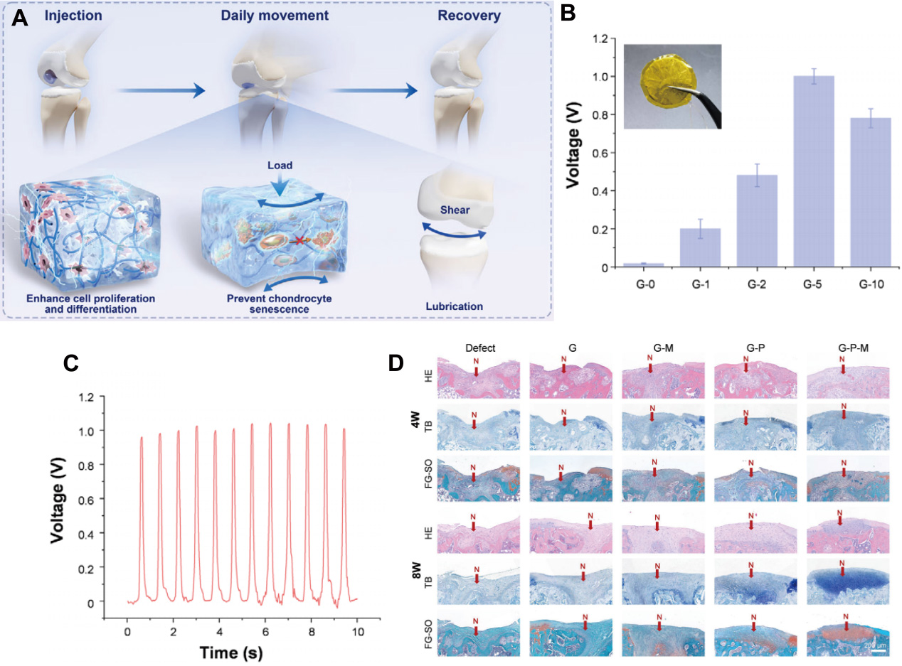
Figure 5. Regenerative rehabilitation synergistic strategies mediated by endogenous piezoelectric micro-electric fields. (A) Therapeutic mechanism in vivo: the injectable scaffold fills the cartilage defect. During daily joint movement, the surface PMPC layer provides lubrication to dissipate shear stress. Concurrently, the piezoelectric signals under compressive load synergistically prevent chondrocyte senescence and enhance cell proliferation and differentiation for osteochondral recovery; (B) Piezoelectric output of GelMA hydrogels doped with different concentrations of PLLA short fibers; (C) The voltage output; (D) Representative H&E, Toluidine Blue (TB), and Safranin O-Fast Green (SO/FG) staining images of regenerated tissues at 4 and 8 weeks (N: newly generated tissue). The figure is reprinted from Ref.[87] with permission. PMPC: PLLA:
As previously discussed, the reconstruction of the endogenous electrophysiological microenvironment heavily relies on the patient’s daily movements (e.g., walking) to provide mechanical energy. However, for late-stage OA patients with severely restricted mobility due to intense pain, patient-generated movement is often insufficient to continuously drive piezoelectric materials to generate adequate therapeutic electrical charges. Under these circumstances, deep-penetrating therapeutic ultrasound serves as an ideal “remote power source.” Vinikoor et al. developed an injectable piezoelectric hydrogel composed of piezoelectric PLLA nanofibers and a collagen matrix, utilizing external ultrasound (40 kHz) to drive the mechanical deformation of the nanofibers and generate localized piezoelectric signals in situ[89]. Without requiring batteries or implanted electrodes, this system achieves a dual physical rehabilitation intervention comprising “ultrasound mechanical energy + micro-electric field,” effectively inducing stem cell migration and promoting hyaline cartilage regeneration[89]. Similarly, Li et al. constructed targeted barium titanate piezoelectric microspheres (BaTiO3-RVG@GA/HA), integrating ultrasound and piezoelectric stimulation to precisely act on pain targets, suppress the Nav1.7 channel, and synergistically achieve analgesia, anti-inflammation, and cartilage protection[28].
PROGRESS IN ANIMAL AND CLINICAL STUDIES
Preclinical evidence supporting regenerative rehabilitation
In preclinical animal studies, destabilization of the medial meniscus (DMM), anterior cruciate ligament transection (ACLT), and chemically induced models remain the most widely used experimental models for evaluating OA interventions, although each model recapitulates only specific facets of the heterogeneous human disease[90,91]. Building on the biological repair strategies and physio-biological coupling mechanisms described in Sections APPLICATION OF BIOLOGICAL REPAIR STRATEGIES IN OSTEOARTHRITIS TREATMENT and MULTIMODAL PHYSIO-BIOLOGICAL COUPLING: SYNERGISTIC MECHANISMS AND FUNCTIONAL INTEGRATION, numerous recent studies have demonstrated that biomaterials can be combined with mechanical, acoustic, optical, or electromagnetic stimuli to regulate cartilage homeostasis, inflammatory signaling, extracellular matrix metabolism, and pain-related pathways[28,57-59,66,75].
Importantly, however, most of these studies remain preclinical and are conducted under highly controlled experimental conditions. The biological response observed in rodents may not fully predict the response of human weight-bearing joints, particularly because human OA is characterized by substantial heterogeneity in disease severity, synovial inflammation, obesity/metabolic status, joint alignment, gait loading, pain sensitization, and psychosocial factors. Therefore, the major translational question is no longer simply whether biological materials or physical stimulation can elicit therapeutic benefits in animal models, but rather which patients, at which disease stage, and under which physical and biological conditions are most likely to benefit from this combinatorial approach.
Clinical evidence for biological repair and physical rehabilitation
Clinical evidence for the individual components of regenerative rehabilitation has increased substantially, although direct clinical validation of the complete physio-biological coupling paradigm remains limited. Current clinical guidelines consistently identify therapeutic exercise as a core component of OA management, whereas pharmacological and intra-articular interventions are generally used to control symptoms and facilitate participation in rehabilitation[92,93]. The 2019 American College of Rheumatology/Arthritis Foundation guideline strongly recommends exercise for patients with knee, hip, and hand OA, while emphasizing that multiple forms of exercise can be beneficial and that there is insufficient evidence to identify a single universally optimal exercise prescription[92]. Similarly, the NICE guideline recommends individually tailored therapeutic exercise for all patients with OA and suggests supervised exercise when appropriate[93].
Recent randomized evidence further underscores the importance of individualized physical rehabilitation rather than a single standardized exercise modality. In a randomized clinical trial involving 117 adults with knee OA, both yoga and strengthening exercise produced clinically meaningful improvements in knee pain over 12 weeks. Notably, yoga was found to be noninferior to strengthening exercise, with a between-group difference in visual analogue scale (VAS) pain change of -1.1 mm (95% CI, -7.8 to 5.7 mm). These findings support the broader concept that physical rehabilitation should be prescribed according to patient preference, functional status, adherence, and mechanical phenotype rather than assuming that one specific exercise modality is optimal for every patient[94].
Clinical translation of cell-based biological repair is also progressing. A 2025 systematic review and meta-analysis of eight randomized controlled trials involving 502 patients with knee OA found that intra-articular MSC therapy significantly improved WOMAC scores at both 6 months (MD 7.44, 95% CI 1.45-13.42) and 12 months (MD 10.31, 95% CI 0.96-19.67), with additional improvements in pain and KOOS outcomes and no significant increase in adverse events compared with controls. Subgroup analyses further suggested that cell source and cell dose may influence therapeutic efficacy, highlighting the importance of product standardization and patient-product matching in future clinical development[95].
More recently, a multicenter, double-blind, placebo-controlled phase III study evaluated pooled allogeneic bone marrow-derived MSCs in 146 patients with Kellgren and Lawrence (KL) grade 2-3 knee OA. Seventy-three patients received 25 million BM-MSCs and 73 received placebo, followed by 20 mg HA in both groups. Among patients completing the 24-month follow-up, the MSC arm demonstrated significantly greater improvement in WOMAC outcomes than placebo (P < 0.0001), while magnetic resonance imaging (MRI) T2 mapping showed no progressive deterioration in the medial femorotibial compartment in the MSC group compared with gradual worsening in the placebo group. Although these findings provide encouraging evidence for the potential of MSC-based disease modification, the concomitant administration of HA in both groups implies that the independent contribution of MSCs cannot be completely separated from the overall treatment regimen[96].
Patient selection appears to be particularly important for cell-based interventions. In a clinical study examining baseline MRI predictors of response to autologous adipose-derived stem cell injection, patients with mild-to-moderate OA responded more favorably than those with severe disease, and smaller bone marrow lesions and less meniscal extrusion were associated with higher response rates. These observations suggest that structural disease burden may represent an important determinant of biological treatment responsiveness and provide a clinically relevant rationale for incorporating MRI-derived structural phenotypes into regenerative rehabilitation trials[97].
Clinical studies combining biological interventions with physical rehabilitation remain considerably less mature. A 2025 monocentric observational study in patients with moderate knee OA evaluated intra-articular HA combined with multimodal physiotherapy and reported improvements in synovial-fluid rheological properties, including enhanced viscoelastic behavior[98]. The study provides mechanistic clinical evidence that HA supplementation and physical rehabilitation may influence the physicochemical environment of the joint, but its observational design does not establish that the combined intervention yields superior disease-modifying effects compared with HA alone. Accordingly, such findings should be interpreted as evidence of clinical feasibility and biological complementarity, rather than definitive proof of synergistic therapeutic efficacy.
Taken together, current clinical evidence supports three key conclusions: first, exercise and rehabilitation are clinically meaningful components of OA management; second, cell-based therapies, particularly MSC-based approaches, are progressing toward more rigorous randomized clinical evaluation; and third, preliminary clinical studies suggest that biological and physical interventions may influence complementary aspects of the joint microenvironment[92-98]. Nevertheless, high-quality clinical trials specifically designed to test whether physical rehabilitation potentiates biological repair - or whether biological therapies modulate the optimal rehabilitation response - remain scarce.
Emerging clinical translation of stimulus-responsive and physical therapies
The clinical translation of physical modalities is increasingly shifting toward more mechanism-based and phenotype-oriented trial designs. Rather than evaluating physical therapy solely as a nonspecific analgesic intervention, recent trials increasingly incorporate imaging, inflammatory phenotyping, and quantitative functional outcomes to identify patient subgroups exhibiting differential responsiveness.
PBM is one example of a modality that is beginning to move toward patient-accessible treatment platforms. ClinicalTrials.gov registry NCT06773676 describes a prospective double-blind randomized sham-controlled trial evaluating a home-use near-infrared PBM laser for knee OA, with pain and functional outcomes assessed during follow-up. The study is particularly relevant to regenerative rehabilitation because it tests whether a non-invasive physical stimulus can be incorporated into routine self-management rather than being restricted to laboratory or supervised settings. However, the trial should be regarded as an ongoing translational evaluation rather than evidence of established disease-modifying efficacy[99].
Similarly, PEMF therapy is increasingly being investigated using more targeted clinical designs. NCT07443462 is a double-blind randomized sham-controlled pilot study specifically enrolling patients with inflammatory knee OA defined by MRI-detected effusion-synovitis[100]. Fifty participants are planned to receive active PEMF or sham treatment twice weekly for 8 weeks, with WOMAC pain as the primary outcome and MRI-based structural and inflammatory measures as secondary outcomes. This study is particularly important from the perspective of regenerative rehabilitation because it explicitly links patient biological phenotype with physical intervention selection, providing a clinically testable framework for physio-biological coupling.
Cell-free regenerative approaches are similarly entering early-phase clinical evaluation. NCT06937528 is a phase I dose-finding study evaluating intra-articular extracellular vesicles in patients with advanced stage III-IV knee OA, with clinical assessments, laboratory investigations, and MRI used during follow-up. Although the study is not designed to establish definitive efficacy, its progression exemplifies the transition of extracellular-vesicle-based regenerative strategies from preclinical models toward controlled clinical development[101].
MSC-based clinical development is also expanding across different cell sources and trial phases. NCT05933434 (STEMJOINT) is a phase I/II double-blind randomized controlled trial evaluating intra-articular allogeneic adipose-derived MSCs vs. saline in patients with mild-to-moderate knee OA, with an estimated enrollment of 80 participants[102]. NCT06463847 is a phase I study investigating UC-MSCs for knee OA, focusing on early clinical safety and tolerability[103]. More recently, NCT07547735 entered a phase II clinical evaluation of UC-MSCs for knee OA, while NCT07542808 is a phase II/III open-label study comparing the allogeneic bone marrow-derived MSC product StromaForte with HA in knee OA[104,105]. Collectively, these trials demonstrate that clinical translation is shifting from small exploratory cell studies toward dose exploration, controlled comparisons, and later-stage efficacy evaluation[102-105].
However, these ongoing trials also highlight a significant translational gap: most investigate biological therapies or physical modalities independently, rather than testing a predefined treatment sequence in which biological repair is deliberately synchronized with rehabilitation. Thus, although the individual components of regenerative rehabilitation are entering clinical evaluation, direct evidence for a fully integrated physio-biological coupling strategy remains insufficient.
Patient stratification as a central challenge for clinical translation
Patient heterogeneity represents one of the most important barriers to translating regenerative rehabilitation from experimental models into clinical practice. OA is not a biologically uniform disease, and patients with similar radiographic grades may differ substantially in synovial inflammation, metabolic status, structural damage, mechanical loading, pain sensitization, and response to treatment.
Evidence for clinically meaningful inflammatory and metabolic phenotypes has emerged from data-driven studies. In a cohort of 168 women with symptomatic knee OA and persistent joint effusion, unsupervised statistical and machine-learning approaches integrating 45 anthropometric, metabolic, systemic inflammatory, and local inflammatory variables identified four distinct phenotypes[106]. These included a metabolic syndrome/obesity-associated phenotype with high local and systemic inflammation, a metabolically healthy phenotype with relatively mild inflammation, a non-metabolic phenotype characterized by high inflammation, and a metabolic phenotype characterized by lower inflammation but cardiovascular risk factors. Importantly, these groups differed in pain, functional disability, radiographic severity, and subsequent radiographic progression, supporting the clinical relevance of biological phenotyping.
Structural phenotyping may be equally important for regenerative cell therapy. MRI-based evidence indicates that lower OA severity, smaller bone marrow lesions, and less meniscal extrusion are associated with a more favorable therapeutic response to intra-articular adipose-derived stem cells[97]. Thus, MRI could potentially be used not only as an outcome measure but also as a patient-selection tool for regenerative interventions.
Inflammatory phenotyping, however, does not automatically guarantee treatment success. The MESKO randomized clinical trial enrolled 215 patients with inflammatory knee OA characterized by MRI-detected effusion-synovitis and compared low-dose methotrexate with placebo. After 52 weeks, no statistically significant differences were observed between groups in either VAS pain or effusion-synovitis area; the between-group difference was 0.3 mm for VAS pain and 0.1 cm² for maximal effusion-synovitis area[107]. This negative result is highly informative because it demonstrates that identifying an inflammatory phenotype alone is insufficient; the biological pathway selected for intervention must also be causally relevant and the therapeutic mechanism must be appropriately matched to the patient subgroup.
Consequently, a clinically meaningful stratification framework for regenerative rehabilitation should incorporate multiple dimensions rather than relying exclusively on KL radiographic grading. These dimensions include (i) the structural domain, such as cartilage loss, bone marrow lesions, and meniscal extrusion; (ii) the inflammatory domain, including synovitis and effusion-synovitis; (iii) the metabolic domain, particularly obesity and metabolic syndrome; (iv) the mechanical domain, including gait loading, alignment, muscle strength, and physical activity; and (v) the pain and psychosocial domain. Such multidimensional stratification could provide the basis for matching the biological intervention, physical stimulus, and rehabilitation dose to individual patients.
From independent therapies toward integrated regenerative rehabilitation trials
The current clinical evidence therefore suggests a transitional rather than definitive stage of regenerative rehabilitation. Biological therapies such as MSCs and extracellular vesicles are increasingly entering controlled clinical trials, while exercise, PBM, PEMF, and other physical modalities have established or emerging clinical applications[92-107]. Nevertheless, these lines of evidence have largely evolved in parallel rather than within a unified clinical trial framework.
The next generation of clinical studies should therefore move beyond conventional comparisons of “treatment versus placebo” and directly evaluate the interaction between biological repair and physical rehabilitation. A clinically meaningful regenerative rehabilitation trial could, for example, randomize patients receiving the same biological intervention to different rehabilitation protocols, thereby determining whether mechanical loading, PBM, PEMF, or ultrasound modifies the biological response. Conversely, physical therapy trials could stratify participants according to inflammatory, structural, metabolic, or mechanical phenotypes to determine whether regenerative interventions can selectively enhance treatment responsiveness.
Such trials should incorporate multimodal outcome measures, including patient-reported pain and function (WOMAC, KOOS), quantitative gait and loading parameters, MRI-based assessments of cartilage and synovitis, biochemical or inflammatory biomarkers, and longitudinal structural outcomes. This design would allow researchers to distinguish symptomatic improvement from genuine modification of joint structure and function.
Overall, the available evidence supports the biological plausibility and clinical feasibility of combining regenerative medicine with physical rehabilitation, yet falls short of establishing regenerative rehabilitation as a validated disease-modifying clinical standard. The primary translational imperative is therefore to develop patient-stratified, mechanism-guided clinical trials in which the biological intervention and rehabilitation stimulus are deliberately matched in space, time, dose, and mechanism. Such a framework would provide a rigorous clinical test of the physio-biological coupling hypothesis proposed in this review.
CHALLENGES AND FUTURE DIRECTIONS
Despite the substantial progress summarized in Sections APPLICATION OF BIOLOGICAL REPAIR STRATEGIES IN OSTEOARTHRITIS TREATMENT-PROGRESS IN ANIMAL AND CLINICAL STUDIES, the translation of regenerative rehabilitation from an experimentally validated concept to a clinically actionable therapeutic paradigm continues to pose significant challenges. Current evidence supports the biological activity of regenerative materials, cell- and extracellular vesicle-based therapies, and several physical rehabilitation modalities; however, these approaches have largely been investigated independently, and direct clinical evidence demonstrating that their strategic integration yields synergistic structural and functional restoration remains limited. The major translational challenge therefore lies not merely in developing more potent biomaterials or more sophisticated physical stimuli, but in defining how, when, for whom, and at what intensity biological repair and physical rehabilitation should be coupled. Several interconnected challenges require systematic investigation to establish a clinically meaningful physio-biological coupling framework.
Spatiotemporal mismatch between biomaterial remodeling, tissue regeneration, and rehabilitation
A fundamental challenge lies in matching biomaterial degradation with the biological and mechanical timescales of tissue regeneration. Intra-articular hydrogels are increasingly designed to provide sustained retention, localized therapeutic activity, and a temporary regenerative microenvironment within the joint. Recent OA studies have demonstrated that modifying hydrogel stability and degradation behavior can influence therapeutic retention and tissue responses. For example, a ROS-scavenging HA-based hydrogel designed for osteoarthritic cartilage repair exhibited substantially greater resistance to enzymatic degradation than conventional HA and was associated with improved protection of transplanted stem-cell bioactivity under oxidative stress[108]. Similarly, an injectable hydrogel microsphere system with MMP-responsive degradation enabled sustained local retention of therapeutic nanoparticles and alleviated cartilage degeneration in a surgically induced OA model[109]. These findings support the importance of controlling hydrogel persistence within the joint; however, they do not establish a universal optimal degradation rate, and the relationship between degradation kinetics, cell survival, and tissue regeneration remains strongly influenced by material composition and the diseased joint microenvironment.
The timing of degradation is also relevant to the transition from biomaterial-supported repair toward endogenous tissue remodeling. In an osteochondral regeneration study, an injectable alginate-based hydrogel was engineered to combine mechanical stability with gradual degradation and sustained release, and the material supported cartilage and bone regeneration over different stages of repair[110]. These observations indicate that degradation should be considered alongside the temporal requirements of matrix deposition and tissue maturation, rather than being optimized solely for prolonged persistence. At the same time, evidence directly linking degradation kinetics to the transmission of physiological mechanical signals in OA joints remains limited. This gap is particularly important for regenerative rehabilitation because biomaterial mechanics and degradation may alter how external mechanical loading is transmitted to encapsulated cells and surrounding tissues.
This problem becomes more complex when rehabilitation is introduced. Physical rehabilitation induces time-dependent changes in joint loading, tissue deformation, and mechanotransduction, whereas the mechanical properties of a biomaterial may change during swelling, degradation, and tissue integration. Consequently, the appropriate timing and magnitude of physical stimulation may change during the course of repair. In the early post-implantation phase, excessive loading could compromise the nascent material-tissue interface, whereas insufficient mechanical stimulation during later stages may fail to provide appropriate cues for matrix maturation and functional adaptation. Future studies should therefore investigate the temporal coordination among material degradation, cellular responses, tissue maturation, and rehabilitation exposure rather than optimizing these variables independently.
Future biomaterials should consequently evolve toward adaptive systems in which mechanical properties, permeability, degradation, and therapeutic release can dynamically respond to the evolving joint microenvironment. MMP-, ROS-, pH-, and mechanically responsive materials offer promising strategies for enabling such temporal control[108,109]. Nevertheless, responsiveness at the material level should not automatically be interpreted as therapeutic benefit. Future studies should establish whether changes in material degradation or release profiles translate into reproducible improvements in cellular viability, tissue integration, joint mechanics, and functional outcomes.
Dose optimization and the need for a quantitative physio-biological coupling framework
A second major challenge is the lack of standardized quantitative relationships linking the parameters of physical stimulation to biological and functional outcomes. Mechanical loading, photobiomodulation/NIR irradiation, ultrasound, PEMF, and mechanoelectric stimulation can influence cellular signaling, metabolism, inflammatory responses, and tissue adaptation, but their effects depend on the characteristics of the stimulus and the biological context in which they are applied. Clinical evidence in knee OA provides a clear example of this dose-dependence. A 2023 systematic review and meta-analysis specifically examining resistance training in patients with knee OA found that the therapeutic effects of resistance exercise varied according to training dose, underscoring the need to define intervention-specific dose-response relationships rather than treating exercise as a binary intervention[111].
Importantly, the relationship between treatment dose and clinical response may not always be linear. A systematic review with meta-regression of resistance exercise in knee OA found that prescribed exercise dose, including volume, frequency, intensity, and duration, did not exhibit a straightforward linear relationship with improvements in pain and physical function[112]. These findings suggest that identifying a clinically relevant therapeutic window may be more informative than assuming that increasing the magnitude or duration of stimulation will continuously improve outcomes.
The treatment context may also influence the biological contribution of a physical modality. For example, a double-blind randomized controlled trial involving 127 patients with knee OA compared strengthening exercise alone with exercise combined with active or placebo photobiomodulation. Although all groups improved over time, adding photobiomodulation did not produce additional improvements in pain, physical function, or quality of life compared with the individualized strengthening program alone[113]. This result does not imply that photobiomodulation is universally ineffective; rather, it illustrates that the therapeutic contribution of a physical stimulus depends on the parameters of the stimulus, the treatment context, and the characteristics of the population receiving the intervention. Such findings are particularly relevant to physio-biological coupling, in which a physical stimulus is expected to interact with a biological therapy or biomaterial rather than act as an isolated treatment.
Therefore, future studies should establish quantitative stimulus-material-cell-tissue response relationships. Rather than reporting only the nominal physical parameters and terminal histological outcomes, studies should, where technically feasible, characterize local mechanical deformation, temperature, acoustic exposure, electromagnetic field distribution, material deformation or degradation, therapeutic release, intracellular signaling, and functional tissue outcomes. Such multidimensional datasets could help define mechanistic dose-response relationships and clinically interpretable treatment windows.
Physical rehabilitation should likewise be conceptualized as a quantifiable therapeutic exposure. Exercise intensity, joint loading, repetition number, frequency, duration, and progression should be systematically characterized and related to biological and functional responses[111,112]. This approach could facilitate a transition from generic prescriptions such as “moderate exercise” toward individualized mechanical dosing and may provide a quantitative basis for integrating rehabilitation with biomaterial- and cell-based interventions.
Patient heterogeneity and the need for multidimensional stratification
The clinical translation of regenerative rehabilitation is further complicated by substantial heterogeneity among patients with OA. Notably, this heterogeneity extends beyond differences in radiographic severity. Recent studies involving patient samples have identified distinct molecular and cellular characteristics within OA joints that may not be adequately captured by conventional radiographic classification. In a study of patients undergoing knee replacement, synovial tissues obtained from painful and non-painful regions displayed distinct fibroblast phenotypes; additionally, single-cell RNA sequencing revealed disease-stage- and pain-associated differences in synovial fibroblast subsets[114]. These findings provide direct evidence that patients with similar structural diagnoses may exhibit different local biological states.
Systemic and metabolic factors can further contribute to this heterogeneity. A 2023 study analyzing synovial tissue from 32 OA patients found that obesity, joint loading, and anatomical site independently influenced the inflammatory landscape of synovial fibroblasts. Integrated proteomic, metabolic, bulk RNA-sequencing, and single-cell RNA-sequencing analyses identified heterogeneous molecular endotypes, including obesity-associated inflammatory fibroblast populations[115]. These findings provide a biological basis for considering obesity and metabolic status when stratifying patients for regenerative interventions.
Accordingly, future regenerative rehabilitation should transition from a conventional one-size-fits-all treatment paradigm toward multidimensional patient phenotyping. Structural features could include cartilage loss, bone marrow lesions, meniscal abnormalities, osteophyte burden, and joint-space narrowing. Inflammatory phenotyping could incorporate synovitis, effusion, and inflammatory biomarkers, whereas mechanical phenotyping may consider gait loading, lower-limb alignment, muscle strength, physical activity, and joint-specific loading patterns. Metabolic characteristics, particularly obesity and metabolic dysfunction, should also be considered because they may modify the inflammatory and mechanical environment of the joint[115]. Pain-related phenotyping may additionally be required because synovial phenotypes associated with painful regions can differ from those observed in non-painful regions[114].
Importantly, patient stratification should not be limited to the retrospective identification of responders after treatment. It should be prospectively incorporated into trial design to determine whether a particular biological intervention should be paired with a specific physical stimulus. For example, patients characterized by prominent inflammatory or metabolically associated phenotypes might require initial microenvironment-modifying strategies, whereas patients with relatively preserved joint structure but abnormal mechanical loading may require greater emphasis on mechanically guided rehabilitation. These treatment-matching strategies should be regarded as testable hypotheses rather than established clinical rules and require prospective validation.
From biomaterial-centered therapy to whole-joint functional reconstruction
Another conceptual challenge is that much of the regenerative literature remains cartilage-centered, whereas accumulating molecular evidence supports the view that OA involves coordinated pathological changes across multiple joint tissues. A 2022 multi-tissue transcriptomic study analyzed articular cartilage, meniscus, synovium, and subchondral bone and identified distinct tissue-specific gene-expression changes together with potential ligand-receptor interactions among these tissues. Several predicted interactions were further reproduced experimentally in chondrocyte-synovial cell co-culture models[116]. These findings provide direct evidence that pathological processes in OA are not restricted to cartilage but involve molecular communication across multiple joint compartments.
This whole-joint-level perspective has significant implications for regenerative rehabilitation. A biomaterial that improves cartilage histology will not necessarily correct synovial inflammation, subchondral bone remodeling, meniscus pathology, or abnormal joint loading. Conversely, symptomatic improvement after physical rehabilitation does not necessarily establish structural regeneration. Therefore, the efficacy of regenerative rehabilitation should be evaluated across multiple hierarchical levels rather than inferred from a single tissue or surrogate endpoint.
Consequently, future regenerative rehabilitation should define therapeutic success at the levels of molecular homeostasis, cellular phenotype, tissue structure, joint biomechanics, and patient-centered function. At the molecular level, successful intervention should reduce pathological inflammatory and catabolic signaling while supporting anabolic and pro-survival pathways. At the tissue level, cartilage matrix organization and osteochondral integrity should be evaluated together with relevant changes in synovium, meniscus, and subchondral bone. At the joint level, synovial inflammation, subchondral bone remodeling, lubrication, and mechanical loading should be considered. Finally, patient-centered outcomes such as pain, mobility, gait, physical activity, and quality of life should determine whether structural changes translate into meaningful functional recovery.
This multidimensional framework is particularly important when evaluating claims of “functional reconstruction.” Histological cartilage regeneration alone should not be equated with restoration of joint function. Future studies should therefore integrate structural imaging, quantitative gait analysis, joint-loading measurements, patient-reported outcomes, and longitudinal assessment of disease progression. Establishing relationships among these outcome levels will be essential for determining whether regenerative rehabilitation produces coordinated restoration of joint structure and function rather than improvement in isolated surrogate markers.
Safety, standardization, and manufacturing remain major translational barriers
The increasing complexity of regenerative rehabilitation systems poses distinct challenges regarding safety, reproducibility, manufacturing, and quality control. For cell-based therapies, product variability can arise from differences in donor characteristics, cell phenotype, culture and expansion conditions, viability, and functional potency. A 2023 experimental study specifically evaluating quality control for mesenchymal stromal cell-based products demonstrated the need to assess cell identity and viability together with immunogenicity, differentiation potential, microbiological contamination, mycoplasma, endotoxin, genomic stability, tumorigenicity, and functional potency. Importantly, the authors emphasized that standardized manufacturing and quality-control procedures are required to reduce variability associated with donor heterogeneity[117]. These findings are directly relevant to regenerative rehabilitation systems containing living cells, in which the biological component cannot be treated as a fixed and chemically defined therapeutic ingredient.
Extracellular vesicle-based therapies introduce additional sources of variability. Experimental comparison of commonly used EV isolation procedures demonstrated substantial differences in particle yield, sample purity, and surface-marker profiles among ultracentrifugation, precipitation-based methods, aqueous two-phase systems, and size-exclusion chromatography. Moreover, conventional particle-count or particle-to-protein measurements did not necessarily correspond to high-resolution measurements of EV surface markers, indicating that the apparent quality of an EV preparation can depend strongly on the analytical method used[118]. These findings demonstrate that differences in EV preparation and characterization can directly complicate comparisons among studies and may affect the reproducibility of therapeutic products.
This issue is particularly relevant to OA because recent work has begun to move EV therapy toward clinical translation. A 2025 study developed reproducible clinical-grade small EV batches derived from umbilical cord MSCs for intra-articular treatment of knee OA and combined standardized manufacturing with characterization of molecular profiles, safety, potency, preclinical efficacy, and an early first-in-human evaluation[119]. Such studies illustrate that successful translation requires control over both the biological activity of EVs and the manufacturing process used to generate the therapeutic product.
Integrating physical responsiveness further increases translational complexity. A clinically translatable system must demonstrate the safety of not only the biomaterial and biological component individually but also the complete material-device-biological intervention. Parameters such as local heating, acoustic exposure, electromagnetic field strength, degradation products, inflammatory responses, and long-term material retention may therefore require systematic evaluation. In addition, complex microfluidic or multi-component manufacturing processes must ensure reproducibility in particle size, mechanical properties, loading efficiency, cell viability, and therapeutic activity at clinically relevant production scales.
Future studies should therefore prioritize design-for-translation principles from the beginning of material development. Simple and reproducible formulations, clinically compatible crosslinking approaches, scalable manufacturing, standardized potency assays, and well-defined quality-control criteria may ultimately facilitate translation more effectively than unnecessary material complexity. The goal should be to identify the minimum level of complexity required to achieve a reproducible therapeutic effect.
Closed-loop monitoring and intelligent regenerative rehabilitation
A further opportunity lies in the development of adaptive regenerative rehabilitation systems. Current treatment strategies are predominantly open-loop: a biomaterial is administered at a predetermined dose, while physical rehabilitation is prescribed according to standardized or semi-individualized protocols. However, the biological and mechanical state of an OA joint changes over time, and treatment response varies among individuals. Static treatment schedules may therefore fail to account for temporal changes in disease activity and individual responsiveness.
Smart biomaterials capable of sensing local biochemical or mechanical changes could potentially provide information regarding pH, ROS, inflammatory enzymes, matrix degradation, or mechanical deformation. When combined with wearable sensors, gait analysis, imaging, and longitudinal clinical assessment, such multimodal datasets could provide a more comprehensive representation of the evolving joint state. Machine-learning approaches may then help identify associations between baseline phenotype, treatment exposure, and subsequent response, potentially supporting adaptive adjustment of rehabilitation intensity or treatment timing.
However, the current evidence base for AI and machine learning in OA also highlights important limitations in reproducibility and generalizability. A systematic literature review of clinical OA machine-learning studies found that most studies focused on knee OA and relied heavily on radiological data, while independent external validation was reported in only 7% of the included studies[120]. The heavy reliance on a limited number of established cohorts further raises concerns about whether models trained within one population can be reliably transported to independent clinical settings. These limitations are illustrated by recent OA-specific model development studies: a 2024 study developed a machine-learning model for radiographic knee OA progression using the Osteoarthritis Initiative cohort and externally validated it in the Multicenter Osteoarthritis Study, highlighting the importance of independent validation when assessing clinical applicability[121].
Accordingly, future studies should prioritize sufficiently large and diverse cohorts, standardized data acquisition and outcome definitions, external validation, transparent reporting, and clinically meaningful endpoints. Model performance should be evaluated not only by discrimination within the development cohort but also by calibration, robustness, and performance across independent populations and clinical settings. AI should ultimately function as a decision-support component within regenerative rehabilitation rather than substitute for mechanistic understanding or clinical judgment.
A particularly promising future direction is the development of adaptive “sense-respond-adapt” regenerative rehabilitation systems. In such a framework, sensors would first characterize the biochemical and mechanical state of the joint; the therapeutic platform would then deliver or activate an appropriate biological intervention; physical rehabilitation would subsequently provide a controlled mechanical or physical stimulus; and treatment parameters could be adjusted according to longitudinal responses. Such an integrated framework could transform regenerative rehabilitation from a static combination of interventions into a more adaptive therapeutic process. Nevertheless, whether such systems can provide clinically meaningful benefits beyond conventional individualized treatment remains to be established in prospective studies.
Future clinical trial design: from combination therapy to true physio-biological coupling
A paramount future challenge is to demonstrate whether physio-biological coupling yields benefits surpassing the independent effects of its individual components. Existing clinical studies have predominantly evaluated biological therapies, exercise, PBM, PEMF, or other physical modalities separately, while direct randomized trials explicitly designed to assess the interaction between biological repair and rehabilitation are scarce. Therefore, future clinical trials should be designed to distinguish additive effects from true synergistic effects.
A suitable design could employ factorial or adaptive randomized controlled trials in which patients receiving the same biological intervention are additionally randomized to different physical rehabilitation protocols. For example, a trial could compare biological therapy alone, rehabilitation alone, combined therapy, and sham/control conditions. Such a design would allow the independent and interaction effects of each component to be statistically evaluated. More importantly, patient stratification could be incorporated prospectively so that structural, inflammatory, metabolic, and mechanical phenotypes can be tested as treatment-effect modifiers.
The timing of intervention should also become a predefined experimental variable. Rather than simply applying rehabilitation throughout the entire treatment period, future trials could investigate whether physical stimulation is most effective before biological administration, during early cell retention and survival, or during later matrix maturation. This temporal dimension is essential for testing the central hypothesis of regenerative rehabilitation: that physical stimulation may actively shape the biological response rather than passively accompany biological therapy.
Finally, future trials should use multimodal endpoints that distinguish symptom relief from structural and functional recovery. Patient-reported outcomes should be integrated with MRI-based structural assessments, inflammatory measurements, gait and loading analyses, and longitudinal functional testing. Long-term follow-up will also be necessary to determine whether early symptomatic improvements translate into delayed structural progression and sustained functional independence.
Outlook
Taken together, the future of regenerative rehabilitation for OA lies not simply in the mere accumulation of biological agents and physical modalities, but in establishing a mechanism-guided, patient-stratified, temporally coordinated, and quantitatively controlled therapeutic system. The central scientific challenge is to determine the conditions under which a physical stimulus can actively reshape the biological response to a regenerative intervention and thereby yield a synergistic effect surpassing that of either component administered in isolation.
A future clinically mature regenerative rehabilitation platform may therefore follow a hierarchical strategy: first, identify the patient’s structural, inflammatory, metabolic, mechanical, and pain-related phenotype; second, select an appropriate biological repair strategy to remodel the pathological biochemical niche; third, apply a precisely dosed physical stimulus to regulate mechanotransduction, drug release, cellular metabolism, or tissue loading; and finally, continuously monitor functional and biological responses to adapt treatment parameters. Such a framework would directly address the major translational barriers identified in Sections APPLICATION OF BIOLOGICAL REPAIR STRATEGIES IN OSTEOARTHRITIS TREATMENT-PROGRESS IN ANIMAL AND CLINICAL STUDIES and bridge the gap between experimental physio-biological coupling and precision regenerative rehabilitation.
Nevertheless, regenerative rehabilitation should currently be regarded as an emerging therapeutic paradigm rather than an established clinical standard. The field now requires a transition from proof-of-concept studies toward rigorous comparative clinical trials, standardized manufacturing and dosing frameworks, prospective patient stratification, and long-term evaluation of structural and functional outcomes. Should these challenges be systematically addressed, the integration of biological repair and physical rehabilitation may ultimately shift OA management from predominantly symptom-oriented treatment toward coordinated restoration of the biochemical, biomechanical, and functional integrity of the whole joint.
CONCLUSIONS
OA is increasingly recognized as a whole-joint disorder characterized by the progressive interplay among cartilage degeneration, synovial inflammation, subchondral bone remodeling, meniscal and ligamentous alterations, and impaired neuromuscular function. Accordingly, therapeutic strategies focused exclusively on symptom relief or isolated tissue repair are unlikely to achieve durable functional recovery. As reviewed in this article, advances in biomaterials, cell- and cell-free therapies, and physical rehabilitation have provided complementary opportunities to modify the biological and mechanical microenvironment of the osteoarthritic joint. Biomaterials can provide spatially controlled delivery, mechanical support, and instructive microenvironments for tissue repair, whereas cells, extracellular vesicles, and other biologically active agents can modulate inflammation and promote endogenous regenerative responses. Furthermore, physical interventions, including photobiomodulation, ultrasound, electromagnetic stimulation, and mechanically mediated therapies, can regulate cellular mechanotransduction, metabolism, inflammation, and tissue remodeling. Importantly, these modalities should not be considered independent therapeutic components. Their therapeutic potential may be substantially enhanced when biological repair and physical rehabilitation are temporally and spatially coordinated.
In this context, we propose regenerative rehabilitation as an emerging framework that integrates regenerative medicine with rehabilitation through deliberate physio-biological coupling. Rather than simply combining a regenerative product with a conventional rehabilitation program, regenerative rehabilitation aims to establish a dynamic therapeutic interaction in which the biological state of the joint, the properties of the regenerative intervention, and the magnitude, timing, and mode of physical stimulation are mutually coordinated to promote tissue remodeling and functional recovery. Current preclinical and early clinical evidence provides encouraging support for this concept, but the field remains at an early stage of translation. Major challenges include spatiotemporal mismatch between biomaterial remodeling and tissue regeneration, insufficiently defined stimulation doses, substantial patient heterogeneity, limited whole-joint outcome assessment, and the absence of standardized clinical protocols and validated biomarkers for patient selection and treatment monitoring. Future studies should therefore move beyond proof-of-concept combination therapies toward mechanism-driven, patient-stratified, and quantitatively optimized regenerative rehabilitation strategies. The integration of multimodal imaging, biomechanical assessment, molecular biomarkers, wearable technologies, and intelligent biomaterials may ultimately enable closed-loop therapeutic systems that dynamically adapt treatment to the changing biological and functional state of individual patients. Such an approach could shift OA management from passive symptom control toward coordinated restoration of joint biology, mechanics, and function, providing a conceptual foundation for the next generation of precision regenerative therapies.
DECLARATIONS
Acknowledgments
The graphical abstract was created with BioRender.com (Created in BioRender. Zhao, W. (2026) https://BioRender.com/t0hz2d6).
Authors’ contributions
Conceptualization: Zhao W, Li L
Literature search and screening: Zhao W, Wang Z
Analysis and interpretation of the literature: Zhao W
Writing - original draft: Zhao W
Preparation of tables and figures: Zhao W, Wang Z, Li Y
Analysis and interpretation of relevant studies: Wang Z
Critical revision of the manuscript: Wang Z, Li Y
Literature search and data collection: Li Y
Organization and synthesis of the literature: Li Y
Supervision: Li L
Development of the overall framework of the review: Li L
Critical revision and editing of the manuscript: Li L
Project administration: Li L
Funding acquisition: Li L
All authors read and approved the final manuscript.
Availability of data and materials
Not applicable.
AI and AI-assisted tools statement
During the preparation of this manuscript, the authors used ChatGPT (GPT-6, OpenAI; released on 2025-08-07) as an AI-assisted tool to support language polishing, organization of written content, and refinement of the manuscript structure. The use of the AI-assisted tool was limited to editorial and writing support. The authors critically reviewed, verified, and revised all AI-assisted outputs and take full responsibility for the accuracy, integrity, originality, and final content of the manuscript. No AI-assisted tool was used to generate, fabricate, or independently validate scientific data, experimental results, or references.
Conflicts of interest
Li L is a Youth Editorial Board Member of Science Orthopedics. Li L was not involved in any steps of editorial processing, notably including reviewer selection, manuscript handling, and decision-making. The other authors declare that there are no conflicts of interest.
Ethical approval and consent to participate
Not applicable.
Consent for publication
Not applicable.
Copyright
© The Author(s) 2026.
REFERENCES
1. Geng R, Li J, Yu C, et al. Knee osteoarthritis: current status and research progress in treatment (review). Exp Ther Med. 2023;26:481.
3. Katz JN, Arant KR, Loeser RF. Diagnosis and treatment of hip and knee osteoarthritis: a review. JAMA. 2021;325:568.
4. Steinmetz JD, Culbreth GT, Haile LM, et al. Global, regional, and national burden of osteoarthritis, 1990-2020 and projections to 2050: a systematic analysis for the Global Burden of Disease Study 2021. Lancet Rheumatol. 2023;5:e508-22.
5. Coppola C, Greco M, Munir A, et al. Osteoarthritis: insights into diagnosis, pathophysiology, therapeutic avenues, and the potential of natural extracts. Curr Issues Mol Biol. 2024;46:4063-105.
6. Ferreira De Meneses S, Rannou F, Hunter DJ. Osteoarthritis guidelines: barriers to implementation and solutions. Ann Phys Rehabil Med. 2016;59:170-3.
7. Van Den Bosch MHJ. Inflammation in osteoarthritis: is it time to dampen the alarm(in) in this debilitating disease? Clin Exp Immunol. 2019;195:153-66.
8. Greene M, Loeser R. Aging-related inflammation in osteoarthritis. Osteoarthr Cartilage. 2015;23:1966-71.
9. Zhang H, Cai D, Bai X. Macrophages regulate the progression of osteoarthritis. Osteoarthr Cartilage. 2020;28:555-61.
10. Kuppa SS, Kim HK, Kang JY, Lee SC, Seon JK. Role of mesenchymal stem cells and their paracrine mediators in macrophage polarization: an approach to reduce inflammation in osteoarthritis. Int J Mol Sci. 2022;23:13016.
11. Yang H, Xie Y, Yang D, Ren D. Oxidative stress-induced apoptosis in granulosa cells involves JNK, p53 and Puma. Oncotarget. 2017;8:25310-22.
12. Ma C, Wang X, He S, et al. Ubiquitinated AIF is a major mediator of hypoxia-induced mitochondrial dysfunction and pulmonary artery smooth muscle cell proliferation. Cell Biosci. 2022;12:9.
13. Liu Y, Zhang Z, Li T, Xu H, Zhang H. Senescence in osteoarthritis: from mechanism to potential treatment. Arthritis Res Ther. 2022;24:174.
14. Lepetsos P, Papavassiliou KA, Papavassiliou AG. Redox and NF-κB signaling in osteoarthritis. Free Radic Biol Med. 2019;132:90-100.
15. Feng W, Zhang Z, Zhao Z, et al. Biomaterial strategies in osteoarthritis: new insights bridging aging phenotypes and hypoxic microenvironment pathology. Adv Healthcare Mater. 2025;14:e01570.
16. Chen J, Deng M, Wang J, et al. Recent advances in injectable hydrogels for osteoarthritis treatments. Front Bioeng Biotechnol. 2025;13:1644222.
17. Perez-Terzic C, Childers MK. Regenerative rehabilitation: a new future? Am J Phys Med Rehab. 2014;93:S73-8.
18. Tan B, Zhang A, Yu L. Regenerative rehabilitation: mechanisms, clinical applications, and translational challenges. Regen Repair Rehabil. 2025;1:1-5.
19. Lu J, Deng S, Cheng H, Zhu J. Stem cell therapy for osteoarthritis: advances and challenges in clinical trials. Int J Surg. 2025;111:4815-7.
20. Gan X, Wang X, Huang Y, Li G, Kang H. Applications of hydrogels in osteoarthritis treatment. Biomedicines. 2024;12:923.
21. Chen J, Du C, Tang B, et al. Application and progress of smart hydrogel microspheres for regulating oxidative stress in osteoarthritis. Chem Eng J. 2025;507:160620.
22. Wang W, Jiang J, Yu H, Sun Y. Application and progress of smart hydrogels in regulating oxidative stress and improving the immune microenvironment in osteoarthritis. Chem Eng J. 2025;522:167273.
23. Lan X, Li L, Jia Q, et al. Physical modalities for the treatment of knee osteoarthritis: a network meta-analysis. Aging Clin Exp Res. 2025;37:121.
24. Jia Y, Le H, Wang X, et al. Double-edged role of mechanical stimuli and underlying mechanisms in cartilage tissue engineering. Front. Bioeng. Biotechnol. 2023;11:1271762.
25. Liu Y, Bai Y, Heng BC, et al. Biomimetic electroactive materials and devices for regenerative engineering. Nat Rev Electr Eng. 2025;2:188-204.
26. Choi SW, Guan W, Chung K. Basic principles of hydrogel-based tissue transformation technologies and their applications. Cell. 2021;184:4115-36.
27. Zhang Y, Lv J, Zhao J, Ling G, Zhang P. A versatile GelMA composite hydrogel: designing principles, delivery forms and biomedical applications. Eur Polym J. 2023;197:112370.
28. Li X, Wang F, Zhang X, et al. Ultrasound-responsive piezoelectric analgesic microspheres alleviate osteoarthritis pain. J Controlled Release. 2025;385:114049.
29. Highley CB, Prestwich GD, Burdick JA. Recent advances in hyaluronic acid hydrogels for biomedical applications. Curr Opin Biotechnol. 2016;40:35-40.
30. Wu D, Yang S, Gong Z, et al. Enhanced therapeutic potential of a self-healing hyaluronic acid hydrogel for early intervention in osteoarthritis. Mater Today Bio. 2024;29:101353.
31. Gu Z, Huang K, Luo Y, et al. Double network hydrogel for tissue engineering. WIREs Nanomed Nanobiotechnol. 2018;10:e1520.
32. Chen Q, Jin Y, Chen T, et al. Injectable nanocomposite hydrogels with enhanced lubrication and antioxidant properties for the treatment of osteoarthritis. Mater Today Bio. 2024;25:100993.
33. Xu Z, Liu J, Hu H, et al. Recent applications of stimulus-responsive smart hydrogels for osteoarthritis therapy. Front Bioeng Biotechnol. 2025;13:1539566.
34. Gou Y, Huang Y, Luo W, et al. Adipose-derived mesenchymal stem cells (MSCs) are a superior cell source for bone tissue engineering. Bioact Mater. 2024;34:51-63.
35. Bartold M, Gronthos S, Haynes D, Ivanovski S. Mesenchymal stem cells and biologic factors leading to bone formation. J Clin Periodontol. 2019;46 Suppl:12-32.
36. Almahasneh F, Abu-El-Rub E, Khasawneh RR. Mechanisms of analgesic effect of mesenchymal stem cells in osteoarthritis pain. World J Stem Cells. 2023;15:196-208.
37. Hamdalla HM, Ahmed RR, Galaly SR, et al. Assessment of the efficacy of bone marrow-derived mesenchymal stem cells against a monoiodoacetate-induced osteoarthritis model in wistar rats. Stem Cells Int. 2022;2022:1-14.
38. Kim J, Kim K, Yoon WK, Song S, Jin W. Intra-articular injection of mesenchymal stem cells after high tibial osteotomy in osteoarthritic knee: two-year follow-up of randomized control trial. Stem Cells Transl Med. 2022;11:572-85.
39. Wang Z, Hu Z, Niu L, Xu Y, Qi Y. Mesenchymal stem cell-derived exosomes for the treatment of knee osteoarthritis: a systematic review and meta-analysis based on rat model. Front Pharmacol. 2025;16:1588841.
40. Liu W, Jiang H, Chen J, et al. High paracrine activity of hADSCs cartilage microtissues inhibits extracellular matrix degradation and promotes cartilage regeneration. Mater Today Bio. 2025;30:101372.
41. Zhang X, Liu T, Ran C, et al. Immunoregulatory paracrine effect of mesenchymal stem cells and mechanism in the treatment of osteoarthritis. Front Cell Dev Biol. 2024;12:1411507.
42. Platas J, Guillén MI, Pérez Del Caz MD, et al. Paracrine effects of human adipose-derived mesenchymal stem cells in inflammatory stress-induced senescence features of osteoarthritic chondrocytes. Aging. 2016;8:1703-17.
43. Huang Y, Wang Y, Yang G, et al. Modular hydrogel niche orchestrates microenvironment detoxification and stem cell redirecting toward precision osteoarthritis therapy. Bioact Mater. 2026;57:660-75.
44. Peng X, Song W, Yan Z, Zhai W, Ren L. Gelatin microcarriers as an effective adipose-derived stem cells delivery strategy in osteoarthritis treatment. Int J Biol Macromol. 2024;283:137524.
45. Selvadoss A, Baby HM, Zhang H, Bajpayee AG. Harnessing exosomes for advanced osteoarthritis therapy. Nanoscale. 2024;16:19174-91.
46. Wu C, He Y, Yao Y, Yang H, Lu F. Exosomes treating osteoarthritis: hope with challenge. Heliyon. 2023;9:e13152.
47. Duan L, Xu X, Xu L, et al. Exosome-mediated drug delivery for cell-free therapy of osteoarthritis. Curr Med Chem. 2021;28:6458-83.
48. Chen M, Wang Q, Wang Y, Fan Y, Zhang X. Biomaterials-assisted exosomes therapy in osteoarthritis. Biomed Mater. 2022;17:022001.
49. Zhang H, Wang G, Chen J, et al. Harnessing Bi-exosome combination alleviates osteoarthritis progression. Bioact Mater. 2026;58:701-18.
50. Deng S, Chen X, Cao H, et al. Self‐assembled bifunctional exosomes simplify chondrocyte-targeted sustained-release systems in decellularized cartilage microgels for synergistic osteoarthritis therapy. Small. 2025;21:e03492.
51. Felician MCP, Belotto R, Tardivo JP, Baptista MS, Martins WK. Photobiomodulation: cellular, molecular, and clinical aspects. J Photoch Photobio A. 2023;17:100197.
52. Liebert A, Capon W, Pang V, et al. Photophysical mechanisms of photobiomodulation therapy as precision medicine. Biomedicines. 2023;11:237.
53. De Freitas LF, Hamblin MR. Proposed mechanisms of photobiomodulation or low-level light therapy. IEEE J Sel Top Quantum Electron. 2016;22:348-64.
54. Lohr NL, Keszler A, Pratt P, Bienengraber M, Warltier DC, Hogg N. Enhancement of nitric oxide release from nitrosyl hemoglobin and nitrosyl myoglobin by red/near infrared radiation: potential role in cardioprotection. J Mol Cell Cardiol. 2009;47:256-63.
55. Sharma SK, Kharkwal GB, Sajo M, et al. Dose response effects of 810 nm laser light on mouse primary cortical neurons. Lasers Surg Med. 2011;43:851-9.
56. Tim CR, Bossini PS, Kido HW, et al. Effects of low level laser therapy on inflammatory and angiogenic gene expression during the process of bone healing: a microarray analysis. J Photoch Photobio B. 2016;154:8-15.
57. Hong Y, Ji M, Su Y, Song C. Cartilage-targeted Au@Pt nanospheres with enhanced photothermal effect for promoting the recovery of osteoarthritis via ROS catalysis. Colloid Surface B. 2025;256:115036.
58. Chen F, Wang W, Zhao H, et al. MXene-based cartilage-adhesive microspheres for photothermal-controlled hydrophobic drug release and mesenchymal stem cell delivery in osteoarthritis. ACS Nano. 2025;19:20502-15.
59. Shi G, Jiang H, Yang F, et al. NIR-responsive molybdenum (Mo)-based nanoclusters enhance ROS scavenging for osteoarthritis therapy. Pharmacol Res. 2023;192:106768.
60. Zhou M, He Y, Zhang N, et al. Ultrasound-triggered diselenide bonds scission for on-demand drug release in osteoarthritis therapy. Adv Funct Mater. 2026;36:e27134.
61. Gong H, Zhang J, Fan Y, et al. Ultrasound-propelled antioxidant nanobowls for the treatment of osteoarthritis. Adv Funct Mater. 2025;36:e23507.
62. Ikenoue T, Trindade MCD, Lee MS, et al. Mechanoregulation of human articular chondrocyte aggrecan and type II collagen expression by intermittent hydrostatic pressure in vitro. J Orthop Res. 2006;21:110-6.
63. Sun HB. Mechanical loading, cartilage degradation, and arthritis. Ann N Y Acad Sci. 2010;1211:37-50.
64. Gilbert SJ, Blain EJ. Cartilage mechanobiology: how chondrocytes respond to mechanical load. In: Mechanobiology in health and disease. Elsevier; 2018. pp. 99-126.
65. Mecchi L, Caron M, Welting T, Stoddart M. The impact of mechanical bioreactors on human mesenchymal stromal cells utilized for articular cartilage repair. Acta Biomater. 2026;210:40-56.
66. Zhou Y, Li M, Lin S, et al. Mechanical sensing protein PIEZO1 controls osteoarthritis via glycolysis mediated mesenchymal stem cells-Th17 cells crosstalk. Cell Death Dis. 2025;16:231.
67. Sun Q, Zhang Y, Hu B, et al. Development of a dual-responsive injectable GelMA/F127DA hydrogel for enhanced cartilage regeneration in osteoarthritis: harnessing MMP-triggered and mechanical stress-induced release of therapeutic agents. Int J Biol Macromol. 2025;304:140823.
68. Wang Y, Jin Z, Jia S, Shen P, Yang Y, Huang Y. Mechanical stress protects against chondrocyte pyroptosis through TGF-β1-mediated activation of Smad2/3 and inhibition of the NF-κB signaling pathway in an osteoarthritis model. Biomed Pharmacother. 2023;159:114216.
69. Alizadeh Sardroud H, Chen X, Eames BF. Applied compressive strain governs hyaline-like cartilage versus fibrocartilage-like ECM produced within hydrogel constructs. Int J Mol Sci. 2023;24:7410.
70. Wang T, Xie W, Ye W, He C. Effects of electromagnetic fields on osteoarthritis. Biomed Pharmacother. 2019;118:109282.
71. Cianni L, Di Gialleonardo E, Coppola D, et al. Current evidence using pulsed electromagnetic fields in osteoarthritis: a systematic review. JCM. 2024;13:1959.
72. Petecchia L, Sbrana F, Utzeri R, et al. Electro-magnetic field promotes osteogenic differentiation of BM-hMSCs through a selective action on Ca2+-related mechanisms. Sci Rep. 2015;5:13856.
73. Varani K, Vincenzi F, Ravani A, et al. Adenosine receptors as a biological pathway for the anti-inflammatory and beneficial effects of low frequency low energy pulsed electromagnetic fields. Mediators Inflamm. 2017;2017:1-11.
74. Wang A, Ma X, Bian J, et al. Signalling pathways underlying pulsed electromagnetic fields in bone repair. Front Bioeng Biotechnol. 2024;12:1333566.
75. Li Y, Li L, Li Y, et al. Enhancing cartilage repair with optimized supramolecular hydrogel-based scaffold and pulsed electromagnetic field. Bioact Mater. 2023;22:312-24.
76. Deng C, Li Z, Lu L, et al. Sophisticated magneto-mechanical actuation promotes in situ stem cell assembly and chondrogenesis for treating osteoarthritis. ACS Nano. 2023;17:21690-707.
77. Zhou J, Ning E, Lu L, Zhang H, Yang X, Hao Y. Effectiveness of low-intensity pulsed ultrasound on osteoarthritis: molecular mechanism and tissue engineering. Front Med. 2024;11:1292473.
78. Jia W, Zhou Z, Zhan W. Musculoskeletal biomaterials: stimulated and synergized with low intensity pulsed ultrasound. JFB. 2023;14:504.
79. Yang S, Kuo C, Chang SJ, et al. Does Low-intensity pulsed ultrasound treatment repair articular cartilage injury? A rabbit model study. BMC Musculoskelet Disord. 2014;15:36.
80. Wei Y, Li Z, Yu T, et al. Ultrasound-activated piezoelectric biomaterials for cartilage regeneration. Ultrason Sonochem. 2025;117:107353.
81. Kooiman K, Roovers S, Langeveld SA, et al. Ultrasound-responsive cavitation nuclei for therapy and drug delivery. Ultrasound Med Biol. 2020;46:1296-325.
82. Marcotulli M, Barbetta A, Scarpa E, et al. Jingle cell rock: steering cellular activity with low-intensity pulsed ultrasound (LIPUS) to engineer functional tissues in regenerative medicine. Ultrasound Med Biol. 2024;50:1973-86.
83. Pan C, Lu F, Hao X, et al. Low-intensity pulsed ultrasound delays the progression of osteoarthritis by regulating the YAP-RIPK1-NF-κB axis and influencing autophagy. J Transl Med. 2024;22:286.
84. Zheng T, Huang Y, Zhang X, Cai Q, Deng X, Yang X. Mimicking the electrophysiological microenvironment of bone tissue using electroactive materials to promote its regeneration. J Mater Chem B. 2020;8:10221-56.
85. Wang X, Stefanello ST, Shahin V, Qian Y. From mechanoelectric conversion to tissue regeneration: translational progress in piezoelectric materials. Adv Mater. 2025;37:2417564.
86. Tandon B, Blaker JJ, Cartmell SH. Piezoelectric materials as stimulatory biomedical materials and scaffolds for bone repair. Acta Biomater. 2018;73:1-20.
87. Yu D, Li L, Cui X, et al. Synergistic piezoelectricity and lubrication in an injectable hydrogel for osteochondral regeneration. Adv Funct Mater. 2026;36:e75561.
88. Jacob J, More N, Kalia K, Kapusetti G. Piezoelectric smart biomaterials for bone and cartilage tissue engineering. Inflamm Regener. 2018;38:2.
89. Vinikoor T, Dzidotor GK, Le TT, et al. Injectable and biodegradable piezoelectric hydrogel for osteoarthritis treatment. Nat Commun. 2023;14:6257.
90. Szponder T, Latalski M, Danielewicz A, et al. Osteoarthritis: pathogenesis, animal models, and new regenerative therapies. JCM. 2022;12:5.
91. Aswathy SH, Narendrakumar U, Manjubala I. An overview of approaches and evaluation methods for tissue-engineered articular cartilage constructs in animal models. Ann Biomed Eng. 2025;53:3009-30.
92. Kolasinski SL, Neogi T, Hochberg MC, et al. 2019 American college of rheumatology/arthritis foundation guideline for the management of osteoarthritis of the hand, hip, and knee. Arthrit Care Res. 2020;72:149-62.
93. Nelligan RK. Appraisal of Clinical Practice Guideline: National Institute for Health and Care Excellence (NICE) Clinical Practice Guideline for osteoarthritis in over 16s: diagnosis and management. J Physiother. 2023;69:196.
94. Abafita BJ, Singh A, Aitken D, et al. Yoga or strengthening exercise for knee osteoarthritis: a randomized clinical trial. JAMA Netw Open. 2025;8:e253698.
95. Cao M, Ou Z, Sheng R, et al. Efficacy and safety of mesenchymal stem cells in knee osteoarthritis: a systematic review and meta-analysis of randomized controlled trials. Stem Cell Res Ther. 2025;16:122.
96. Gupta PK, Maheshwari S, Cherian JJ, et al. Two years efficacy and safety outcomes of using allogeneic, pooled mesenchymal stromal cells for osteoarthritis of knee in a double-blind randomized placebo-controlled phase 3 study. Osteoarthr Cartil Open. 2026;8:100723.
97. Yamagami R, Terao T, Kasai T, et al. Baseline magnetic resonance imaging findings associated with short-term clinical outcomes after intraarticular administration of autologous adipose-derived stem cells for knee osteoarthritis. Regen Ther. 2025;28:227-34.
98. Iordan DA, Coman M, Hrisca-Eva O, Stavrică-George A, Gherghin A, Onu I. Enhanced synovial fluid rheology in moderate knee osteoarthritis through combined intra-articular hyaluronic acid and multimodal physiotherapy: a monocentric observational study. JCM. 2025;14:6051.
99. ClinicalTrials. gov. NCT06773676: Evaluating the effectiveness of using a home laser device to treat patients with knee osteoarthritis.
100. ClinicalTrials. gov. NCT07443462: Evaluation of the effects of pulsed electromagnetic field on inflammatory knee osteoarthritis.
101. ClinicalTrials. gov. NCT06937528: Use of extracellular vesicles (EV) for knee osteoarthrosis.
102. ClinicalTrials. gov. NCT05933434: Allogenic mesenchymal stem cell intraarticular injection for knee osteoarthritis therapy (STEMJOINT).
103. ClinicalTrials. gov. NCT06463847: Phase I clinical trial of UC-MSCs in the treatment of knee osteoarthritis.
104. ClinicalTrials. gov. NCT07547735: Phase II CLINICAL Trial of UC-MSCs in the treatment of knee osteoarthritis.
105. ClinicalTrials. gov. NCT07542808: An open-label, double-treatment group study to evaluate the efficacy and safety of human allogeneic bone-marrow-derived mesenchymal stromal cell product Stromaforte compared with hyaluronic acid for the treatment of knee osteoarthritis.
106. Calvet J, García-Manrique M, Berenguer-Llergo A, et al. Metabolic and inflammatory profiles define phenotypes with clinical relevance in female knee osteoarthritis patients with joint effusion. Rheumatology. 2023;62:3875-85.
107. Zhu Z, Yu Q, Leng X, et al. Low-dose methotrexate for the treatment of inflammatory knee osteoarthritis: a randomized clinical trial. JAMA Intern Med. 2025;185:808.
108. Li H, Xiang D, Gong C, Wang X, Liu L. Naturally derived injectable hydrogels with ROS-scavenging property to protect transplanted stem cell bioactivity for osteoarthritic cartilage repair. Front Bioeng Biotechnol. 2023;10:1109074.
109. Li B, Wang F, Hu F, et al. Injectable “nano-micron” combined gene-hydrogel microspheres for local treatment of osteoarthritis. NPG Asia Mater. 2022;14:1.
110. Feng J, Deng X, Hao P, et al. Intra-articular injection of platinum nanozyme-loaded silk fibroin/pullulan hydrogels relieves osteoarthritis through ROS scavenging and ferroptosis suppression. Int J Biol Macromol. 2024;280:135863.
111. Wang H, Ma B, Wang G, et al. Dose-response relationships of resistance training in adults with knee osteoarthritis: a systematic review and meta-analysis. J Geriatr Phys Ther. 2023.
112. De Wit J, Haber T, Hall M, et al. Association between prescribed dosage of resistance exercise and change in pain and physical function in knee osteoarthritis: a systematic review with meta-regression. Musculoskeletal Care. 2025;23:e70110.
113. Jorge AES, Dantas LO, Aburquerque-Sendín F, et al. Photobiomodulation does not provide incremental benefits to patients with knee osteoarthritis who receive a strengthening exercises program: a randomized controlled trial. Braz J Phys Ther. 2023;27:100519.
114. Nanus DE, Badoume A, Wijesinghe SN, et al. Synovial tissue from sites of joint pain in knee osteoarthritis patients exhibits a differential phenotype with distinct fibroblast subsets. eBioMedicine. 2021;72:103618.
115. Wijesinghe SN, Badoume A, Nanus DE, et al. Obesity defined molecular endotypes in the synovium of patients with osteoarthritis provides a rationale for therapeutic targeting of fibroblast subsets. Clin Transl Med. 2023;13:e1232.
116. Wang M, Tan G, Jiang H, et al. Molecular crosstalk between articular cartilage, meniscus, synovium, and subchondral bone in osteoarthritis. Bone Joint Res. 2022;11:862-72.
117. Rebelatto CLK, Boldrini-Leite LM, Daga DR, et al. Quality control optimization for minimizing security risks associated with mesenchymal stromal cell-based product development. Int J Mol Sci. 2023;24:12955.
118. Williams S, Fernandez-Rhodes M, Law A, Peacock B, Lewis MP, Davies OG. Comparison of extracellular vesicle isolation processes for therapeutic applications. J Tissue Eng. 2023;14:20417314231174609.
119. Figueroa-Valdés AI, Luz-Crawford P, Herrera-Luna Y, et al. Clinical-grade extracellular vesicles derived from umbilical cord mesenchymal stromal cells: preclinical development and first-in-human intra-articular validation as therapeutics for knee osteoarthritis. J Nanobiotechnol. 2025;23:13.
120. Binvignat M, Pedoia V, Butte AJ, et al. Use of machine learning in osteoarthritis research: a systematic literature review. RMD Open. 2022;8:e001998.
Cite This Article
 Open Access
How to Cite
Zhao W, Wang Z, Li Y, Li L. Synergistic regenerative rehabilitation for osteoarthritis: from biochemical microenvironment remodeling to multimodal physio-biological coupling. Sci Orthop. 2026;1:6. https://dx.doi.org/10.20517/so.2026.10
Download Citation
Export Citation File:
Type of Import
Tips on Downloading Citation
Citation Manager File Format
Type of Import
Direct Import: When the Direct Import option is selected (the default state), a dialogue box will give you the option to Save or Open the downloaded citation data. Choosing Open will either launch your citation manager or give you a choice of applications with which to use the metadata. The Save option saves the file locally for later use.
Indirect Import: When the Indirect Import option is selected, the metadata is displayed and may be copied and pasted as needed.
About This Article
Copyright

Data & Comments
Data
0




Comments
Comments must be written in English. Spam, offensive content, impersonation, and private information will not be permitted. If any comment is reported and identified as inappropriate content by OAE staff, the comment will be removed without notice. If you have any queries or need any help, please contact us at support@oaepublish.com.