


fig2
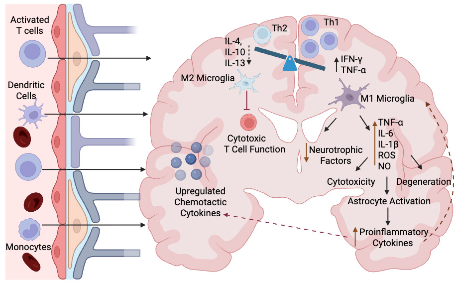
Figure 2. Pathophysiology of neuroinflammatory disease. Infiltration of peripheral activated T cells, dendritic cells, and monocytes across the blood-brain barrier initiate the transition toward a proinflammatory environment in the brain. This proinflammatory state is dominated by Th1 helper cells, proinflammatory cytokines and M1 proinflammatory microglia. Activation of M1 microglia results in a reduction in neurotrophic factors and increased production of proinflammatory cytokines and other factors which cause cytotoxicity, degeneration and astrocyte activation. Astrocyte activation contributes to further increase in proinflammatory cytokines which stimulate the M1 microglia and the upregulation of chemotactic cytokines to further recruit inflammatory cells into the brain. Reduction in Th2 helper cells, anti-inflammatory cytokines, and M2 microglia reduces the inhibition of cytotoxic T cell function, further perpetuating injury. Created in BioRender. Windsor R (2026) https://BioRender.com/tc9tymv. M1: Proinflammatory M1 microglia subtype; M2: anti-inflammatory M2 microglia subtype; TNF-α: tumor necrosis factor-alpha; IL: interleukin; ROS: reactive oxygen species; IFN-γ: interferon gamma. NO: nitric oxide.


