
Stomatocytes as multifunctional therapeutic vehicles: opportunities and challenges for next generation nanomedicine
 0
0 
Abstract
Stomatocytes, bowl-shaped nanostructures derived from polymer vesicles, have been extensively investigated as multifunctional nanocarriers for application in nanomedicine. Their asymmetric morphology, tunable polymer composition, controllable membrane permeability, and tailorable surface functionalization enable efficient encapsulation of therapeutic agents, imaging probes, and stimuli-responsive components, positioning them as versatile vehicles for advanced biomedical applications. Recent progress in engineering stomatocytes has further enhanced their capability to achieve improved targeting specificity toward diseased sites, as well as increased therapeutic precision and efficacy. This Review highlights recent advances in stomatocyte-based nanocarriers, summarizes their key functional advantages, and discusses the major bottlenecks that currently hinder their clinical translation. Finally, we outline future directions for the development of intelligent, multifunctional, and clinically translatable stomatocyte-based platforms for next generation nanomedicine.
Keywords
INTRODUCTION
Nanocarriers are nanoscale delivery systems designed to transport therapeutic and diagnostic cargos, including small molecule drugs, proteins, peptides, DNA, mRNA, siRNA, other nucleic acids, imaging and contrast agents, and various bioactive molecules such as growth factors, enzymes, antibodies, and cell signaling molecules, to specific cells or tissues[1-4]. Conventional nanocarriers are typically constructed from liposomes, lipid nanoparticles, polymeric vesicles, inorganic nanoparticles (e.g., mesoporous silica nanoparticles), micelles, and dendrimers, and generally rely on a passive delivery mechanism[5-8]. Despite significant advances in the design and fabrication of nanocarriers, several fundamental limitations continue to restrict their clinical translation and broader application. Consequently, the development of next generation nanocarriers has become a major focus in nanomedicine.
The term “drug delivery” refers to technologies that transport therapeutic cargos to specific cells or tissues in the body[9]. Drug delivery relies heavily on nanocarriers, which encapsulate payloads to deliver them to desired sites of action. Conventional nanocarriers often rely on passive targeting mechanisms, including the enhanced permeability and retention (EPR) effect, while their responsiveness to specific biological stimuli can be relatively limited. The major limitations of these conventional nanocarriers [Figure 1] include: (1) Low targeting efficiency and off-target accumulation. Only a small fraction of the administered cargo reaches the target tissue, while substantial accumulation occurs in off-target organs, particularly the liver, spleen, and kidneys[10,11]. (2) Rapid clearance by the immune system. Many conventional nanocarriers are recognized as “foreign” by the body’s mononuclear phagocyte system (MPS)[12,13]; Following intravenous administration, they are rapidly coated with plasma proteins (opsonization), which promotes their uptake and clearance by immune cells. This process shortens circulation time, reduces drug accumulation at the disease site, and often necessitates higher therapeutic doses; (3) Limited penetration across biological barriers. Several physiological barriers significantly restrict nanocarrier delivery, including the blood-brain barrier (BBB), tumor penetration barriers, and mucus barriers. Even when nanocarriers reach the target tissue, they often fail to effectively cross the BBB, penetrate deeply into tumor tissues, or traverse mucus layers to reach epithelial surfaces[14,15]; (4) Insufficient drug loading and premature cargo leakage. Insufficient drug loading refers to the inability of nanocarriers to encapsulate therapeutically relevant amounts of cargo. This limitation may arise from weak interactions between the drug and carrier matrix or poor physicochemical compatibility between them. Furthermore, some nanocarriers inherently possess low loading capacities. Premature cargo leakage, defined as the unintended release of encapsulated therapeutics before reaching the target site, further compromises delivery efficiency[16,17]. Together, these limitations may lead to increased dosing frequency, reduced therapeutic efficacy, and difficulties in delivering large biomolecules such as proteins and nucleic acids; (5) Lack of spatiotemporal control over drug release. Most conventional nanocarriers release their payloads by passive mechanisms, including diffusion, carrier degradation, or unintended leakage during circulation. They generally lack responsiveness to internal stimuli, such as pH, temperature, or redox conditions, as well as external triggers including light, ultrasound, or magnetic fields[18,19]. Consequently, drug release may occur at undesired locations or times, resulting in reduced therapeutic efficacy, increased exposure of healthy tissues, elevated risk of adverse effects, and the need for repeated administration; (6) Manufacturing and scale-up challenges. The large-scale production of nanocarriers remains challenging due to batch-to-batch variability, limited storage stability, complex manufacturing processes, and high production costs[20]. These factors present significant obstacles to regulatory approval and commercial translation.
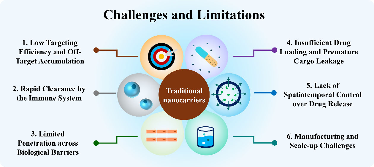
Figure 1. Challenges and limitations of conventional nanocarriers.
Collectively, these limitations have driven the development of the next generation of nanocarriers. These advanced platforms are designed to possess enhanced targeting capabilities, stimulus responsiveness, biomimetic properties, and multifunctionality, with the goal of improving therapeutic efficacy and addressing the unmet needs of modern nanomedicine.
STOMATOCYTES: FROM POLYMERIC VESICLES TO MULTIFUNCTIONAL NANO-PLATFORMS
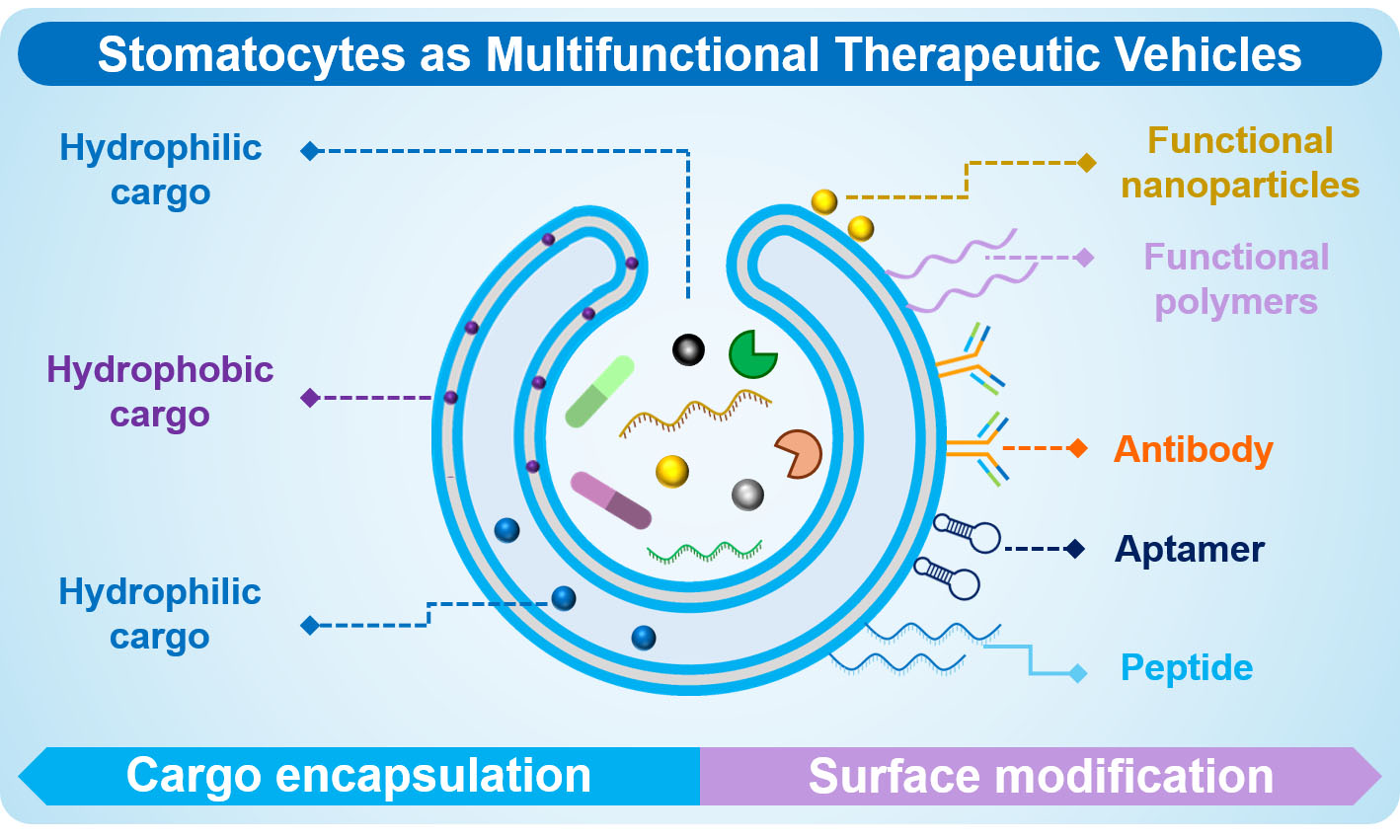
Bowl-shaped polymersomes, commonly known as stomatocytes, represent a unique class of vesicular nanostructures formed through the self-assembly of amphiphilic block copolymers in solution, which are subsequently shape-changed by dialysis. These structures feature a hollow internal cavity and a tunable opening that connects the cavity to the surrounding environment[21]. The term “stomatocyte” originates from the Greek word stoma (“mouth”), reflecting the characteristic opening of the structure. Stomatocytes are a subclass of polymersomes with interesting potential for a wide range of biomedical applications [Figure 2].
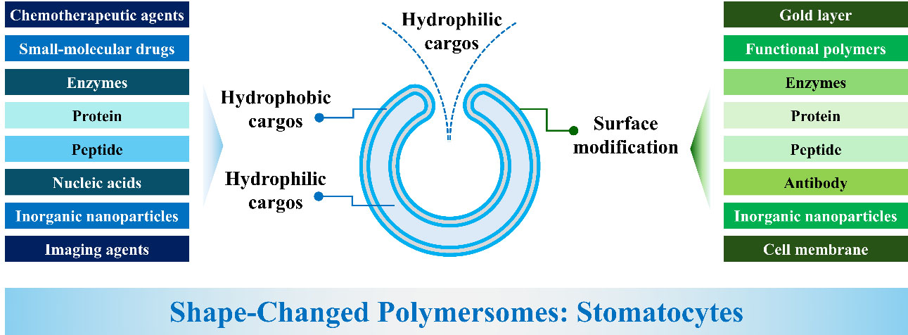
Figure 2. Schematic illustration of stomatocytes with their capacity for cargo encapsulation and surface modification.
Structural design and formation principles
Stomatocytes are typically fabricated from polymersomes through an osmotic-pressure-driven shape transformation process[22]. Spherical polymersomes are formed by the addition of water to a solution of amphiphilic block copolymers in an organic solvent. Subsequently, the organic solvent is removed from inside the polymersome’s lumen and exchanged by water through dialysis. This leads to osmotic stress and a difference in influx and efflux of solvent. The decreased luminal volume triggers a shape change. In the case of stomatocytes, this shape change follows an oblate pathway, going from discs to the bowl-shaped morphology until the polymer membrane has lost its fluidity due to the removal of the organic solvent and the structure is kinetically trapped. The oblate route is achieved when the membrane displays a negative spontaneous curvature to induce inward folding[23]. There are some key physicochemical factors that determine the formation of stomatocytes. The composition of block copolymers strongly influences stomatocyte formation. Changing hydrophobic block chemistry alters membrane rigidity and curvature preferences. The membrane must be flexible enough to deform but stable enough to maintain the concave shape. Furthermore, composition of the dialysis solution not only determines actual stomatocyte formation but also the size of the neck region[24]. Consequently, the combination of the correct polymer composition, membrane curvature and osmotic pressure enables the transformation of a spherical vesicle into a stable bowl-shaped structure.
Amphiphilic block copolymers for stomatocyte formation
Amphiphilic block copolymers serve as the fundamental building blocks for the formation of stomatocytes. Through self-assembly, these polymers form well-defined membranes that provide the structural basis for the characteristic bowl-shaped morphology and tunable opening of stomatocytes. The first stomatocytes were reported in 2010 using poly(ethylene glycol)-block-polystyrene (PEG-b-PS)[21]. In this approach, PEG-b-PS was initially self-assembled in a solvent mixture containing 50% vol % organic solvent (THF : dioxane = 1:1, v/v) and 50% vol % Milli-Q water, followed by dialysis against pure water at room temperature. The organic solvent mixture acts as a plasticizing medium, increasing the conformational mobility of the PS chains and maintaining sufficient membrane permeability to facilitate the shape-transformation process[25]. Subsequently, biodegradable block copolymers, such as poly(ethylene glycol)-block-poly(D,L-lactide) (PEG-PDLLA), were investigated for the preparation of stomatocytes with improved potential for biomedical applications. In a typical preparation, PEG-PDLLA was dissolved in an organic solvent mixture (THF : dioxane = 1:4, v/v), followed by the addition of an equal volume of water. The resulting vesicles were subsequently subjected to dialysis against a 50 mM NaCl solution at 5 ℃ to induce shape transformation. During dialysis, the change in solvent composition across the vesicular membrane generates an osmotic pressure that promotes solvent diffusion from the vesicle lumen to the external medium. The resulting decrease in the internal volume relative to the membrane area induces membrane buckling and inward deformation, ultimately leading to the formation of the characteristic stomatocyte morphology. The resulting bowl-shaped structures have been characterized by scanning electron microscopy (SEM) and cryogenic transmission electron microscopy (cryo-TEM), as shown in Figure 3.
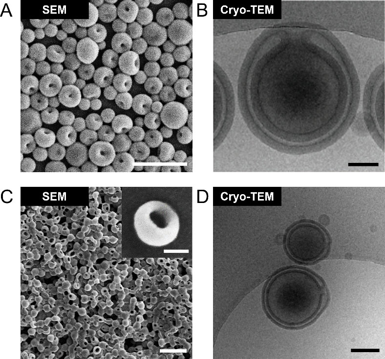
Figure 3. Representative microscopic images of stomatocytes showing their characteristic bowl-shaped morphology and distinctive concave cavity. (A) Morphological characterization of stomatocytes prepared by PEG-b-PS using SEM. Scale bar = 500 nm. Adapted from Shao et al., under the terms of the Creative Commons Attribution 4.0 International License[26]; (B) Cryo-TEM image of PEG-b-PS stomatocytes. Scale bar = 100 nm. Adapted from Wu et al., under the terms of the Creative Commons Attribution 4.0 International License[27]; (C) SEM image of stomatocytes prepared by PEG-PDLLA. Scale bar = 1 μm, insert = 100 nm. Adapted from Shao et al., under the terms of the Creative Commons Attribution 4.0 International License[28]; (D) Bowl-shaped morphology of PEG-PDLLA stomatocytes characterized by cryo-TEM. Scale bar = 200 nm. Adapted from Wang et al., under the terms of the Creative Commons Attribution 4.0 International License[29]. SEM: Scanning electron microscopy; cryo-TEM: cryogenic transmission electron microscopy; PEG-b-PS: poly(ethylene glycol)-block-polystyrene; PEG-PDLLA: poly(ethylene glycol)-block-poly(D,L-lactide).
Importantly, the morphology and physicochemical properties of stomatocytes are strongly influenced by the molecular characteristics of the constituent block copolymers. Key parameters include the hydrophilic-to-hydrophobic block ratio, polymer molecular weight, solvent mixture ratio, and dialysis conditions. Accordingly, careful control over polymer composition and assembly conditions enables the formation of stomatocytes with tunable cavity dimensions, membrane characteristics, and opening sizes.
Structural features enabling functionality
Unlike conventional spherical polymersomes, stomatocytes exhibit asymmetric morphology that endows them with distinct structural and functional advantages[30]. Their compartmentalized structure enables the encapsulation of a wide range of therapeutic, diagnostic, and functional cargos. In addition, both the internal cavity and the membrane opening can be engineered to regulate cargo loading, retention, and release, thereby providing control over cargo transport and delivery. Compared with conventional vesicular systems, this asymmetric architecture offers additional opportunities for structural and functional diversification. The key differences between polymersomes and stomatocytes are summarized in Table 1. In particular, the unique open cavity configuration not only facilitates cargo encapsulation and controlled release, but also provides a confined microenvironment that can be further exploited for the development of nanomotors and nanoreactors, expanding the potential applications of stomatocytes in biomedicine and nanotechnology. Importantly, the tunable opening size provides an additional means of regulating molecular exchange and limiting the premature leakage of encapsulated cargos. Cargo retention can be further enhanced through strategies such as enzyme clustering or in situ growth of functional components within the cavity, which physically confines active cargos and reduces their diffusion-driven loss. These approaches are particularly attractive for maintaining a high local concentration of therapeutic or catalytic payloads and for sustaining their activity within the confined cavity. By integrating the compartmentalization and tunable permeability characteristic of vesicular carriers with additional structural and functional capabilities, stomatocytes may offer potential advantages over some conventional nanocarrier systems, thereby offering significant potential for applications in nanomedicine.
Comparison of features of polymersomes and stomatocytes
| Feature | Polymersomes | Stomatocytes | Ref. |
| Shape | Spherical | Bowl-shaped | [29-37] |
| Morphology | Symmetric | Asymmetric | [29-37] |
| Internal structure | Fully enclosed cavity | Cavity with tunable opening size | [30,31,33,36-38] |
| Cargo loading | Encapsulation during self-assembly | Encapsulation during self-assembly and within the cavity | [35-38] |
| Surface engineering | Different functional components | Different functional components | [29,31,32,34,37] |
| Control release | Mainly diffusion / Stimulus-driven | Stimulus-driven / tunable opening size | [31,32,36] |
| Functionalization | Limited spatial compartmentalization | Separate modification of inner and outer surfaces | [31,34,36,37] |
| Motility | Generally passive / motility via Janus coating | Covert to nanomotors with different driving force | [31,32,34,36,37] |
Functional advantages of stomatocytes for nanomedicine
Owing to their asymmetric morphology and internal cavities, stomatocytes offer several unique functional advantages for nanomedicine applications. These advantages include high-capacity and multifunctional cargo loading, enhanced protection of sensitive payloads, controlled and stimuli-responsive release, compartmentalization of multiple therapeutics, improved biological interactions, active transport and enhanced tissues penetration, tunable surface engineering, and the ability to serve as platforms for artificial organelles.
High-capacity and multifunctional cargo loading
One of the most significant advantages of stomatocytes in nanomedicine is their capacity for highly efficient and multifunctional cargo loading. Their internal cavity, which is in direct contact with the outside environment, provides a large loading volume for the encapsulation of a broad range of therapeutic and functional agents[38]. This unique architecture offers a substantially larger effective loading volume than many conventional nanocarriers of comparable size. Unlike solid nanocarriers, stomatocytes possess a bilayer membrane that can accommodate hydrophobic cargo, while their broad aqueous lumen enables the encapsulation of considerable amounts of hydrophilic compounds[30]. As a result, different types of agents can be selectively incorporated into distinct regions of the nanostructure, facilitating multifunctionality and combination therapies. These features make stomatocytes highly attractive and versatile platforms for advanced drug delivery and theranostic applications. The comparison of stomatocytes with several major classes of nanocarriers is summarized in Table 2.
Comparative overview of stomatocytes and representative major classes of nanocarriers
| Typical nanocarriers | Illustrative scheme | Main composition | Size / nm | Suited cargo | Key advantages | Main limitation | Ref. |
| Stomatocytes |  | Polymers | 300- 500 | Hydrophilic Hydrophobic | Dual compartments, cargo protection, and motility | Challenges in precise shape control and large scale production | [21-24] |
| Polymersomes |  | Polymers | 50- 500 | Hydrophilic Hydrophobic | High structural stability and tunable surface properties | Batch-to-batch variability and low membrane permeability | [39-41] |
| Liposomes |  | Phospholipids | 20- 1000 | Hydrophilic Hydrophobic | Natural Origin, Safety, and dual-cargo encapsulation | Membrane instability and rapid immune clearance | [42,43] |
| Micelles |  | Amphiphilic Surfactants | 10- 200 | Hydrophobic | Enhanced tumor penetration and tunable composition | Limited cargo loading capacity and premature cargo leakage | [44,45] |
| Dendrimers |  | Branched Polymers | 1-10 | Small molecules | Tunable size, surface charge, and chemical properties | High-dose toxicity and complex, costly synthesis | [46-48] |
| Nanoparticles |  | Metallic materials or polymers | 100-500 | Drugs or imaging agents | High structural stability and broad diversity in materials and functions | Limited internal structure and potential toxicity | [49-51] |
Improved protection of sensitive cargos
In addition to their high loading capacity, stomatocytes possess several structural characteristics that enhance the protection of sensitive cargo from degradation and premature loss during transport. This feature is particularly important for fragile biological payloads, such as enzymes, antibodies, and RNA-based therapeutics. The hollow internal cavity provides a dedicated compartment for cargo encapsulation, while the tunable neck opening can regulate molecular diffusion and minimize premature leakage of encapsulated agents. Furthermore, the polymeric membrane acts as a protective physical barrier against external factors and helps shield hydrophobic cargo from the surrounding environment. Most importantly, this compartmentalized architecture isolates sensitive payloads from potentially harmful extracellular conditions, thereby preserving their biological activity and functionality. Collectively, these structural attributes create a protected microenvironment that enhances cargo stability, prolongs circulation, reduces premature degradation, and improves overall delivery efficiency. Recent studies demonstrate the versatility of stomatocytes in enzyme protection and regulation. Proteins can be dynamically loaded and unloaded within polymeric stomatocytes, enabling the construction of enzyme-loaded supramolecular nanoreactors with controllable catalytic activity[52]. Furthermore, compartmentalized cross-linked enzymatic nanoaggregates in the stomatocyte’s cavity (c-CLEnA) have shown remarkable performance in in-flow biocatalysis, where spatial confinement and structural stabilization significantly enhance enzyme activity and operational stability under dynamic conditions[53]. In addition, the loading of enzymes within stomatocytes has endowed the nanoparticles with sustained autonomous motion, as chemical gradients were created due to the enzymatic reaction, which led to a diffusiophoresis process[54].
Controlled and stimuli-responsive release
Precisely controlled and stimuli-responsive payload release from stomatocytes can be achieved by employing responsive amphiphilic block copolymers as building blocks. This strategy enables on-demand cargo release in response to specific physiological conditions or external stimuli, including pH, glutathione (GSH) redox conditions, and temperature. Recent studies have demonstrated the versatility of this approach by exploiting different stimuli-responsive chemistries and polymers to regulate stomatocyte structure and function [Figure 4]. For example, Wang et al. reported GSH-responsive stomatocyte nanomotors for osteoarthritis treatment, in which GSH-triggered structural changes facilitated drug activation in inflamed tissues[36]. This study suggests that the redox-responsive architecture of stomatocytes may be exploited for site-selective therapeutic activation.
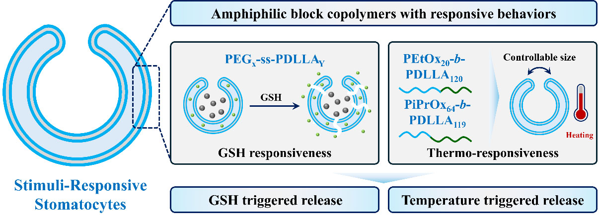
Figure 4. Schematic illustration of GSH-responsive and thermo-responsive stomatocytes. GSH-responsive stomatocytes were prepared by introducing disulfide bonds into PEG-PDLLA copolymers, followed by methotrexate (MTX) loading into the hydrophilic domain and in situ growth of MnO2 nanoparticles within the stomatocyte cavity. Thermo-responsive stomatocytes were obtained by co-assembling PEtOx-b-PDLLA and PiPrOx-b-PDLLA block copolymers (4:1, w/w) via a solvent-switch method, followed by dialysis. Upon increasing temperature, the stomatocyte opening progressively narrowed and eventually underwent irreversible closure. GSH: Glutathione; PEtOx-b-PDLLA: poly(2-ethyl-2-oxazoline)-block-poly(D,L-lactide); PiPrOx-b-PDLLA: poly(2-isopropyl-2-oxazoline)-block-poly(D,L-lactide); PEG-PDLLA: poly(ethylene glycol)-block-poly(D,L-lactide).
Terracciano et al. developed thermo-responsive stomatocytes based on poly(2-oxazoline), which exhibited temperature-dependent changes in shape and permeability and consequently enabled temperature-modulated cargo release[55]. Although these findings demonstrate the feasibility of using thermo-responsive stomatocytes for externally regulated delivery, the practical utility of this mechanism may depend strongly on the magnitude, spatial uniformity, and physiological relevance of the applied temperature stimulus. More broadly, these studies suggest that integrating stimuli responsiveness with functional features such as propulsion and imaging could provide additional control over cargo delivery. However, the extent to which such multifunctionality translates into improved therapeutic precision or reduced off-target effects remains dependent on the specific stimulus, biological environment, and performance requirements, and therefore warrants further validation under physiologically relevant conditions.
Compartmentalization of multiple therapeutics
Unlike conventional nanocarriers, stomatocytes possess a unique compartmentalized structure that enables the spatial segregation and co-encapsulation of multiple therapeutic agents with distinct physicochemical properties[56]. This capability allows the simultaneous delivery of drugs, proteins, nucleic acids, imaging agents, or catalytic components while minimizing undesirable interactions between cargos[38]. Such compartmentalization is particularly advantageous for combination therapies, where synergistic agents can be protected, transported, and function in a coordinated manner. For example, biomimetic “nano-red blood cells” were designed by exploiting the dual-compartmentalized structure of PEG-PDLLA stomatocytes to co-encapsulate oxygen-carrying hemoglobin within the inner lumen and the hydrophobic photosensitizer chlorin e6 within the polymer membrane. This spatial organization enabled simultaneous oxygen delivery and photodynamic therapy, effectively alleviating tumor hypoxia and significantly enhancing therapeutic efficacy[28]. Beyond this proof-of-concept, the ability of stomatocytes to precisely organize multiple functional components within a single nano-system offers exciting opportunities for the development of cascade therapeutic systems, multimodal therapies, and theranostic nanomedicines, where distinct cargos can work synergistically.
Active transport and enhanced tissue penetration
As discussed above, the internal cavity of stomatocytes provides a confined nanoreactor-like space for the incorporation of catalytic components, enabling in situ chemical reactions or fuel conversion processes. In particular, the encapsulation of catalytic enzymes, such as catalase and glucose oxidase, or catalytic inorganic nanoparticles, such as platinum (Pt) or manganese dioxide (MnO2), can endow stomatocytes with autonomous propulsion. Surface functionalization with metallic components, such as Pt or Au, can further introduce bubble-driven or light-driven motion, respectively. These design strategies have been demonstrated in various stomatocyte-based nanomotors, including biodegradable systems containing MnO2 nanoparticles within the stomatocyte cavity and Pt nanoparticles immobilized on the outer surface [Figure 5].
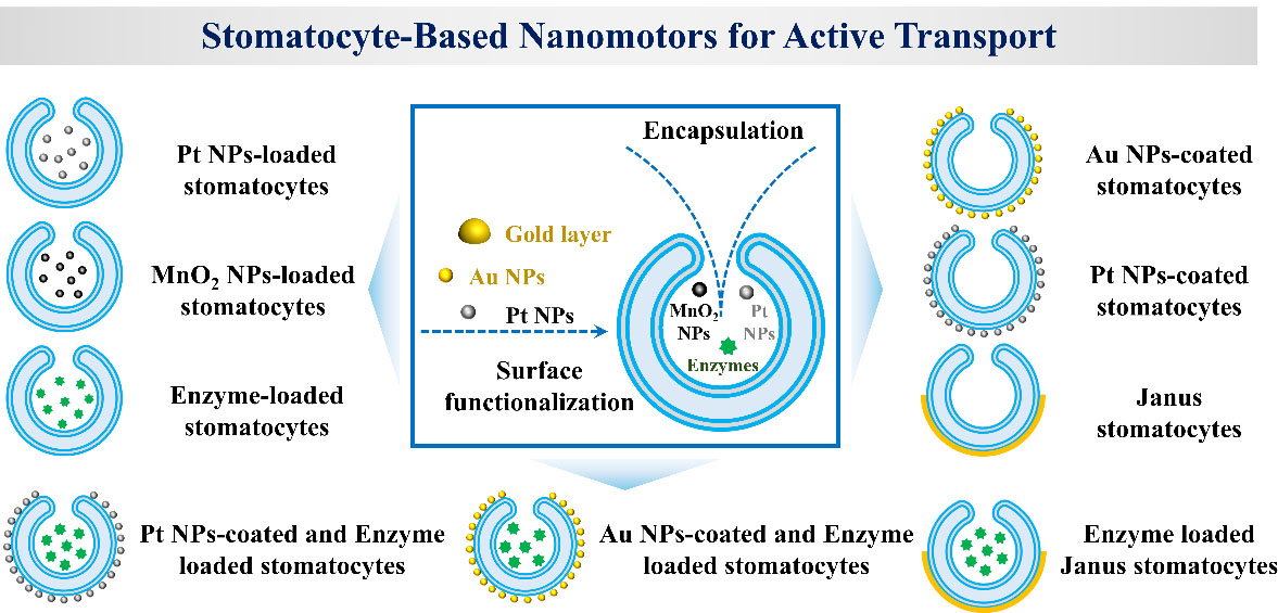
Figure 5. Schematic illustration of stomatocyte-based nanomotors and their propulsion mechanisms. Stomatocytes can encapsulate catalytic components, including Pt or MnO2 nanoparticles and enzymes such as catalase or urease, to generate chemically driven propulsion. Alternatively, surface functionalization with Pt nanoparticles enables bubble-driven motion, while Au nanoparticles or a hemispherical Au coating can confer light-driven propulsion. Combining catalytic nanoparticles or Au-based components with enzymes within a single stomatocyte enables multimodal propulsion. Pt NP: Platinum nanoparticle; Au NP: Gold nanoparticle; MnO2 NP: Manganese dioxide nanoparticle.
Such catalytic stomatocyte nanomotors have shown enhanced active transport and intracellular delivery efficiency[57-60]. Moreover, the integration of catalytic components with asymmetric metallic coatings enables more sophisticated propulsion and motion control, including Janus-type and “twin-engine” architectures with counterbalanced or “seesaw” motion[26]. Collectively, these advances highlight the versatility of stomatocytes as programmable nanomotor platforms for active cargo delivery. Beyond chemically driven propulsion, surface incorporation of photothermal or plasmonic materials such as gold nanoparticles enables externally controlled and light-driven motion, providing an additional means to overcome the limitations of passive diffusion. For example, gold nanoparticles (Au NPs)-functionalized stomatocytes have been developed as ultrafast light-activated nanomotors, exhibiting enhanced transport efficiency and tissue penetration[29].
This strategy enhances navigational adaptability in complex biological environments and further improves deep tissue penetration and intracellular, particularly cytoplasmic delivery. Light-driven nanomotors transform external optical energy into directional propulsion, facilitating traversal through heterogeneous biological environments and markedly improving penetration across physiological barriers[32]. Similarly, nitric oxide-driven nanogel motors generate in situ gaseous propulsion, enabling autonomous motion that enhances intratumoral diffusion depth and promotes more homogeneous distribution within dense extracellular matrices, ultimately improving therapeutic outcomes[61]. Guo et al. developed oxygen- and heat-dual-driven stomatocyte nanomotors that converted endogenous and photothermal energy into sustained propulsion, enabling efficient deep tissue penetration[62]. This dual-driven mechanism overcame diffusion limitations in dense tumor matrices, reduced transport resistance, and promoted deeper and more uniform intratumoral distribution compared with passive nanocarriers[62].
Platforms for artificial organelles
Artificial organelles have emerged as a promising strategy for mimicking the functions of natural subcellular compartments and restoring or enhancing cellular activities[63-65]. Owing to their unique bowl-shaped morphology, well-defined internal cavities, and tunable membrane properties, stomatocytes provide an attractive platform for the construction of artificial organelles[66]. Their spacious internal compartment offers a high loading capacity for functional biomacromolecules, including enzymes, proteins, and catalytic nanoparticles, enabling spatially confined biochemical reactions that closely resemble those occurring within natural organelles[67]. By incorporating single enzymes or multi-enzyme cascades, stomatocyte-based artificial organelles have been developed to replicate essential cellular functions[68-70]. Furthermore, the modular nature of stomatocyte fabrication allows precise control over membrane composition, particle size, catalytic cargo loading, and responsiveness to external stimuli, facilitating the development of programmable artificial organelles with tailored functionalities. Beyond serving as biomimetic models of natural organelles, stomatocyte-based artificial organelles hold promise for intracellular therapeutic applications[66,71]. Upon cellular internalization, these nanoscale compartments can function as synthetic intracellular reactors, locally generating therapeutic molecules, regulating metabolite levels, or modulating cellular signaling pathways[72,73]. Such capabilities are particularly attractive for the treatment of diseases associated with enzyme deficiencies, oxidative stress, and metabolic platforms for intracellular catalysis, cellular engineering, and the development of cell-mimicking nanomedicines.
THERAPEUTIC OPPORTUNITIES
Cancer therapy
Stomatocytes have been effectively applied in cancer therapy [Figure 6]. Their unique structure enables the simultaneous encapsulation of both hydrophilic and hydrophobic therapeutic agents, while their chemically addressable surface allows precise conjugation of targeting ligands, polymers, or stimulus-responsive moieties. These features collectively enhance tumor selectivity, cellular uptake, and spatiotemporal control over therapeutic delivery. One of the earliest and most extensively investigated applications of stomatocytes in nanomedicine is their use as nanocarriers for chemotherapeutic drug delivery[74,75]. For example, mannose-functionalized stomatocyte nanomotors have demonstrated improved dynamic targeting and cellular uptake in cancer cells[76]. Beyond conventional chemotherapy, stomatocytes can be used as versatile carriers for a broad range of cancer treatments, including photothermal therapy (PTT), photodynamic therapy (PDT), gene therapy, and immunotherapy. By encapsulating photothermal agents or photosensitizers, stomatocytes enable externally triggered tumor ablation upon near-infrared laser irradiation, offering precise spatial and temporal control over therapeutic activation[74]. A representative example is the recent study reporting the design and preparation of targeted peptide nanofiber-loaded stomatocytes for combined PDT and PTT treatment, demonstrating the strong potential of stomatocyte-based nanoplatforms for synergistic therapy[77]. Similarly, nucleic acid therapeutics such as siRNA, miRNA, or mRNA are protected from enzymatic degradation and delivered into tumor cells, enabling gene regulation-based cancer interventions.
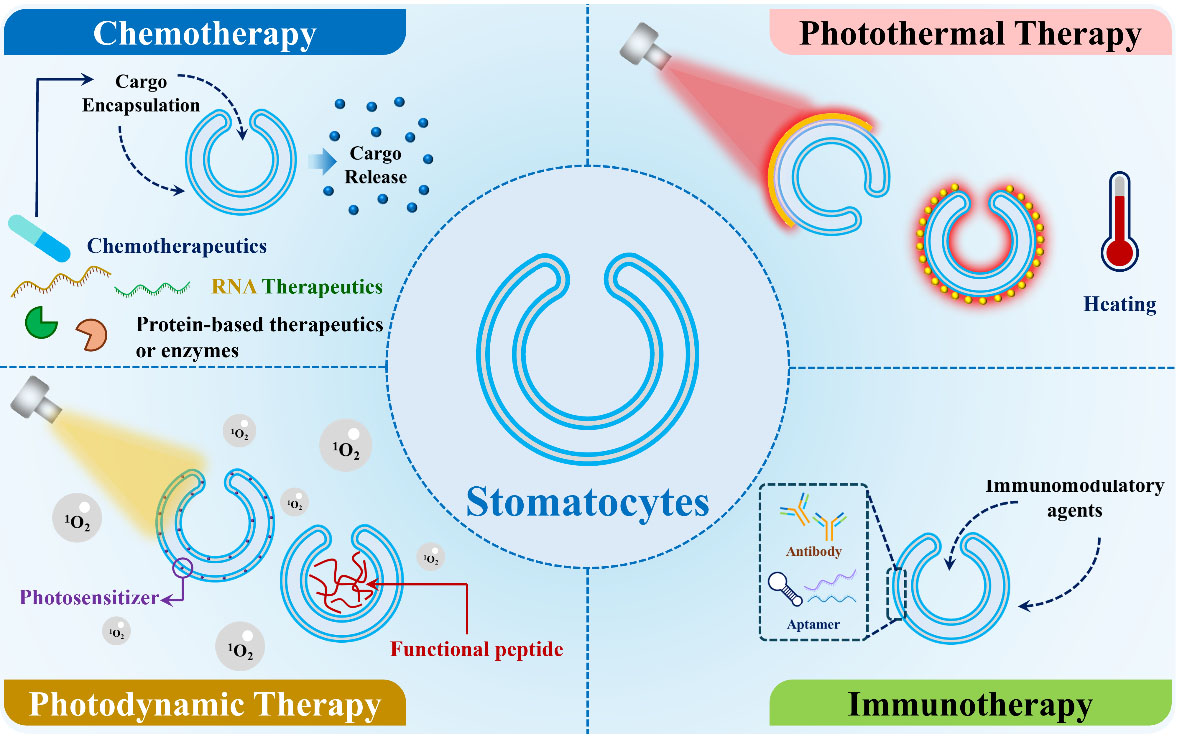
Figure 6. Illustration of the applications of stomatocytes in cancer therapy, including chemotherapy, photothermal therapy, photodynamic therapy, and immunotherapy.
In the context of immunotherapy, stomatocytes function as delivery vehicles for immunomodulatory agents, thereby enhancing tumor immune recognition and promoting antitumor immune response[78,79]. Furthermore, the inherently multicompartmental feature of stomatocytes enables the co-encapsulation of multiple therapeutic payloads within a single nanocarrier system. This includes small-molecule chemotherapeutics (e.g., paclitaxel, cisplatin, irinotecan), nucleic acid therapeutics (e.g., mRNA, siRNA, miRNA), and protein-based therapeutics or enzymes (e.g., L-asparaginase, caspase, and L-arginase). Such versatility allows the rational design of synergistic combination therapies, including chemo-gene therapy, chemo-immunotherapy, and chemo-catalytic therapy, which are particularly promising for overcoming tumor heterogeneity, multidrug resistance, and limited treatment efficacy associated with monotherapies.
Antibacterial and anti-biofilm therapy
Beyond cancer treatment, stomatocytes have recently shown promising potential for antibacterial and anti-biofilm applications. A broad spectrum of antimicrobial payloads, including conventional antibiotics (e.g., vancomycin, ciprofloxacin, and rifampicin), antimicrobial peptides, metal-based antibacterial agents (e.g., silver, copper, and ZnO nanoparticles), bacteriolytic enzymes (e.g., lysozyme), as well as catalytic nanozymes capable of generating reactive oxygen species (ROS) were effectively encapsulated in stomatocytes[80-82]. Compared with free antimicrobial agents, stomatocyte-mediated delivery offers several significant advantages, including prolonged blood circulation, improved pharmacokinetic profiles, enhanced stability against premature degradation or enzymatic inactivation, reduced systemic toxicity, and increased accumulation at infected tissues through passive or active targeting mechanisms. Furthermore, surface functionalization with bacterial-targeting ligands, antibodies, antimicrobial peptides, or cell membrane coating enables selective recognition of pathogenic microorganisms while minimizing off-target effects on microbiota and healthy tissues[83,84]. One of the most attractive features of stomatocyte lies in their potential to penetrate bacterial biofilms, which remain one of the major causes of chronic infections. Biofilms consist of densely packed bacterial communities embedded within an extracellular polymeric substance (EPS) matrix that severely limits antibiotic penetration and protects bacteria from host immune responses[85]. The unique morphology and tunable physicochemical properties of stomatocytes facilitate enhanced penetration into biofilm matrices, thereby improving local drug delivery. Moreover, multifunctional stomatocytes can be engineered to disrupt biofilms through multiple strategies, including enzymatic degradation of EPS components, catalytic ROS generation, photothermal or photodynamic therapy, and localized release of antimicrobial agents.
Recent advances in stomatocyte engineering have enabled the development of next generation antibacterial systems by integrating stimuli-responsive and self-propelled functionalities. For example, pH-responsive stomatocytes selectively release antibacterial agents within the acidic microenvironment of infected tissues, while enzyme-responsive systems exploit bacterial enzymes, such as lipases or proteases, to trigger site-specific drug release. Catalytic stomatocyte nanoreactors capable of producing ROS or other bactericidal species provide an antibiotic-independent strategy for bacterial elimination. Furthermore, autonomously propelled stomatocyte-based nanomotors could potentially facilitate transport through viscous biological fluids and dense biofilm matrices, thereby improving their accumulation and therapeutic efficacy at infection sites. This strategy provides a promising opportunity for overcoming the diffusion limitations encountered by conventional nanocarriers. Collectively, stomatocytes offer a versatile nanoplatform for antibacterial and anti-biofilm applications. Their high loading capacity, tunable structure, and surface engineering provide opportunities to integrate targeted drug delivery, stimuli-responsive release, catalytic biofilm disruption, and multimodal antimicrobial strategies within a single nanoplatform.
Other potential applications
Although polymeric stomatocytes have been predominantly explored as drug delivery vehicles for cancer treatment and antimicrobial/anti-biofilm applications, their unique bowl-shaped morphology and compartmentalized structure may also offer opportunities in several emerging biomedical fields. One promising direction is the development of stomatocyte-based systems for cardiovascular therapy. The tunable polymeric membrane and accessible internal cavities allow the encapsulation of a broad range of therapeutic agents, including anti-inflammatory, antithrombotic, and vascular-protective molecules[86,87]. Moreover, surface functionalization with ligands targeting activated endothelial cells, platelets, or atherosclerotic plaques could potentially enhance site-specific accumulation while minimizing off-target effects. The characteristic cavity and opening of stomatocytes provide opportunities for cargo loading and retention, as well as stimuli-responsive release, which may offer additional flexibility for localized therapeutic delivery. In addition, incorporation of catalytic enzymes or inorganic nanoparticles within the cavity could facilitate the in situ conversion of endogenous substrates into therapeutic products, which may be particularly relevant for cardiovascular disorders associated with oxidative stress and dysregulated metabolic microenvironments[88]. Another intriguing application arises from the structural resemblance of stomatocytes to biological cells, particularly in the development of artificial red blood cell (RBC)-mimicking systems. A representative example was reported by Shao et al., who developed therapeutic nano-RBCs based on biodegradable PEG-PDLLA stomatocytes[28]. In this design, hemoglobin was encapsulated within the stomatocyte cavity as an oxygen-carrying component, while the photosensitizer chlorin e6 was co-loaded to enable oxygen-enhanced photodynamic therapy. To further improve biomimicry and circulation behavior, erythrocyte-derived cell membranes were coated onto the stomatocyte surface, imparting selected biological characteristics of native RBCs. The resulting membrane-coated stomatocytes exhibited prolonged blood circulation and enhanced tumor accumulation in vivo, suggesting that biomimetic interfaces may complement the multifunctional and compartmentalized architecture of stomatocytes to improve their therapeutic potential. Ocular drug delivery represents another emerging application for stomatocytes. Recent studies have highlighted the potential of polymersomes as ocular nanocarriers, owing to their prolonged drug-release profiles, enhanced ocular retention, and tunable transport properties within ocular tissues[89]. Compared with conventional spherical vesicles, stomatocytes feature a confined internal cavity connected to the external environment through a tunable opening. This architecture may facilitate the encapsulation and spatial organization of diverse therapeutic cargos, including small-molecule drugs, proteins, antioxidants, and neuroprotective agents. Furthermore, tuning the opening size could modulate cargo diffusion and release kinetics within the complex ocular microenvironment, potentially enabling sustained and localized drug delivery.
Collectively, these examples suggest that the distinctive morphology and compartmentalized structure of stomatocytes may broaden their applications beyond cancer therapy and antimicrobial applications. However, most of these emerging applications remain at an early stage of development, and further studies are needed to determine whether the structural advantages of stomatocytes translate into meaningful improvements in therapeutic efficacy, targeting, and clinical performance compared with conventional polymersomes and other established nanocarrier platforms.
TRANSLATIONAL CHALLENGES TOWARD CLINICAL NANOMEDICINE
Scalable and reproducible manufacturing
Despite significant progress in the development of stomatocyte nanocarriers, their translation into clinical applications remains limited by manufacturing challenges[90]. Most current preparation methods, including solvent-switch and osmotic-induced approaches, are optimized for laboratory-scale production and often involve multiple processing steps that are difficult to standardize. As production is scaled up, maintaining consistent particle characteristics, such as uniform hydrodynamic size, morphology, and the neck opening, becomes increasingly challenging. A major obstacle is achieving batch-to-batch consistency, as even slight variations in synthesis conditions, such as solvent composition, osmotic conditions, temperature, and processing time, can lead to differences in particle size, morphology, membrane thickness, and encapsulation efficiency. Such variability may in turn affect drug loading, therapeutic efficacy, and biodistribution. Therefore, the development of robust, scalable, reproducible, and economically feasible manufacturing processes that comply with Good Manufacturing Practice (GMP) standards is essential for future commercialization. Recent advances in manufacturing techniques, such as microfluidic-assisted self-assembly, flash nanoprecipitation, and automated in-line process monitoring, offer promising strategies to improve the large-scale and reliable production of stomatocytes[91-93]. Addressing these challenges will be a critical step toward the clinical translation and commercialization of stomatocyte-based nanomedicines.
Structural stability and cargo retention
Maintaining the structural integrity of stomatocytes during storage and under physiological conditions is essential for preserving cargo retention and functional performance. However, compared with the extensive literature on stomatocyte formation and morphological transformation, systematic studies of their long-term stability remain limited. Available evidence suggests that environmental conditions can affect membrane organization, colloidal stability, and morphology, but the effects of temperature, osmotic stress, protein adsorption, and storage conditions have not been systematically evaluated across different stomatocyte formulations. Potential instability, including membrane rearrangement, aggregation, shape transformation, or premature cargo release, should therefore be considered as possible but system-dependent risks rather than universal characteristics. Temperature and osmotic conditions may be particularly important because changes in membrane fluidity, polymer-chain organization, and transmembrane osmotic pressure could alter stomatocyte morphology and cargo retention. Nevertheless, quantitative evidence linking these parameters to long-term stability remains scarce. Similarly, although lyophilization is widely used to improve nanocarriers’ storage stability, its applicability to stomatocytes requires system-specific validation, as dehydration and rehydration may affect morphology, aggregation, and cargo retention. Thus, low-temperature storage and lyophilization should not be assumed to be universally suitable preservation strategies. Following systemic administration, stomatocytes encounter a complex physiological environment involving ionic strength, osmolarity, plasma proteins, and hydrodynamic forces, which may affect surface interactions, colloidal stability, membrane properties, and cargo retention. Surface engineering strategies, including optimized PEGylation, zwitterionic modification, and biomimetic coatings, may reduce nonspecific protein adsorption and immune recognition, although their long-term immunological compatibility requires systematic evaluation, particularly for repeated administration. Future studies should therefore establish standardized stability assessment frameworks encompassing morphology, size distribution, membrane integrity, aggregation, and cargo retention under controlled variations in temperature, osmolarity, ionic strength, protein exposure, and storage duration. For lyophilized formulations, these parameters should be compared before and after freezing, drying, and rehydration. Importantly, structural stability should also be correlated with functional performance, including cargo release and, where applicable, catalytic or propulsion activity. Such systematic evaluation will help establish quantitative stability criteria, identify formulation-specific failure modes, and determine whether the distinctive stomatocyte architecture can be reliably maintained during storage and under physiologically relevant conditions.
Long-term bio-safety assessment
Although stomatocytes prepared from different amphiphilic block copolymers have exhibited favorable biocompatibility, comprehensive evaluation of their long-term biosafety remains relatively limited[94]. As stomatocyte-based nanocarriers advance toward clinical translation, establishing a comprehensive long-term safety profile will become increasingly important, particularly for chronic diseases, requiring repeated administration. Beyond conventional cytotoxicity assays, future investigations should systematically address chronic toxicity, immunogenicity, inflammatory responses, and the biological consequences of repeated dosing to better predict their long-term clinical performance. A major knowledge gap concerns the long-term in vivo fate of stomatocytes and their degradation products. Although many amphiphilic block copolymers are designed to be biodegradable under physiological conditions, their degradation kinetics, metabolic pathways, and clearance mechanisms remain incompletely characterized. In addition, prolonged retention or accumulation in organs associated with the mononuclear phagocyte system, such as the liver and spleen, may lead to chronic tissue burden or subtle functional impairment. Consequently, long-term studies integrating biodistribution, organ histopathology, biodegradation, and metabolic analyses are important for establishing a comprehensive understanding of chronic toxicity. Another important consideration is the potential interaction between stomatocytes and the immune system. Upon systemic administration, nanocarriers inevitably encounter plasma proteins and immune cells, which may trigger protein corona formation. Detailed profiling of pro-inflammatory cytokines and immune cell activation patterns is therefore essential to evaluate their immuno-inflammatory compatibility. Repeated exposure to nanocarriers may alter pharmacokinetics, biodistribution, and clearance behavior over time. Potential issues include accumulation in major organs, altered immune recognition after first exposure, and changes in therapeutic efficiency. Long-term repeated-dose studies are necessary to assess cumulative toxicity and to validate dosing regimens for safe translational application.
Translating nanomotors into living systems
Despite substantial progress in demonstrating the motion of nanomotors in vitro and in vivo, their translation into physiologically relevant and clinically applicable systems remains challenging[95]. General barriers include limited fuel availability, the complexity of physiological environments, immune recognition and clearance, and interference from blood flow. For stomatocyte-based nanomotors, however, these challenges may be further influenced by the distinctive interplay among polymer membrane integrity, stomatocyte morphology, confined cavity geometry, and propulsion behavior. In particular, the stability and degradation of the polymer membrane will determine propulsion performance over time. Changes in polymer composition, membrane thickness, surface properties, or overall stomatocyte integrity during degradation could alter the accessibility and spatial organization of catalytic components, as well as the transport of fuels and reaction products. These changes may, in turn, affect catalytic efficiency and propulsion behavior. Establishing quantitative correlations between polymer degradation, structural integrity, and propulsion characteristics will therefore be important for determining whether the propulsion performance observed under controlled experimental conditions can be maintained over relevant biological timescales. The confined cavity of stomatocytes represents a potentially important but comparatively underexplored feature that may distinguish these nanomotors from other motor architectures. Although the cavity and opening provide a defined spatial configuration for integrating catalytic components, confinement may either facilitate or hinder the diffusion and exchange of fuels and reaction products, depending on cavity dimensions, opening size, membrane permeability, and the surrounding medium. Consequently, the compartmentalized architecture that enables functional integration may also introduce mass-transfer limitations under certain physiological conditions. For example, confinement could potentially enhance local fuel conversion by increasing reactant residence time, but it could also reduce catalytic turnover by restricting molecular exchange. These context-dependent effects should therefore be considered when interpreting propulsion behavior and optimizing stomatocyte geometry for specific biological environments. Systematic studies correlating cavity architecture, catalytic activity, and propulsion kinetics under physiologically relevant conditions are needed to determine whether confinement represents a functional advantage, a limitation, or both depending on the operating environment.
Several strategies may help address these challenges. Fuel-independent propulsion driven by magnetic, acoustic, or light fields could reduce reliance on exogenous chemical fuels, while biologically available substrates such as glucose or urea may provide more physiologically compatible alternatives. Surface engineering with antifouling or biomimetic coatings may reduce protein adsorption and immune recognition, but their potential effects on membrane stability, cavity accessibility, and propulsion should also be carefully evaluated. External actuation and guidance may further help overcome hemodynamic forces and improve spatiotemporal control. Ultimately, the translation of stomatocyte-based nanomotors will require not only improved propulsion efficiency but also a clearer understanding of how polymer degradation, cavity confinement, and physiological interactions collectively influence propulsion stability and performance over relevant biological timescales.
Regulatory and GMP considerations
The successful clinical translation of stomatocyte-based nanomedicine will rely not only on their therapeutic performance but also on their ability to satisfy increasingly strict regulatory and manufacturing requirements[96]. As complex polymeric nanocarriers, stomatocytes must demonstrate consistent quality, safety, reproducibility, and scalability throughout their development. Consequently, establishing standardized manufacturing processes, validated quality control strategies, GMP compliant production, and well-defined regulatory pathways will be indispensable for their clinical and commercial implementation. A primary challenge lies in the standardization of manufacturing and characterization protocols. Reliable batch-to-batch reproducibility requires tight control over critical process parameters during stomatocyte fabrication, together with standardized analytical methods to evaluate physicochemical properties, drug loading efficiency, encapsulation stability, and release behavior. Sterility represents another important element. Conventional sterilization methods may compromise the structural integrity and biological functionality of stomatocytes, particularly for formulation encapsulating enzymes or other fragile therapeutics. Aseptic manufacturing using sterile raw materials is likely to represent the most practical strategy for clinical production. Importantly, sterilization procedures should be validated to ensure that particle morphology, cargo loading, catalytic activity, and therapeutic performance remain unaffected. Beyond laboratory-scale fabrication, the transition to GMP manufacturing requires production processes that are scalable, reproducible, and readily validated. Traditional batch-based preparation methods frequently suffer from limited scalability and process variability, presenting significant barriers to industrial translation. Continuous manufacturing approaches, such as microfluidic assembly and automated solvent exchange systems, offer enhanced process control, improved reproducibility, and greater manufacturing consistency, making them promising candidates for large-scale GMP production of stomatocyte formulations. Regulatory approval of stomatocyte-based nanomedicines should follow existing frameworks established for liposomes formation, polymeric nanoparticles, or other nanomedicines.
CONCLUSION AND OUTLOOK
AI-guided design and optimization
The rapid advancement of artificial intelligence (AI) and machine learning (ML) is reshaping the development of nanomedicine by enabling data-driven optimization of formulation design, predictive modeling of nanocarrier performance, reduction of experimental workload, and accelerated clinical translation[97,98]. Recent advances in machine learning have demonstrated the potential to accelerate the design and optimization of polymeric nanocarriers by linking formulation parameters with physicochemical and biological outcomes. For example, machine-learning models have been applied to predict nanoparticle size, drug-loading efficiency, and cellular uptake based on polymer composition and formulation parameters, thereby enabling data-driven optimization of polymeric delivery systems[99-102]. Similar approaches could be extended to stomatocytes by integrating descriptors such as block-copolymer composition, molecular weight, hydrophilic-to-hydrophobic balance, opening size, cavity dimensions, cargo characteristics, preparation conditions, and surface functionalization. Such models may ultimately facilitate the prediction and optimization of cargo encapsulation and retention, stimulus-responsive release, colloidal stability, and biodistribution. However, AI-guided design of stomatocytes remains at an early stage and will require sufficiently large, standardized, and experimentally validated datasets to achieve reliable predictive performance.
Beyond formulation optimization, AI is expected to transform the characterization and manufacturing of stomatocyte nanocarriers. Advanced image analysis algorithms can automatically quantify morphological features, identify structural defects, and classify heterogeneous stomatocyte populations with greater accuracy and reproducibility than manual analysis[103]. When integrated with automated synthesis platforms and high-throughput experimentation, these approaches may establish closed-loop optimization systems capable of continuously refining fabrication parameters and improving production consistency. AI also provides an unprecedented opportunity to establish quantitative structure-function relationships for stomatocyte-based nanocarriers. By integrating experimental characterization, molecular simulations and datasets, ML models can identify the structural determinants that govern pharmacokinetics, biodistribution, tumor accumulation, intracellular delivery, and therapeutic efficacy. Most importantly, combining AI with molecular dynamics simulations, finite-element analysis, and computational fluid dynamics could enable multiscale prediction of stomatocyte behavior across molecular, cellular and physiological levels, providing mechanistic insights that are difficult to obtain experimentally.
Looking forward, the integration of AI may support the development of personalized stomatocyte therapeutics. Predictive models incorporating nanocarrier characteristics together with patient-specific clinical information could facilitate the rational design of individualized formulations with optimized targeting efficiency, drug loading, and dosing strategies. As these techniques continue to mature, AI-guided design is expected to be a driving force for the next generation of stomatocytes in nanomedicine, accelerating their translation from laboratory research to clinical applications.
Biomimetic and biohybrid stomatocytes
The next generation of stomatocyte-based nanocarriers may increasingly incorporate biomimetic and biohybrid design strategies to address biological barriers that continue to limit the clinical translation of conventional nanocarriers[104,105]. By integrating naturally derived biological components with engineered stomatocytes, these hybrid systems could combine the structural programmability of synthetic polymeric nanocarriers with selected biological functions of living cells. Such integration may help modulate immune recognition, prolong systemic circulation, and improve tissue-specific interactions, potentially expanding the therapeutic applications of stomatocytes. One promising strategy is the functionalization of stomatocytes with cell membranes[106,107]. These biomimetic interfaces provide membrane proteins, lipids, and glycocalyx components that can influence immune recognition and cell-nanocarrier interactions, thereby offering opportunities to improve circulation behavior and facilitate targeted delivery. Beyond cell membrane coating, exosome-inspired engineering represents another promising direction for stomatocyte design. As naturally occurring extracellular vesicles involved in intercellular communication, exosomes exhibit favorable biocompatibility and cellular uptake properties and can carry diverse biomolecular cargos. Future stomatocyte platforms could incorporate selected structural and functional features of exosomes while retaining the architectural and engineering advantages of stomatocytes. Incorporation of exosomal membrane proteins, extracellular vesicle-derived lipids, or bioactive targeting ligands may facilitate interactions with specific cell types and potentially improve transport across biological barriers. Such hybrid systems may be particularly relevant for the delivery of nucleic acids, gene-editing components, therapeutic proteins, and immunomodulatory agents, for which efficient cellular uptake and controlled immune interactions are important. However, whether exosome-inspired modifications can provide consistent advantages over conventional stomatocytes remains to be systematically established.
Engineering clinically relevant active stomatocytes
Translating active nanomotors into clinical applications remains limited because most propulsion strategies rely on exogenous chemical fuels that are either absent from or incompatible with physiological environments. Rather than continuing to maximize propulsion speed or efficiency, the next generation of stomatocyte-based nanomotors should prioritize clinically translatable actuation strategies that are biocompatible, externally controllable, and readily integrated with existing medical technologies[108,109]. From this review, fuel-free propulsion, magnetic actuation, ultrasound-driven locomotion, and intelligent in vivo motion regulation represent particularly promising directions. A major challenge is the replacement of chemically fueled propulsion systems, which commonly depend on hydrogen peroxide or other nonphysiological reactants with limited biocompatibility and potential toxicity. Future designs should instead exploit endogenous energy sources available within biological systems. Examples include propulsion driven by naturally occurring biochemical gradients, enzyme-mediated metabolic reactions, or biohybrid systems powered by living cells or microorganisms. Such approaches not only eliminate the need for exogenous fuels but also improve compatibility with complex physiological environments.
Among externally powered strategies, magnetic actuation is particularly attractive for clinical translation. Magnetic fields penetrate deep tissues noninvasively and are already widely used in clinical imaging and interventional medicine. Incorporating magnetic nanoparticles into stomatocytes enables remote guidance, directional navigation, and enhanced retention at disease sites while simultaneously allowing integration with magnetic resonance imaging (MRI) for real-time tracking and image-guided therapy. Ultrasound provides another highly promising actuation modality owing to its deep tissue penetration and widespread clinical availability. Acoustic propulsion, generated through mechanisms such as acoustic streaming, radiation forces, or microbubble oscillation, enables efficient locomotion without chemical fuels and is therefore well suited for applications in deep or otherwise inaccessible tissues. Looking forward, the development of active stomatocytes should extend beyond the pursuit of increasingly sophisticated propulsion mechanisms toward the creation of intelligent nanomotors capable of adaptive and programmable motion. By integrating responsiveness to external physical cues (e.g., magnetic fields or ultrasound) with endogenous biological signals associated with disease microenvironments, stomatocyte nanomotors could achieve on-demand activation, autonomous behavioral adaptation, and spatiotemporally precise navigation.
Precision and personalized nanomedicine
The merger of nanotechnology and precision medicine is driving a shift from conventional “one-size-fits-all” therapeutics toward highly individualized treatment tailored to patient-specific biological characteristics[110]. In this context, stomatocyte-based nanocarriers offer a versatile platform that can be engineered with multifunctionality and stimuli-responsiveness to meet the demands of precision and personalized medicine. These systems can be readily functionalized with targeting ligands, antibodies, peptides, or aptamers to recognize disease-associated biomarkers expressed in a patient-specific manner on diseased tissues or cells. Beyond molecular targeting, personalization can also be achieved by leveraging interpatient variability in tumor microenvironment features, thereby enhancing therapeutic selectivity, reducing off-target toxicity, and addressing the biological heterogeneity that often limits the efficacy of conventional treatments[111]. The integration of companion diagnostics is therefore essential to identify patient subpopulations most likely to benefit from stomatocyte-based therapies. By incorporating imaging agents or molecular probes into stomatocytes, it becomes possible to classify patients based on biomarker expression profiles, biodistribution behavior, or microenvironmental features[112]. Such theranostic capabilities further enable real-time feedback to optimize dosing schedule and delivery method in a patient-specific manner. Overall, the integration of targeted delivery, companion diagnostics, and theranostic functionality positions stomatocyte-based systems as a promising platform for advancing precision and personalized nanomedicine.
DECLARATIONS
Authors’ contributions
Supervision and revision: van Hest JCM
Literature review, scheme design and preparation, and writing: Shao J.
Availability of data and materials
Not applicable.
AI and AI-assisted tools statement
Not applicable.
Financial support and sponsorship
This work was supported by the Gravitation Program Interactive Polymer Materials; Gravitation Program Materials Driven Regeneration; Spinoza premium (No. 024.005.020; No. 024.003.013; SPI 72-259); the European Union’s Horizon 2020 research and innovation program Marie Sklodowska-Curie Innovative Training Networks (ITN) Nanomed (No. 676137); and the European Research Council grant PRO-ARTIS (No. 101141866).
Conflicts of interest
van Hest JCM is an Associate Editor of Nanomedicine Therapeutics. van Hest JCM was not involved in any steps of editorial processing, notably including reviewers’ selection, manuscript handling, or decision-making. Shao J declares that there are no conflicts of interest.
Ethical approval and consent to participate
Not applicable.
Consent for publication
Not applicable.
Copyright
© The Author(s) 2026.
REFERENCES
1. Mitchell MJ, Billingsley MM, Haley RM, Wechsler ME, Peppas NA, Langer R. Engineering precision nanoparticles for drug delivery. Nat Rev Drug Discov. 2021;20:101-24.
2. Mendes BB, Conniot J, Avital A, et al. Nanodelivery of nucleic acids. Nat Rev Methods Primers. 2022;2:24.
3. Cheng H, Liao J, Ma Y, Sarwar MT, Yang H. Advances in targeted therapy for tumor with nanocarriers: A review. Mater Today Bio. 2025;31:101583.
4. Tenchov R, Hughes KJ, Ganesan M, et al. Transforming medicine: cutting-edge applications of nanoscale materials in drug delivery. ACS Nano. 2025;19:4011-38.
5. Hou X, Zaks T, Langer R, Dong Y. Lipid nanoparticles for mRNA delivery. Nat Rev Mater. 2021;6:1078-94.
6. Oh JY, Villaseñor KE, Kian AC, Cormode DP. Advances in ultrasmall inorganic nanoparticles for nanomedicine: from diagnosis to therapeutics. ACS Appl Mater Interfaces. 2025;17:28982-9001.
7. Vallet-Regí M, Schüth F, Lozano D, Colilla M, Manzano M. Engineering mesoporous silica nanoparticles for drug delivery: where are we after two decades? Chem Soc Rev 2022;51:5365-451.
8. Manzano M, Vallet-Regí M. Mesoporous silica nanoparticles in biomedicine: advances and prospects. Adv Mater. 2025;:e12433.
9. Bittner B, Sánchez-Félix M, Lee D, et al. Drug delivery breakthrough technologies - a perspective on clinical and societal impact. J Control Release. 2023;360:335-43.
10. Hashida M. Advocation and advancements of EPR effect theory in drug delivery science: A commentary. J Control Release. 2022;346:355-7.
11. Dasgupta A, Sofias AM, Kiessling F, Lammers T. Nanoparticle delivery to tumours: from EPR and ATR mechanisms to clinical impact. Nat Rev Bioeng. 2024;2:714-6.
12. Fang RH, Zhang L. Nanoparticle-based modulation of the immune system. Annu Rev Chem Biomol Eng. 2016;7:305-26.
13. Zhang T, Tai Z, Cui Z, Chai R, Zhu Q, Chen Z. Nano-engineered immune cells as “guided missiles” for cancer therapy. J Control Release. 2022;341:60-79.
14. Liu J, Wang T, Dong J, Lu Y. The blood-brain barriers: novel nanocarriers for central nervous system diseases. J Nanobiotechnology. 2025;23:146.
15. Qian H, Wang J, Dai Z, et al. Pressure-driven microinjection (PMI) of porous-coated balloon for ultrafast endoluminal drug delivery across biological barriers. Sci Adv. 2025;11:eadv1182.
16. Irie T, Das S, Negishi Y. Covalent organic frameworks as precision nanocarriers for targeted drug delivery: developments, hurdles, and horizons. Nanoscale Horiz. 2026;11:928-52.
17. Khalid-Salako F, Salimi Khaligh S, Fathi F, et al. The nanocarrier landscape - evaluating key drug delivery vehicles and their capabilities: a translational perspective. ACS Appl Mater Interfaces. 2025;17:37383-403.
18. Rahoui N, Jiang B, Taloub N, Huang YD. Spatio-temporal control strategy of drug delivery systems based nano structures. J Control Release. 2017;255:176-201.
19. Xu X, Tong X, Shi Y, et al. Spatiotemporally controlled drug release via a click-release system utilizing mono-alkyl-hydroxylamine and cyclooctyne chemistry. Nat Commun. 2026;17:1794.
20. Mahmud MM, Pandey N, Winkles JA, Woodworth GF, Kim AJ. Toward the scale-up production of polymeric nanotherapeutics for cancer clinical trials. Nano Today. 2024;56:102314.
21. Kim KT, Zhu J, Meeuwissen SA, et al. Polymersome stomatocytes: controlled shape transformation in polymer vesicles. J Am Chem Soc. 2010;132:12522-4.
22. Meeuwissen SA, Kim KT, Chen Y, Pochan DJ, van Hest JC. Controlled shape transformation of polymersome stomatocytes. Angew Chem Int Ed Engl. 2011;50:7070-3.
23. Pijpers IAB, Abdelmohsen LKEA, Williams DS, van Hest JCM. Morphology under control: engineering biodegradable stomatocytes. ACS Macro Lett. 2017;6:1217-22.
24. Rikken RS, Engelkamp H, Nolte RJ, et al. Shaping polymersomes into predictable morphologies via out-of-equilibrium self-assembly. Nat Commun. 2016;7:12606.
25. Li W, Zhang S, Sun M, Kleuskens S, Wilson DA. Shape transformation of polymer vesicles. Acc Mater Res. 2024;5:453-66.
26. Shao J, Cao S, Che H, et al. Twin-engine Janus supramolecular nanomotors with counterbalanced motion. J Am Chem Soc. 2022;144:11246-52.
27. Wu H, Sun H, Oerlemans RAJF, et al. Understanding, mimicking, and mitigating radiolytic damage to polymers in liquid phase transmission electron microscopy. adv mater. 2024;36:e2402987.
28. Shao J, Pijpers IAB, Cao S, et al. Biomorphic engineering of multifunctional polylactide stomatocytes toward therapeutic nano-red blood cells. Adv Sci (Weinh). 2019;6:1801678.
29. Wang J, Wu H, Zhu X, et al. Ultrafast light-activated polymeric nanomotors. Nat Commun. 2024;15:4878.
30. Santos SC, Santos JM. An overview of polymersomes and their shape transformation into stomatocytes for use as nanocarriers and nanoreactors. WSEAS Trans Bio Biomed. 2025;22:248-67.
31. Ortiz-Rivera I, Mathesh M, Wilson DA. A supramolecular approach to nanoscale motion: polymersome-based self-propelled nanomotors. Acc Chem Res. 2018;51:1891-900.
32. Shao J, Cao S, Williams DS, Abdelmohsen LKEA, van Hest JCM. Photoactivated polymersome nanomotors: traversing biological barriers. Angew Chem Int Ed Engl. 2020;59:16918-25.
33. Wong CK, Stenzel MH, Thordarson P. Non-spherical polymersomes: formation and characterization. Chem Soc Rev. 2019;48:4019-35.
34. Song S, Han H, Wang J, et al. Polymersome-based nanomotors: preparation, motion control, and biomedical applications. Chem Sci. 2025;16:7106-29.
35. Hossainy S, Kang S, Gómez Medellín JE, et al. Thermoreversibly assembled polymersomes for highly efficient loading, processing and delivery of protein and siRNA biologics. Nat Biomed Eng. 2026;10:427-44.
36. Wang J, Zhan M, Wu H, et al. Bio-propelled stomatocyte nanomotors with glutathione-responsiveness for osteoarthritis treatment. Angew Chem Int Ed Engl. 2026;65:e3640627.
37. Wang J, Polyviou A, Scheerstra JF, et al. Dual-driven biodegradable nanomotors for enhanced cellular uptake. J Mater Chem B. 2025;13:2820-5.
38. van Rhee PG, Rikken RS, Abdelmohsen LK, et al. Polymersome magneto-valves for reversible capture and release of nanoparticles. Nat Commun. 2014;5:5010.
39. Feng C, Jiang X, Shao Q, Pang Q, Jing X, Yan L. Preparation of Permeable Polymersomes for Biomedical Applications. Biomacromolecules. 2026;27:4155-83.
40. Leong J, Teo JY, Aakalu VK, Yang YY, Kong H. Engineering Polymersomes for Diagnostics and Therapy. Adv Healthc Mater. 2018;7:e1701276.
41. Negut I, Bita B. Polymersomes as innovative, stimuli-responsive platforms for cancer therapy. Pharmaceutics. 2024;16:463.
42. Chelliah R, Rubab M, Vijayalakshmi S, Karuvelan M, Barathikannan K, Oh D. Liposomes for drug delivery: classification, therapeutic applications, and limitations. Next Nanotechnology. 2025;8:100209.
43. Liu P, Chen G, Zhang J. A review of liposomes as a drug delivery system: current status of approved products, regulatory environments, and future perspectives. Molecules. 2022;27:1372.
44. Ghezzi M, Pescina S, Padula C, et al. Polymeric micelles in drug delivery: an insight of the techniques for their characterization and assessment in biorelevant conditions. J Control Release. 2021;332:312-36.
45. Figueiras A, Domingues C, Jarak I, et al. New advances in biomedical application of polymeric micelles. Pharmaceutics. 2022;14:1700.
46. Sarode RJ, Mahajan HS. Dendrimers for drug delivery: an overview of its classes, synthesis, and applications. J Drug Deliv Sci Technol. 2024;98:105896.
47. Ni Q, Tomalia DA, Wu J, Galanakou C, Peng L. Precision chemical engineering of dendrimers for nucleic acid delivery. Nat Rev Chem. 2026;10:620-36.
48. Fadaei MR, Fadaei MS, Kheirieh AE, et al. Overview of dendrimers as promising drug delivery systems with insight into anticancer and anti-microbial applications. Int J Pharm X. 2025;10:100390.
49. Beach MA, Nayanathara U, Gao Y, et al. Polymeric nanoparticles for drug delivery. Chem Rev. 2024;124:5505-616.
50. Kumar V, Jha SK, Gupta G, Sahebkar A, Kesharwani P. Unveiling the power of polymeric nanoparticles: Pioneering advances in breast cancer treatment. Adv Colloid Interface Sci. 2025;344:103606.
51. Sreena R, Nathanael AJ. Biodegradable biopolymeric nanoparticles for biomedical applications-challenges and future outlook. Materials (Basel). 2023;16:2364.
52. Abdelmohsen LK, Nijemeisland M, Pawar GM, et al. Dynamic loading and unloading of proteins in polymeric stomatocytes: formation of an enzyme-loaded supramolecular nanomotor. ACS Nano. 2016;10:2652-60.
53. De Martino MT, Tonin F, Yewdall NA, et al. Compartmentalized cross-linked enzymatic nano-aggregates (c-CLEnA) for efficient in-flow biocatalysis. Chem Sci. 2020;11:2765-9.
54. Nijemeisland M, Abdelmohsen LK, Huck WT, Wilson DA, van Hest JC. A compartmentalized out-of-equilibrium enzymatic reaction network for sustained autonomous movement. ACS Cent Sci. 2016;2:843-9.
55. Terracciano R, Liu Y, Varanaraja Z, et al. Poly(2-oxazoline)-based thermoresponsive stomatocytes. Biomacromolecules. 2024;25:6050-9.
56. Peng F, Tu Y, van Hest JC, Wilson DA. Self-guided supramolecular cargo-loaded nanomotors with chemotactic behavior towards cells. Angew Chem Int Ed Engl. 2015;54:11662-5.
57. Pijpers IAB, Cao S, Llopis-Lorente A, et al. Hybrid biodegradable nanomotors through compartmentalized synthesis. Nano Lett. 2020;20:4472-80.
58. Che H, Zhu J, Song S, et al. ATP-mediated transient behavior of stomatocyte nanosystems. Angew Chem Int Ed Engl. 2019;58:13113-8.
59. Tu Y, Peng F, André AA, Men Y, Srinivas M, Wilson DA. Biodegradable hybrid stomatocyte nanomotors for drug delivery. ACS Nano. 2017;11:1957-63.
60. Fusi AD, Li Y, Llopis-Lorente A, Patiño T, van Hest JCM, Abdelmohsen LKEA. Achieving control in micro-/nanomotor mobility. Angew Chem Int Ed Engl. 2023;62:e202214754.
61. Wang J, Liu J, Sümbelli Y, Shao J, Shi X, van Hest JCM. Nanogel-based nitric oxide-driven nanomotor for deep tissue penetration and enhanced tumor therapy. J Control Release. 2024;372:59-68.
62. Guo Y, Wang J, Li G, et al. Oxygen and heat dual-driven stomatocyte nanomotors for highly efficient inflammation-relieved breast tumor photothermal therapy. Adv Funct Mater. 2025;35:2500113.
63. Giaveri S, Abil Z, Kohyama S, et al. Building a synthetic cell together. Nat Commun. 2025;16:7488.
64. Peng H, Zhao M, Liu X, et al. Biomimetic materials to fabricate artificial cells. Chem Rev. 2024;124:13178-215.
65. Tang TD, Huang X. Artificial cells: from basic assembly to directed functionality. Small Methods. 2023;7:e2301446.
66. Schoonen L, van Hest JC. Compartmentalization approaches in soft matter science: from nanoreactor development to organelle mimics. Adv Mater. 2016;28:1109-28.
67. Wang L, Song S, van Hest J, Abdelmohsen LKEA, Huang X, Sánchez S. Biomimicry of cellular motility and communication based on synthetic soft-architectures. Small. 2020;16:e1907680.
68. Mason AF, Yewdall NA, Welzen PLW, et al. Mimicking cellular compartmentalization in a hierarchical protocell through spontaneous spatial organization. ACS Cent Sci. 2019;5:1360-5.
69. Peters RJRW, Louzao I, van Hest JCM. From polymeric nanoreactors to artificial organelles. Chem Sci 2012;3:335-42.
70. Peters RJ, Marguet M, Marais S, Fraaije MW, van Hest JC, Lecommandoux S. Cascade reactions in multicompartmentalized polymersomes. Angew Chem Int Ed Engl. 2014;53:146-50.
71. Han H, Song S, Pu Y, Ivanov T, Cao S. Polymeric artificial cells: from interfacial membranization to cytomimetic architecture engineering. Biomacromolecules. 2026;27:87-111.
72. Maffeis V, Heuberger L, Nikoletić A, Schoenenberger CA, Palivan CG. Synthetic cells revisited: artificial cells construction using polymeric building blocks. Adv Sci (Weinh). 2024;11:e2305837.
73. Deng K, Zhang Y, Jiang W, et al. Metabolism-based artificial organelles: from precise construction to smart theranostics. Mater Today Bio. 2025;35:102573.
74. Zhang P, Wu G, Zhao C, Zhou L, Wang X, Wei S. Magnetic stomatocyte-like nanomotor as photosensitizer carrier for photodynamic therapy based cancer treatment. Colloids Surf B Biointerfaces. 2020;194:111204.
75. Scheerstra J, Wauters A, Tel J, Abdelmohsen L, van Hest J. Polymersomes as a potential platform for cancer immunotherapy. Materials Today Advances. 2022;13:100203.
76. Liu Y, Terracciano R, Scheerstra J, et al. Fast and autonomous mannosylated nanomotors for dynamic cancer cell targeting. Angew Chem Int Ed Engl. 2025;64:e202505717.
77. Liu Y, Le DHT, Yilmaz G, et al. Targeted peptide nanofiber-loaded stomatocytes for combined photodynamic and photothermal therapy. ACS Appl Mater Interfaces. 2025;17:35230-9.
78. Wauters AC, Scheerstra JF, van Leent MMT, et al. Polymersomes with splenic avidity target red pulp myeloid cells for cancer immunotherapy. Nat Nanotechnol. 2024;19:1735-44.
79. Sun H, Zhong Z. Bioresponsive polymeric nanoparticles: from design, targeted therapy to cancer immunotherapy. Biomacromolecules. 2025;26:33-42.
80. Porges E, Jenner D, Taylor AW, et al. Antibiotic-loaded polymersomes for clearance of intracellular Burkholderia thailandensis. ACS Nano. 2021;15:19284-97.
81. Wang T, Qin J, Cheng J, Li C, Du J. Intelligent design of polymersomes for antibacterial and anticancer applications. Wiley Interdiscip Rev Nanomed Nanobiotechnol. 2022;14:e1822.
82. Landa G, Mendoza G, Irusta S, Arruebo M. Bacteria-responsive nanostructured drug delivery systems for targeted antimicrobial therapy. Adv Mater. 2025;:e10355.
83. Caselli L, Malmsten M. Combining functionalities-nanoarchitectonics for combatting bacterial infection. Adv Colloid Interface Sci. 2025;337:103385.
84. Smerkova K, Dolezelikova K, Bozdechova L, Heger Z, Zurek L, Adam V. Nanomaterials with active targeting as advanced antimicrobials. Wiley Interdiscip Rev Nanomed Nanobiotechnol. 2020;12:e1636.
85. Flemming HC, van Hullebusch ED, Little BJ, et al. Microbial extracellular polymeric substances in the environment, technology and medicine. Nat Rev Microbiol. 2025;23:87-105.
86. Yang W, Chen M, Li J, et al. Polymer platforms for cardiovascular disease applications. Prog Polym Sci. 2026;180:102154.
87. Patel R, Patel D, Dani U, Kuperkar K. Nanoparticle-driven drug delivery system for cardiovascular treatment. J Cardiol. 2026;87:23-37.
88. Wang Y, Fu J, Zou M, et al. Programming the beating heart with polymer catalysis: a therapeutic microenvironment revolution. J Nanobiotechnol. 2026;24.
89. Lai WF. Design and applications of polymersomes for oral drug administration. ACS Appl Mater Interfaces. 2025;17:30423-35.
90. Matoori S, Leroux J. Twenty-five years of polymersomes: lost in translation? Mater Horiz 2020;7:1297-309.
91. Shen Y, Gwak H, Han B. Advanced manufacturing of nanoparticle formulations of drugs and biologics using microfluidics. Analyst. 2024;149:614-37.
92. Ristroph KD, Pinkerton NM, Markwalter CE, D’Addio SM, Gindy ME, Pagels RF. 20 years of flash nanoprecipitation - from controlled precipitation to global medicine. Adv Drug Deliv Rev. 2025;227:115700.
93. Abeykoon C. Sensing technologies for process monitoring in polymer extrusion: a comprehensive review on past, present and future aspects. Meas: Sens. 2022;22:100381.
94. Wang Z, Wang X, Xu W, et al. Translational challenges and prospective solutions in the implementation of biomimetic delivery systems. Pharmaceutics. 2023;15:2623.
95. Santiago I. Nanoscale active matter matters: challenges and opportunities for self-propelled nanomotors. Nano Today. 2018;19:11-5.
96. Desai N, Rana D, Patel M, Bajwa N, Prasad R, Vora LK. Nanoparticle Therapeutics in clinical perspective: classification, marketed products, and regulatory landscape. Small. 2025;21:e2502315.
97. Shahin MH, Liu Q. Artificial intelligence in clinical and translational science: from bench insights to bedside impact. Clin Transl Sci. 2025;18:e70383.
98. Pourbaghi M, Bradbury MS. Atomic precision in personalized oncology: ai-designed nanomedicines enabling n-of-1 cancer therapy. ACS Nano Med 2026;1:784-90.
99. McDonald SM, Augustine EK, Lanners Q, Rudin C, Catherine Brinson L, Becker ML. Applied machine learning as a driver for polymeric biomaterials design. Nat Commun. 2023;14:4838.
100. Wang Q, Liu Y, Li C, Xu B, Xu S, Liu B. Machine learning-enhanced nanoparticle design for precision cancer drug delivery. Adv Sci (Weinh). 2025;12:e03138.
101. Ge W, De Silva R, Fan Y, Sisson SA, Stenzel MH. Predictive modelling of solvent effects on drug incorporation into polymeric nanocarriers: a machine learning approach. Macromol Rapid Commun. 2026;47:e00251.
102. Azimi S. Intelligent nanoparticle design: unlocking the potential of AI for transformative drug delivery. Curr Opin Biomed Eng. 2025;36:100625.
103. Khalifa M, Albadawy M. AI in diagnostic imaging: revolutionising accuracy and efficiency. Comput Methods Programs Biomed Update. 2024;5:100146.
104. Guo R, Zhang Z, Zhang S, et al. Innovative nanocarriers: synthetic and biomimetic strategies for enhanced drug delivery. Mater Today Bio. 2025;34:102180.
105. Chennamsetty S, Keerikkadu M, Shetty A, Bangera PD, Tippavajhala VK, Rathnanand M. Biohybrid nanocarriers for targeted drug delivery by integrating biological interfaces with synthetic platforms for site-specific therapeutics. Int J Pharm. 2026;687:126400.
106. Fang RH, Gao W, Zhang L. Targeting drugs to tumours using cell membrane-coated nanoparticles. Nat Rev Clin Oncol. 2023;20:33-48.
107. Zhou K, Zhang Z, Wang B, et al. Engineered cell membrane-coated nanoparticles: a new strategy for pancreatic cancer therapy. ACS Pharmacol Transl Sci. 2026;9:1345-64.
108. Su Y, Feng H, Yuan X, et al. Progress of micro/nanomotors for in vivo biomedical applications. Adv Healthc Mater. 2026;15:e03935.
109. Chen S, Fan DE, Fischer P, et al. A roadmap for next-generation nanomotors. Nat Nanotechnol. 2025;20:990-1000.
110. Wen F, Wang L, Li X, et al. Precision nanomedicine for cancer: innovations, strategies, and translational challenges. Onco Targets Ther. 2025;18:1125-48.
111. Baig MS, Ahmad A, Pathan RR, Mishra RK. Precision nanomedicine with bio-inspired nanosystems: recent trends and challenges in mesenchymal stem cells membrane-coated bioengineered nanocarriers in targeted nanotherapeutics. J Xenobiot. 2024;14:827-72.
Cite This Article
 Open Access
How to Cite
Shao J, van Hest JCM. Stomatocytes as multifunctional therapeutic vehicles: opportunities and challenges for next generation nanomedicine. Nanomed Ther. 2026;1:6. https://dx.doi.org/10.20517/nmt.2026.06
Download Citation
Export Citation File:
Type of Import
Tips on Downloading Citation
Citation Manager File Format
Type of Import
Direct Import: When the Direct Import option is selected (the default state), a dialogue box will give you the option to Save or Open the downloaded citation data. Choosing Open will either launch your citation manager or give you a choice of applications with which to use the metadata. The Save option saves the file locally for later use.
Indirect Import: When the Indirect Import option is selected, the metadata is displayed and may be copied and pasted as needed.
About This Article
Copyright

Data & Comments
Data
0




Comments
Comments must be written in English. Spam, offensive content, impersonation, and private information will not be permitted. If any comment is reported and identified as inappropriate content by OAE staff, the comment will be removed without notice. If you have any queries or need any help, please contact us at support@oaepublish.com.