


fig3
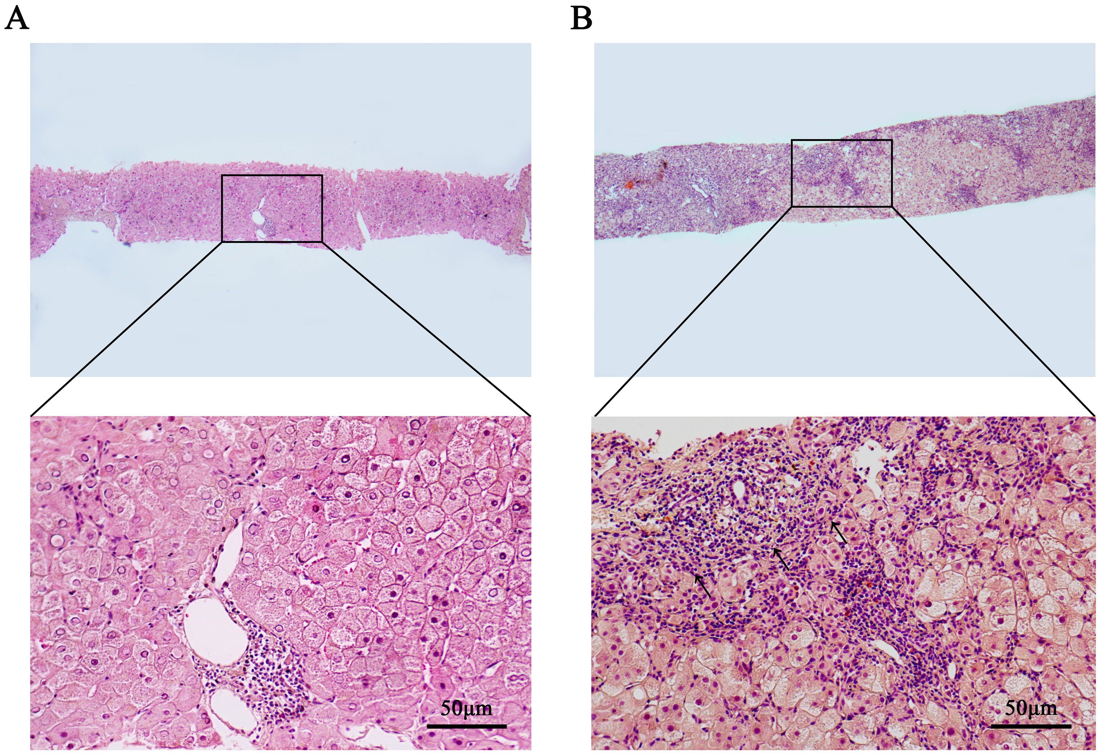
Figure 3. Histopathological features in liver biopsies of NAP-WD and AP-WD patients. (A) NAP-WD patient: no obvious interface hepatitis or plasma cell infiltration was observed (HE staining, 40×/200×); (B) AP-WD patients: marked interface hepatitis and plasma cell infiltration were observed (HE staining, 40×/200×). Diffuse hepatocyte enlargement, with moderate to severe inflammatory cell infiltration within the portal tracts, extensive fibrous tissue proliferation. Disorganized hepatic lobular architecture, with widespread hyaline degeneration of hepatocytes (HE staining, 40×/200×). Generated using Adobe Photoshop 2025. NAP-WD: Wilson’s disease without autoimmune phenomena; AP-WD: Wilson’s disease with autoimmune phenomena; HE: hematoxylin and eosin.


