



The temporal state-dependent fibrotic niche: a paradigm shift or a refinement of metastatic complexity? Examining the Areg-EGFR-Tnc-TLR4 circuit as a case study
 0

INTRODUCTION
A persistent challenge in lung metastasis biology is explaining the landing-to-outgrowth decision. Tumor cells can reach a distant organ, yet only some establish persistent lesions. How the host microenvironment converts an engraftment event into a stable tumor-supportive program remains unclear. Although the “pre-metastatic niche” concept is influential, its precise molecular and cellular architects remain poorly defined.
Fibrotic lung regions correlate with poor outcomes and may harbor permissive features-matrix remodeling, inflammatory cues, and altered immune homeostasis. However, “fibrosis” is not uniform. Different fibroblast programs co-exist across anatomical niches and injury timelines, potentially yielding distinct outputs for tumor adhesion, survival, and immune evasion. Thus, which fibroblast state(s) and temporal windows maximize early colonization?
In this context, Cardoso et al.[1] delineate how specific molecular circuits within these early fibrotic niches dictate permissiveness, moving beyond correlation toward mechanism. Their work bridges fibrotic niche biology with metastatic fate control, building on alveolar fibroblasts in pulmonary fibrosis and matrix remodeling[2]. This commentary explores the “temporal state-dependent fibrotic niche”, using the Amphiregulin (Areg)-Epidermal Growth Factor Receptor (EGFR)-Tenascin C (Tnc)-Toll-like Receptor 4 (TLR4) circuit as an example. However, does this framework represent a paradigm shift or a granular refinement of existing concepts, raising questions about therapeutic leverage?
THE AREG-EGFR-TNC-TLR4 CIRCUIT: A CENTRAL MECHANISM OR ONE NODE?
Cardoso et al.[1] showed that early fibrotic niches become tumor-permissive through an epithelial-stromal-immune signaling axis. Mutated epithelial programs activate EGFR in neighboring fibroblasts via Areg. This signaling drives fibroblast reprogramming and Tnc secretion. Tnc then signals through TLR4 on alveolar macrophages. This shifts immune behavior and creates a permissive microenvironment that suppresses antitumor immunity[1]. However, detailed myeloid/T-cell phenotypes and downstream effects on T-cell function require deeper investigation. These events can occur before overt malignancy-driven dissemination, aligning with pre-metastatic niche theory while emphasizing local early niche construction[3]. Whether this circuit is a master regulator or one of several parallel/redundant pathways remains unclear. Although hepatocellular carcinoma (HCC)-derived exosomal signals can pre-condition the microenvironment[4], direct evidence that Areg-EGFR-Tnc-TLR4 orchestrates this in HCC is limited. Mechanistically, this circuit exemplifies how fibroblast state transitions are co-opted to generate a tumor-compatible immune-matrix environment[1,2].
Extracellular-vesicle-mediated communication is another major mechanism reshaping stromal and immune compartments during niche formation[5], but its interplay with this axis remains largely unexplored.
DEFINING AND OPERATIONALIZING THE COLONIZATION WINDOW: BEYOND A TEMPORAL CONCEPT?
Disseminated tumor cells entering lung tissue face converging constraints: adhesion/growth initiation, survival under immune pressure, and durable niche architecture. A permissive microenvironment must satisfy these demands simultaneously, with timing matching early metastatic kinetics [Figure 1].
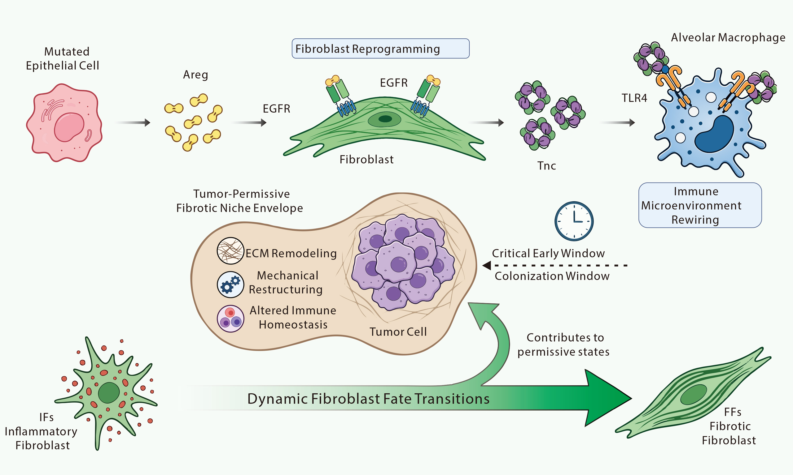
Figure 1. The Areg-EGFR-Tnc-TLR4 Signaling Axis Orchestrates a Temporal State-Dependent Fibrotic Niche for Lung Colonization. This schematic illustrates the intricate molecular and cellular mechanisms by which early fibrotic niches establish tumor-permissive microenvironments, facilitating lung colonization by disseminated tumor cells. The process initiates with mutated epithelial cells secreting amphiregulin (Areg), which then binds to and activates epidermal growth factor receptors (EGFR) on adjacent Fibroblasts. This triggers fibroblast reprogramming, leading to the robust secretion of Tenascin C (Tnc). Subsequently, Tnc engages Toll-like Receptor 4 (TLR4) on alveolar macrophages, resulting in profound immune microenvironment rewiring that favors tumor progression; This orchestrated signaling cascade culminates in the formation of a tumor-permissive fibrotic niche envelope around invading tumor cells. This niche is characterized by critical alterations including ECM remodeling, significant mechanical restructuring of the extracellular matrix, and an altered immune homeostasis that suppresses antitumor immunity. These events occur within a Critical Early Window, also termed the Colonization Window, which is crucial for successful tumor engraftment and survival. Underlying this process are dynamic fibroblast fate transitions, where distinct fibroblast states, such as inflammatory fibroblasts (IFs) and fibrotic fibroblasts (FFs), contribute to the permissive conditions of the niche. This highlights that the specific state and timing of the fibrotic microenvironment are pivotal determinants of metastatic success. ECM: Extracellular matrix.
The “colonization window” is the critical interval after dissemination during which cells either establish a persistent lesion or are eradicated.
1. Start Point: The arrival of circulating tumor cells (CTCs) in the pulmonary microvasculature and their initial extravasation. However, precise clinical detection of this “start point” presents significant diagnostic hurdles, challenging its real-world applicability.
2. End Point: Successful formation of a stable, self-sustaining micrometastasis or complete clearance of tumor cells by host defenses. Tumor cell dormancy and outgrowth dynamics heterogeneity complicates a universal “end point” definition.
3. Relationship With Dormancy/Reactivation: The colonization window likely precedes or critically influences subsequent dormancy or reactivation phases. Successful colonization might lead to rapid outgrowth or dormant lesions; failure leads to elimination. Microenvironmental cues dictate cellular fate. Clear conceptual demarcation from the broader dormancy-outgrowth continuum is required.
Within an inflammatory-fibrotic continuum, permissiveness plausibly emerges through at least three mechanisms, orchestrated by the Areg-EGFR-Tnc-TLR4 circuit identified by Cardoso et al.[1]:
1. Early Inflammatory Signaling: Inflammatory fibroblast states produce chemokine- and interferon-associated cues that promote engraftment and local growth initiation.
2. Extracellular Matrix (ECM) Quantity/Quality and Matricellular Support: Fibrotic differentiation remodels ECM composition and mechanics, affecting integrin engagement and survival. Matricellular proteins, such as Tnc[6], translate ECM remodeling into tumor-compatible signaling[1]. However, Tnc’s context-dependent pro- or antitumorigenic roles raise questions about its universal pro-metastatic nature.
3. Immune Homeostasis Reprogramming: Fibrotic niche programs reduce cytotoxic constraints and promote immune-compatible survival. Thus, inflammatory-fibrotic coupling may be an immune-ECM integrated switch governing progression from “landing” to sustained survival[7]. Tnc-TLR4 signaling on alveolar macrophages[1] exemplifies this reprogramming[8].
DISTINGUISHING FIBROTIC STATES: A CLINICALLY TRACTABLE FRAMEWORK?
Fang et al.[9] showed Runt-related transcription factor 2 (RUNX2) promotes fibrosis via an alveolar-to-pathological fibroblast transition. This reinforces a critical principle - fibroblast permissiveness depends on which transition route is selected, not on fibrosis presence alone. Upon Areg-EGFR activation, distinct fibroblast states could orchestrate the permissive microenvironment[1].
Together, Fang et al. and the detailed circuit defined by Cardoso et al. imply that the “fibrotic niche” should be treated as a set of distinguishable lineage states along an inflammatory-fibrotic continuum[1,2,9]. Metastatic timing may determine which state is encountered by tumor cells during the colonization window, modulating whether fibrosis acts as a barrier, a bystander, or a permissive organizer.
1. Inflammatory-Phase Stroma: Characterized by early fibroblast activation, inflammatory cells (e.g., M1 macrophages), elevated pro-inflammatory cytokines, and initial ECM remodeling. This phase may create an early permissive environment by providing growth factors, promoting angiogenesis, and initiating immune tolerance. Candidate markers include specific fibroblast activation markers (e.g., Alpha-smooth muscle actin) in conjunction with inflammatory cytokines (e.g., Interleukin-6).
2. Mature Scar Fibrotic Stroma: Defined by extensive ECM deposition (e.g., high collagen I/III ratio), reduced cellularity, and decreased vascularization, potentially creating an immunosuppressive or tumor-suppressive environment due to physical barriers and nutrient limitations. This phase might challenge tumor cells, driving dormancy or requiring stromal remodeling for outgrowth. Candidate markers include high levels of mature collagen and reduced cellularity.
This view also resonates with dormancy/reactivation models in which ECM composition and senescence-associated stromal programs regulate the time window for tumor outgrowth[8,10]. Fibrosis may be pro-metastatic only when its dominant state synchronizes with early tumor phase demands. This synchronization hypothesis highlights therapeutic timing challenges: knowing the dominant state and early tumor-phase demand simultaneously for each patient is a major diagnostic hurdle. Table 1 briefly compares the studies by Cardoso et al.[1], Zhu et al.[2], and Fang et al.[9].
Comparative overview of key studies on fibrotic niches and lung metastasis.
| Study | Core finding | Research model | Key cell types | Impact on niche formation/metastasis |
| Cardoso et al.[1] | Identified the Areg-EGFR-Tnc-TLR4 circuit as a key epithelial-stromal-immune signaling axis establishing early tumor-permissive fibrotic niches | Mouse models of lung metastasis, cell line-based assays | Mutated epithelial cells, fibroblasts, alveolar macrophages | Provides a mechanistic wiring diagram for early niche permissiveness |
| Zhu et al.[2] | Emphasized the crucial role of alveolar fibroblasts and their transitions (e.g., inflammatory-to-fibrotic) in driving pulmonary fibrosis and influencing tumor outcomes | In vitro fibroblast studies, pulmonary fibrosis models | Alveolar fibroblasts | Highlights dynamic fibroblast fate decisions as critical determinants of microenvironmental context |
| Fang et al.[9] | Uncovered RUNX2 as a transcriptional regulator promoting fibrosis via an alveolar-to-pathological fibroblast transition, influencing the permissiveness of fibrotic niches | Mouse models of lung fibrosis, genetic mouse models | Alveolar fibroblasts, pathological fibroblasts | Reveals specific fibroblast lineage transitions that dictate niche permissiveness through transcriptional reprogramming |
KEY QUESTIONS FOR THE TEMPORAL STATE-DEPENDENT FIBROTIC NICHE
Three testable questions emerge:
1. Which Fibroblast State Is Required for Colonization? Early metastasis may rely on inflammatory fibroblast dominance (supporting engraftment) or fibrotic consolidation (supporting survival and stable niches). Determining whether this dependency is time-dependent would refine the “colonization window” concept[2,9]. Identifying which fibroblast states are most susceptible to Areg-EGFR co-option is critical[1]. However, isolating transient fibroblast states in vivo remains technically challenging. If inflammatory versus fibrotic states carry distinct signatures, state-defined fibroblast biomarkers may predict early metastatic lesion formation. Yet, robust non-invasive detection of subtle, localized, heterogeneous fibroblast signatures is questionable, and defining permissive timing retrospectively offers little therapeutic advantage.
2. How To Block Permissive Remodeling Without Creating Alternative Routes? Targeting pathway nodes (e.g., Transforming growth factor-beta nodes from lineage transitions[2], or Areg-EGFR-Tnc-TLR4[1]) may reduce permissive outputs but could reconfigure inflammation and immune balance[1,2]. Interventions must inhibit permissive state transitions without compensatory inflammatory-to-fibrotic trajectories that restore permissiveness. This demands exhaustive understanding of compensatory and redundant pathways.
Beyond targeted inhibitors, innovative therapies such as the oncolytic virus VG161 - which reshapes the immunosuppressive and stromal microenvironment in advanced HCC - highlight the feasibility of converting a permissive niche into an antitumor immune response[11]. Areg is an attractive translational node because it bridges fibrosis-associated signaling and tumor-related permissiveness[1]. Yet interventions must be timed and state-matched to the colonization window to ensure net suppression rather than niche rewiring. Increasing evidence suggests microenvironmental components evolve through temporally distinct states, making stage-matched targeting important[12].
LIMITATIONS AND FUTURE PERSPECTIVES
1. Reliance On Animal Models: Much evidence, including Cardoso et al.[1], derives from in vitro studies and mouse models. Human validation using samples, patient-derived organoids, and clinical cohorts is needed.
2. Compensatory Pathways: Biological systems are redundant. Targeting Areg-EGFR-Tnc-TLR4 might trigger compensatory pathways in epithelial, stromal, or immune compartments, causing resistance or alternative pro-metastatic mechanisms. Systems biology approaches are needed to anticipate and mitigate these responses.
3. Clinical Translation Challenges: Developing specific, safe inhibitors, stratifying patients by niche state, and timing interventions within the dynamic colonization window remain difficult. Tumor and microenvironment heterogeneity further complicates one-size-fits-all approaches.
4. Detailed Immunosuppression Mechanisms: Although Tnc-TLR4 signaling on alveolar macrophages promotes immunosuppression, precise downstream myeloid and T-cell phenotypes remain conceptual. Future work should define immune cell subsets, functional states, and molecular mediators to identify immunotherapy targets[13]. The context-dependent nature of TLR4 signaling implies that simple blockade may have unintended systemic consequences or alter beneficial immune responses. A nuanced understanding of the pro-tumorigenic outcome of Tnc-TLR4 signaling is paramount before considering it broadly actionable.
CONCLUSION: FATE CONTROL -DISTANT HORIZON OR A REDEFINED PATH?
Cardoso et al.[1] provide a mechanistic cornerstone, defining Areg-EGFR-Tnc-TLR4 as a central driver of tumor-permissive early fibrotic niches. This moves lung colonization biology from correlation to a molecular and cellular wiring diagram. Integrated with fibroblast fate programs[2] and RUNX2-regulated pathological transitions[9], a more operational picture emerges: lung colonization is governed by lineage-specific, time-resolved niche wiring, not by fibrosis as a binary attribute. This commentary proposes the “temporal state-dependent fibrotic niche” as a unifying framework: the timing and specific state of the fibrotic microenvironment dictate its role in metastatic progression. Yet its impact depends on clinical actionability. If future studies validate lineage-resolved fibroblast states and enable targeted interventions, the field may move toward controlling early metastasis. However, formidable diagnostic hurdles and biological complexity demand realistic appraisal. The promise of “fate control” remains distant, requiring not only mechanistic elucidation but a fundamental leap in monitoring and manipulating these microenvironmental states in real time. Without such advances, this mechanistic understanding, while invaluable for basic science, risks remaining a testament to biological complexity rather than a direct path to clinical triumph.
DECLARATIONS
Authors’ contributions
Critically reviewed the literature and take responsibility for the integrity and accuracy of the analysis and interpretation presented in this commentary: Zhou X, Cui X, Zhang H
Study concept and design, supervised the study: Zhang H
Drafted the manuscript: Zhou X, Cui X
All authors have read and approved the final manuscript.
Availability of data and materials
Not applicable.
AI and AI-assisted tools statement
During the preparation of this manuscript, the AI tool ChatGPT (version GPT-6.1 Sol, released 2026-09-29) was used solely for language editing. The tool did not influence the study design, data collection, analysis, interpretation, or the scientific content of the work. All authors take full responsibility for the accuracy, integrity, and final content of the manuscript.
Financial support and sponsorship
This work was supported by the National Natural Science Foundation of China (81401988), China Postdoctoral Science Foundation (2019M661907), Jiangsu Postdoctoral Science Foundation (2019K159, 2019Z153), General Project of Jiangsu Provincial Health Committee (H2023136), General Project of Nantong Municipal Health Committee (MS2023013), Jiangsu Provincial Research Hospital (YJXYY202204-YSB28), Project of the Nantong Municipal Health Commission (MS2024010), and Nantong Social Livelihood Science and Technology Program (MSZ2025007).
Conflicts of interest
Zhang H is a Youth Editorial Board Member of the Journal of Translational Genetics and Genomics. Zhang H was not involved in any steps of editorial processing, including reviewer selection, manuscript handling, and decision-making. The other authors declare that there are no conflicts of interest.
Ethical approval and consent to participate
Not applicable.
Consent for publication
Not applicable.
Copyright
© The Author(s) 2026.
REFERENCES
1. Cardoso EC, Lee H, England FJ, et al. Early fibrotic niches establish tumour-permissive microenvironments. Nature. 2026;653:254-64.
2. Zhu H, Yi M, Song S, et al. The crucial role of alveolar fibroblasts in pulmonary fibrosis. J Transl Genet Genom. 2025;9:261-267.
3. Kong J, Tian H, Zhang F, et al. Extracellular vesicles of carcinoma-associated fibroblasts creates a pre-metastatic niche in the lung through activating fibroblasts. Mol Cancer. 2019;18:175.
4. Zhao Y, Yu H, Li J, et al. A glucose-enriched lung pre-metastatic niche triggered by matrix stiffness-tuned exosomal miRNAs in hepatocellular carcinoma. Nat Commun. 2025;16:1736.
5. Jahangiri L. The impact of extracellular vesicles on breast cancer metastasis and therapeutics: genetic considerations. Cancer Genet. 2025;298-299:1-9.
6. Oskarsson T, Massagué J. Extracellular matrix players in metastatic niches. EMBO J. 2012;31:254-6.
7. Cohen N, Mundhe D, Deasy SK, et al. Breast cancer-secreted factors promote lung metastasis by signaling systemically to induce a fibrotic premetastatic niche. Cancer Res. 2023;83:3354-67.
9. Fang Y, Chung SSW, Xu L, et al. RUNX2 promotes fibrosis via an alveolar-to-pathological fibroblast transition. Nature. 2025;640:221-30.
10. Di Martino JS, Nobre AR, Mondal C, et al. A tumor-derived type III collagen-rich ECM niche regulates tumor cell dormancy. Nat Cancer. 2022;3:90-107.
11. Hong YC, Chang R, Song S, et al. Oncolytic virus VG161 brings new hope for advanced HCC. Hepatoma Res. 2025;11:22.
12. Wang Y, Jia J, Wang F, et al. Pre-metastatic niche: formation, characteristics and therapeutic implication. Signal Transduct Target Ther. 2024;9:236.
Cite This Article
 Open Access
How to Cite
Zhou X, Cui X, Zhang H. The temporal state-dependent fibrotic niche: a paradigm shift or a refinement of metastatic complexity? Examining the Areg-EGFR-Tnc-TLR4 circuit as a case study. J Transl Genet Genom. 2026;10:592-8. https://dx.doi.org/10.20517/jtgg.2026.112
Download Citation
Export Citation File:
Type of Import
Tips on Downloading Citation
Citation Manager File Format
Type of Import
Direct Import: When the Direct Import option is selected (the default state), a dialogue box will give you the option to Save or Open the downloaded citation data. Choosing Open will either launch your citation manager or give you a choice of applications with which to use the metadata. The Save option saves the file locally for later use.
Indirect Import: When the Indirect Import option is selected, the metadata is displayed and may be copied and pasted as needed.
About This Article
Copyright

Data & Comments
Data
0






Comments
Comments must be written in English. Spam, offensive content, impersonation, and private information will not be permitted. If any comment is reported and identified as inappropriate content by OAE staff, the comment will be removed without notice. If you have any queries or need any help, please contact us at support@oaepublish.com.