fig6

Extraembryonic mesoderm in primate and rodent embryos and models

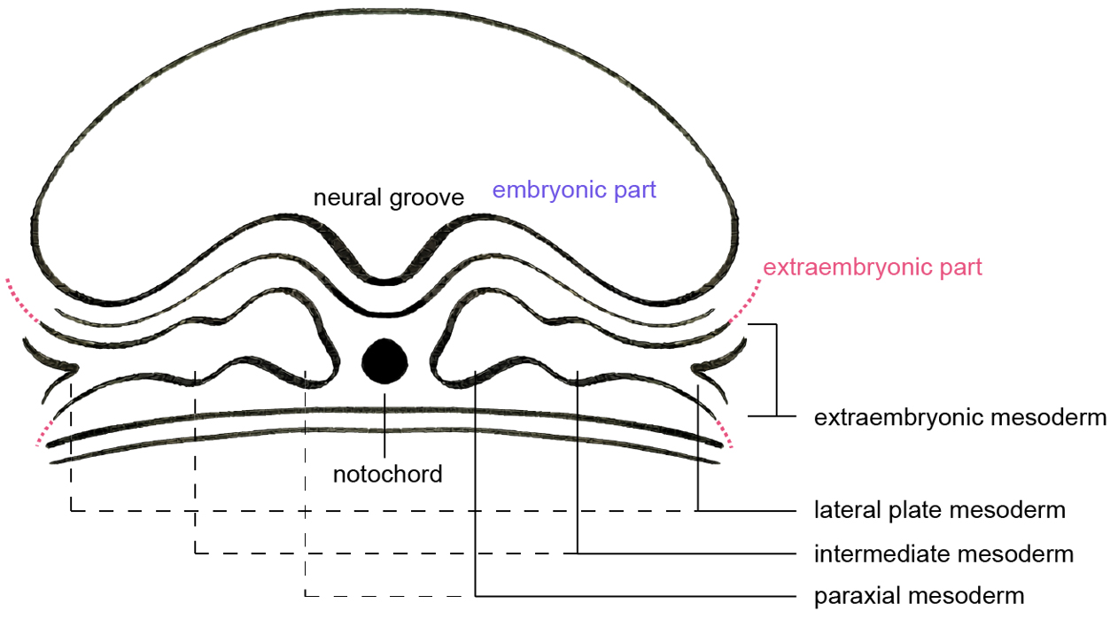
Figure 6. Schematic of a transverse plane through the post-gastrulation three-germ-layer structure, showing the mesoderm differentiating into paraxial, intermediate, lateral plate, and extraembryonic mesoderm. Image created by Adobe Illustrator.

Journal of Translational Genetics and Genomics
ISSN 2578-5281 (Online)
Follow Us

Portico

All published articles are preserved here permanently:

https://www.portico.org/publishers/oae/

Portico

All published articles are preserved here permanently:

https://www.portico.org/publishers/oae/