


fig5
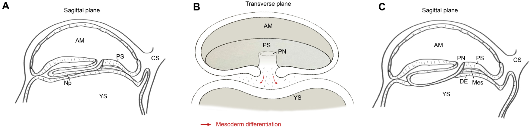
Figure 5. Schematic diagrams in sagittal (A and C) and transverse (B) planes illustrating the generation of nascent mesoderm and definitive endoderm during gastrulation, after which the bilaminar structure transforms into a trilaminar structure. Following gastrulation, the mesoderm migrates outward and differentiates into EXMC. The red arrow indicates that the differentiation of the mesoderm during gastrulation originates from the proliferation and differentiation of the epiblast towards the ventral side. AM: Amnion; PN: primitive node; PS: primitive streak; YS: yolk sac; CS: connecting stalk; Np: notochordal process; DE: definitive endoderm; Mes: mesoderm. Image created by Adobe Illustrator.


