


fig1
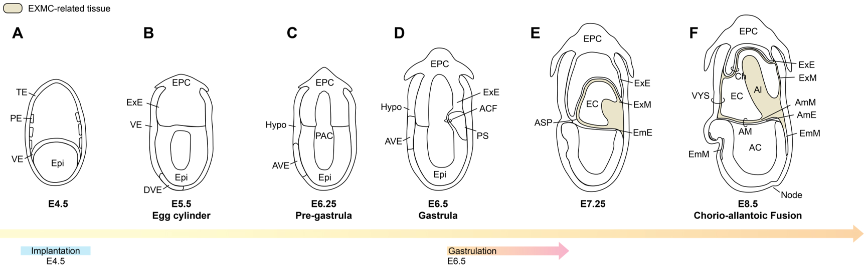
Figure 1. Developmental patterns from the implantation stage (A) to gastrulation (D), and to the early post-gastrulation phase (E and F) in rodent embryos. TE: Trophectoderm; PE: parietal endoderm; VE: visceral endoderm; Epi: epiblast; Hypo: hypoblast; EPC: ectoplacental cone; DVE: distal visceral endoderm; AVE: anterior visceral endoderm; PAC: pro-amniotic cavity: ACF: amniochorionic fold; PS: primitive streak; ASP: anterior separation point; ExE: extraembryonic ectoderm; ExM: extraembryonic mesoderm; AmE: amniotic ectoderm; AmM: amniotic mesoderm; EmE: embryonic ectoderm; EmM: embryonic mesoderm; AM: amnion; AC: amniotic cavity; VYS: visceral yolk sac; EC: exocoelomic cavity; Ch: chorion; Al: allantois. Image created by Adobe Illustrator.


