


fig1
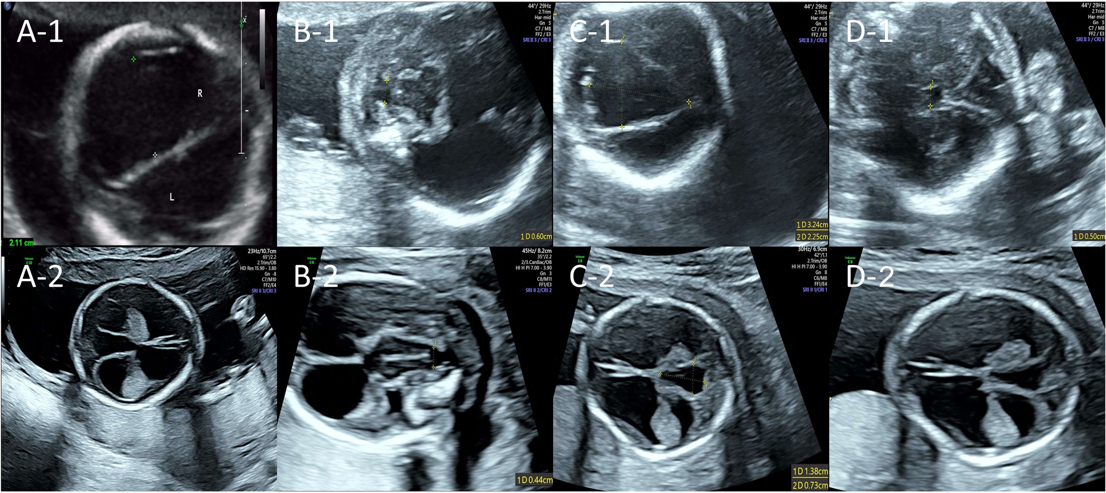
Figure 1. Fetal brain ultrasound findings of two consecutive pregnancies. The upper row shows the prenatal ultrasound from the first pregnancy at 22+4 weeks of gestation; the lower row shows that from the second pregnancy at 17+4 weeks of gestation. (A-1,2) Ultrasound suggested hydrocephalus; (B-1,2) Ultrasound suggested cerebellar dysplasia with vermian agenesis; (C-1,2) Ultrasound suggested a cystic midline cerebellar lesion (suspected arachnoid cyst); (D-1,2) Ultrasound suggested a complete absence of corpus callosum and cavum septum pellucidum.


