



Circulating biomarkers for the diagnosis, risk stratification, and translational assessment of fulminant myocarditis
 0
0 Abstract
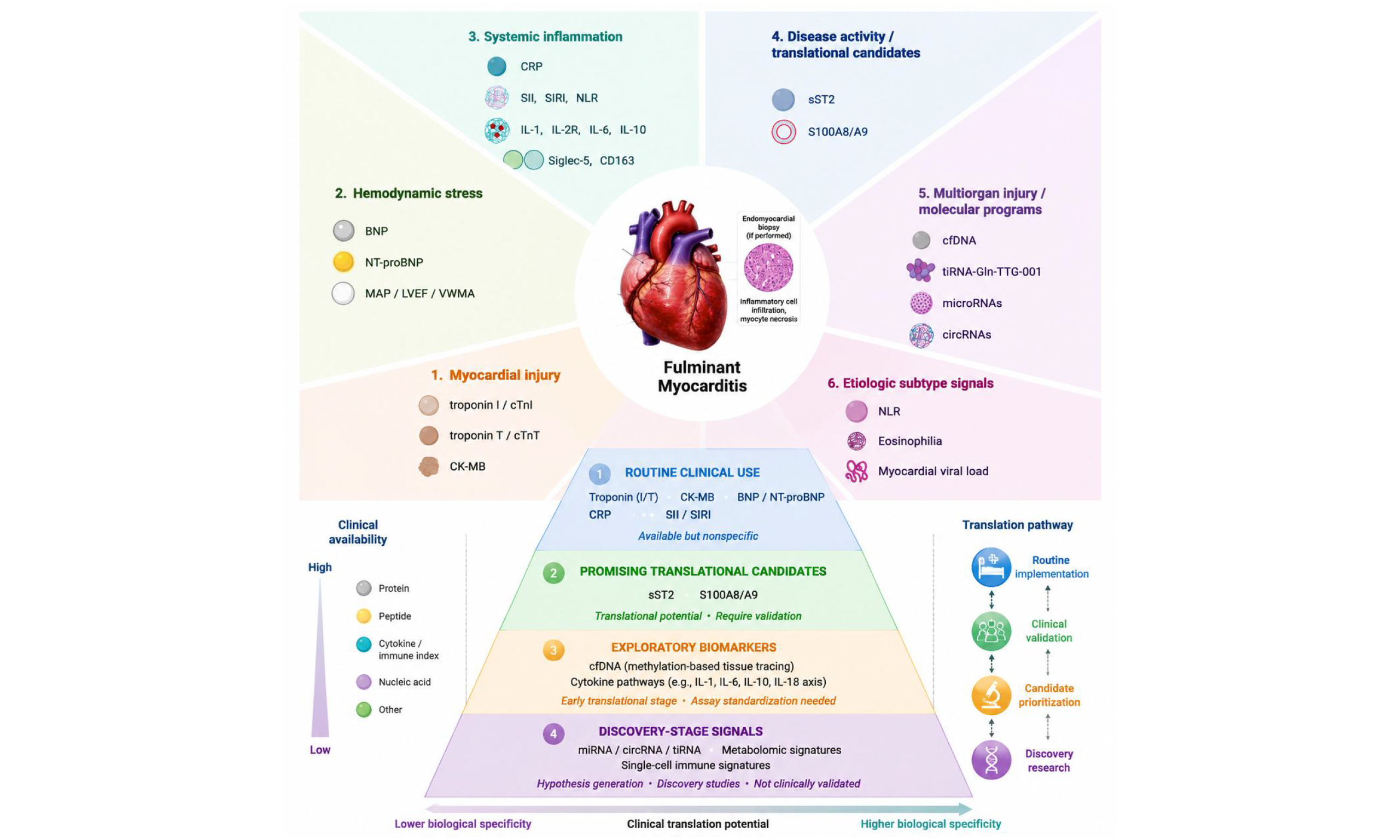
Fulminant myocarditis (FM) is the most severe and urgent type of myocarditis, characterized by rapid hemodynamic deterioration, malignant arrhythmias, and frequent multiorgan dysfunction. In this case, timely diagnostic decisions are critical. Because endomyocardial biopsy or cardiac magnetic resonance is time-consuming and requires specialized equipment and expertise, plasma biomarkers play an important role between initial clinical suspicion and later pathological confirmation. This review summarizes plasma biomarkers for FM according to their predominant biological relevance, including myocardial injury- and stress-related biomarkers, inflammation-related biomarkers, novel molecular biomarkers, and other etiologically informative signals. Conventional cardiac function biomarkers, including cardiac troponin, creatine kinase-MB, and natriuretic peptides, as well as inflammation-related indicators such as C-reactive protein, the systemic immune-inflammation index, and the systemic inflammatory response index, may provide initial information on myocardial injury, hemodynamic stress, and inflammatory activation in FM. Among novel molecular biomarkers, soluble ST2 (sST2) and S100A8/A9 are associated with the severity of FM and could also be used to evaluate the prognosis of patients with FM. Other emerging candidates, including cell-free DNA, tiRNAs, microRNAs, circRNAs, and metabolomic signals, remain promising but still require further validation. Current evidence also suggests that selected subtype-related biomarkers may contribute to etiologic refinement, which may in turn inform treatment selection. Overall, combined plasma biomarkers are needed for the diagnosis and stratified assessment of FM.
Keywords
INTRODUCTION
Fulminant myocarditis (FM) is the most severe and urgent type of myocarditis[1,2]. It is characterized by cardiac inflammation, rapid hemodynamic deterioration, malignant arrhythmias, and frequent multiorgan dysfunction[3-5]. Clinical deterioration often unfolds within a very short time window, leaving limited opportunity for prolonged diagnostic consideration[3,6]. This temporal profile distinguishes FM from less severe types of myocarditis and places a premium on early recognition and rapid risk assessment[7,8].
The clinical burden of FM remains substantial, even in the era of intensive care and temporary mechanical circulatory support[3,9]. In experienced tertiary centers, novel treatment has reduced mortality from more than 50% to less than 5%[3]. Yet these outcomes are not uniformly generalizable, and delayed recognition or limited access to advanced support may still contribute to high mortality in less-resourced settings[10-12]. The recently published 2025 ESC Guidelines for the management of myocarditis and pericarditis further emphasize a multimodal diagnostic and management framework for inflammatory myocardial and pericardial syndromes[13]. Plasma evaluation of suspected FM therefore remains clinically decisive, not because it replaces subsequent etiologic work-up, but because it can accelerate triage, hemodynamic surveillance, and timely escalation of care.
Cardiac magnetic resonance (CMR) is a major noninvasive tool for myocarditis[1]. Endomyocardial biopsy (EMB) also remains the histological reference standard[14,15], especially when further etiologic evaluation is needed. In practice, however, patients with FM are often hemodynamically unstable, mechanically ventilated, in shock, or already receiving vasoactive agents or extracorporeal support[2,9]. Under these circumstances, plasma biomarkers are particularly valuable because they can be obtained rapidly, repeated serially, and used to track dynamic changes in disease activity, thereby supporting timely clinical evaluation in patients with FM[16]. Routine markers of myocardial injury, ventricular stress, and systemic inflammation remain the foundation of initial laboratory assessment, but recent translational studies have expanded the candidate landscape to include pathway-linked protein biomarkers and circulating nucleic-acid signals. Recent evidence has highlighted molecules such as soluble ST2, S100A8/A9, and cell-free DNA (cfDNA) as biologically informative candidates that may capture disease activity, inflammatory burden, organ injury, and short-term risk in FM.
This review classifies FM biomarkers according to their predominant biological relevance, including myocardial injury- and stress-related biomarkers, inflammation-related biomarkers, novel molecular biomarkers, and other etiologically informative signals. Within each category, the available evidence is examined for diagnostic value, association with disease severity, prognostic implications, and current limitations. This biology-oriented approach also helps distinguish candidates supported by relatively strong evidence from exploratory signals that still require translational validation.
SEARCH STRATEGY AND SELECTION CRITERIA
To improve the transparency and reproducibility of this narrative review, we searched PubMed/MEDLINE and Web of Science from database inception to March 2026. The search strategy combined disease-related terms, including “fulminant myocarditis”, “acute myocarditis”, “viral myocarditis”, and “myocarditis”, with biomarker-related and clinical terms, including “biomarker”, “circulating marker”, “plasma marker”, “serum marker”, “diagnosis”, “risk stratification”, “prognosis”, and “severity”. Because the term “fulminant myocarditis” is not used consistently across the literature and is sometimes interchanged with acute or viral myocarditis, potentially relevant studies were further assessed according to whether the reported population met the current clinical concept of FM, including acute onset, rapid clinical deterioration, hemodynamic instability or cardiogenic shock, malignant arrhythmias, severe heart failure, multiorgan dysfunction, intensive care requirement, or the need for mechanical circulatory support. Studies were included when the population was clearly diagnosed as FM or when FM-related data could be reasonably identified from the clinical description or subgroup analysis. Original clinical studies, mechanistic or translational studies, guidelines, consensus documents, and high-quality reviews were considered when they were relevant to circulating biomarkers, diagnostic assessment, severity stratification, prognostic evaluation, etiologic refinement, or clinical translation in FM. Priority was given to original studies reporting diagnostic performance, prognostic or severity-related estimates, dynamic biomarker changes, assay feasibility, or mechanistic evidence. Publications were excluded if they were unrelated to myocarditis or biomarker research, did not provide relevant circulating or clinically applicable biomarker evidence, duplicated data from another report, or provided only tangential background information. Case reports and animal-only studies were cited selectively when they provided clinically important subtype context or mechanistic support for key candidate biomarkers. Because this article is a narrative review rather than a systematic review or meta-analysis, no quantitative pooling was performed.
BIOMARKERS OF MYOCARDIAL INJURY AND HEMODYNAMIC STRESS
The biomarkers discussed in this section are classified according to their predominant relation to cardiomyocyte injury or ventricular stress. In FM, these markers remain clinically important because they are rapidly obtainable, widely available, and routinely measured at admission[1,9]. Their major limitation, however, is limited disease specificity. Overall, they provide an initial biochemical profile of myocardial injury and hemodynamic burden, thereby helping clinicians recognize a severe inflammatory cardiac syndrome during the initial evaluation.
Cardiac troponin and CK-MB
Cardiac troponin and creatine kinase-MB (CK-MB) remain the core routine biomarkers of myocardial injury in FM[17,18]. Their practical value lies in their accessibility and rapid turnaround time. In particular, troponin directly reflects cardiomyocyte damage and is therefore central to the initial laboratory assessment of suspected FM. CK-MB, although less specific, still contributes to the conventional injury profile and may be useful in routine emergency evaluation where extended molecular assays are not available.
These injury markers should not be overinterpreted. Elevated troponin levels may also occur in other acute cardiovascular conditions, particularly acute coronary syndromes[19,20]. Therefore, in FM, abnormal troponin or CK-MB values are best understood as evidence of substantial myocardial injury within the appropriate clinical context, rather than as independent proof of a FM diagnosis. Their major role is to support suspicion, quantify the extent of injury, and prompt timely integration with electrocardiographic, hemodynamic, and imaging findings[17,18].
Natriuretic peptides
Natriuretic peptides, including B-type natriuretic peptide (BNP) and N-terminal pro-B-type natriuretic peptide (NT-proBNP), complement myocardial injury markers by reflecting ventricular wall stress and acute hemodynamic load[17,18]. In FM, BNP and NT-proBNP are often elevated because fulminant disease is frequently accompanied by rapidly evolving ventricular dysfunction, circulatory instability, and heart failure physiology. As a result, natriuretic peptide elevation may provide useful information on the stress imposed on the failing ventricle, especially when interpreted together with troponin rather than in isolation.
As with troponin, natriuretic peptides are not specific for FM. Their levels may also rise in acute heart failure and other settings of ventricular dysfunction[21,22]. Their value in FM therefore lies less in etiologic discrimination than in physiologic characterization. A combined pattern of marked myocardial injury and ventricular stress may be more informative than any single conventional marker alone, because it better reflects the coexistence of inflammatory myocardial damage and acute hemodynamic decompensation that typifies fulminant presentation.
Routine variable-based composite discrimination models
The discriminatory value of routine biomarkers may increase when they are interpreted together with bedside physiologic and organ-injury variables. In a retrospective study of adults, discrimination between fulminant and non-FM improved when routine admission variables were analyzed in combination rather than separately[23]. The final model included mean arterial pressure, creatinine, blood urea nitrogen, aspartate aminotransferase, troponin I, and ventricular wall motion abnormality[23]. This observation is conceptually important because it suggests that FM is often recognized more accurately as a combined syndrome of myocardial injury, hemodynamic compromise, and systemic organ stress than as an isolated elevation in a single cardiac biomarker.
This composite approach is best viewed as a clinical discrimination model rather than a molecular biomarker panel. Its main value is pragmatic: it may help identify which patients with suspected myocarditis are more likely to have a fulminant presentation using information already available at admission[23]. In this context, routine injury- and stress-related biomarkers also show measurable but nonspecific discriminatory value. In a young-adult acute myocarditis cohort, troponin I discriminated FM with an area under the curve (AUC) of 0.772 at a cutoff of 0.48 ng/mL, whereas NT-proBNP showed stronger performance, with an AUC of 0.969 at a cutoff of
BIOMARKERS OF INFLAMMATION AND IMMUNE ACTIVATION
Inflammation-related biomarkers in FM include conventional acute-phase reactants, blood count-derived inflammatory indices, and inflammation-linked protein candidates. Rather than representing a separate temporal stage of testing, these markers reflect the intensity and pattern of systemic and myocardial immune activation that accompany fulminant disease.
CRP and conventional inflammatory markers
C-reactive protein (CRP) remains one of the simplest and most widely available inflammatory markers in the evaluation of suspected FM[17,18]. In clinical practice, its value lies in indicating that severe myocardial injury is occurring in an inflammatory milieu rather than in a purely ischemic or hemodynamic setting[25]. When interpreted together with troponin, natriuretic peptides, clinical presentation, and hemodynamic findings, CRP may therefore strengthen suspicion that the syndrome is inflammatory rather than purely mechanical in nature.
Its limitations, however, are substantial. CRP may also increase in infection[26], sepsis[27], and other inflammatory states[28]. Accordingly, CRP should be regarded as a supportive marker of inflammatory context rather than a discriminator of FM etiology or a standalone determinant of fulminant presentation.
Blood count-derived inflammatory biomarkers
Routine blood count-derived inflammatory indices provide a simple and readily available approach to inflammatory assessment in FM. The systemic immune-inflammation index (SII) and the systemic inflammatory response index (SIRI) are attractive because they can be calculated rapidly after admission and do not require new assays[29,30]. In pediatric and young-adult myocarditis cohorts, SII and SIRI demonstrated potential diagnostic value for FM. In one pediatric cohort, SII showed an AUC of 0.760 with a cutoff value of 1,050, whereas SIRI showed an AUC of 0.640 with a cutoff value of 1.9. In a young-adult myocarditis cohort, SII showed stronger diagnostic performance, with an AUC of 0.911 at a cutoff value of 1020, 91% sensitivity, and 83% specificity, and remained an independent predictor of FM[29]. Across available studies, reported SII performance varied, with AUC values ranging from approximately 0.760 to 0.920, cutoff values from 1,020 to 1,378, sensitivity from 68.8% to 91%, and specificity from 83% to 94.4%. Similarly, SIRI showed variable diagnostic performance, with reported AUC values from approximately 0.640 to 0.710 and cutoff values from 1.9 to 2.058. These variations suggest that age group, cohort design, sampling time, and differences in FM definitions may influence the observed diagnostic performance of these inflammatory indices[29]. The main appeal of SII and SIRI is not molecular specificity but bedside immediacy: they translate a routine complete blood count into an estimate of systemic immune activation at essentially no additional cost. This makes them especially relevant in settings where advanced biomarker assays are unavailable or delayed.
These indices should nonetheless be interpreted cautiously. Their biological plausibility in FM is strong because fulminant disease is often accompanied by marked innate immune activation[31,32], but they are not myocarditis-specific tools. They may also rise in sepsis[33], infective endocarditis[34], and other inflammatory conditions. SII and SIRI are therefore better framed as inflammatory enrichment markers or triage aids rather than as disease-specific FM biomarkers. In the current literature, SII appears more promising than SIRI, but both still require broader validation across more rigorously phenotyped FM cohorts[35]. Accordingly, reported AUC values for SII and SIRI should be interpreted as preliminary cohort-specific estimates rather than stable diagnostic thresholds ready for routine clinical use.
S100A8/A9
S100A8/A9 is the most compelling inflammation-related protein biomarker currently studied in FM. S100A8 and S100A9 are calcium-binding S100 family proteins that form the heterodimeric complex calprotectin and are mainly expressed by activated neutrophils and monocytes/macrophages[36]. Extracellular S100A8/A9 can function as an alarmin and amplify innate immune responses through pattern-recognition receptors such as Toll-like receptor 4 and the receptor for advanced glycation end products[37]. Plasma proteomic studies identified S100A8/A9 as a candidate biomarker and possible therapeutic target[37]. In validation analyses, admission S100A8/A9 distinguished FM from non-FM, acute myocardial infarction, and acute decompensated heart failure[37]. This is clinically important because these conditions are common acute-care mimics of FM.
In a two-center validation cohort, plasma S100A8/A9 measured at admission differentiated FM from non-FM with an AUC of 0.923, from acute myocardial infarction with an AUC of 0.892, and from acute decompensated heart failure with an AUC of 0.978[37]. The heterodimer also performed better than S100A8 or S100A9 alone, supporting the view that the combined signal is biologically and diagnostically more informative than either subunit measured separately.
S100A8/A9 was also associated with disease severity. Higher levels were linked to lower left ventricular ejection fraction and greater inflammatory burden[37]. These findings suggest that S100A8/A9 may reflect both the intensity of myocardial injury and the strength of the inflammatory response. Its biological relevance further supports its candidacy, because S100A8/A9, also known as calprotectin, functions within a damage-associated molecular pattern pathway. In the same clinical dataset, higher S100A8/A9 expression was accompanied by higher circulating IL-1, IL-2R, IL-6, and IL-10 levels, reinforcing the view that this marker tracked a broader hyperinflammatory state rather than isolated tissue injury alone[37]. Experimental evidence has further strengthened this candidate. In a coxsackievirus B3-induced FM model, blockade of the S100A8/A9 pathway improved survival, reduced inflammatory cell infiltration, and improved cardiac function[37]. Pharmacologic blockade with ABR-238901 reduced mortality and mitigated the decline in cardiac function in myocarditis mice, thereby providing interventional support for a mechanistic role rather than a purely associative signal[37]. Thus, S100A8/A9 may represent more than a passive inflammatory biomarker. Experimental intervention studies suggest that it can act as an active inflammatory mediator in FM, partly through Toll-like receptor 4 and receptor for advanced glycation end products signaling. However, whether S100A8/A9 is causally involved in human FM remains unproven. It should therefore be regarded as a mechanistically plausible biomarker and potential therapeutic target, rather than a clinically validated causal factor.
Overall, S100A8/A9 currently represents a high-priority translational candidate among inflammation-related biomarkers in FM, because its evidence spans discovery proteomics, human diagnostic validation, association with functional severity and cytokine activation, and experimental therapeutic rescue. However, it should not yet be regarded as a clinically established diagnostic marker. Further prospective multicenter studies are needed to validate its diagnostic cutoffs, clarify the influence of sampling time and assay platform, and determine whether S100A8/A9 adds incremental value beyond conventional inflammatory and myocardial injury markers.
Other exploratory inflammatory biomarkers
Beyond S100A8/A9, additional inflammation-linked protein candidates have emerged in recent literature. In a cytokine-focused investigation, plasma Siglec-5 and CD163 were reported to have diagnostic and severity-related associations with FM[38]. Siglec-5 is an inhibitory sialic acid-binding immunoglobulin-like lectin expressed mainly on myeloid cells[39], whereas CD163 is a scavenger receptor predominantly expressed on monocytes and macrophages and is commonly regarded as a marker of macrophage activation[40]. These biological features provide a rationale for evaluating circulating Siglec-5 and CD163 as inflammation-related biomarkers in FM. However, compared with S100A8/A9 and sST2, the available evidence remains less mature, and independently validated predictive metrics, stable cutoffs, and externally confirmed sensitivity or specificity estimates are still limited. Their levels were also negatively correlated with left ventricular ejection fraction[38]. These findings are noteworthy because both molecules are biologically consistent with dysregulated innate immune activation and macrophage-related inflammatory responses. Unlike S100A8/A9, however, these candidates remain supported mainly by early exploratory datasets rather than by layered validation across independent cohorts and mechanistic intervention studies. Siglec-5 and CD163 are therefore best presented at this stage as hypothesis-generating inflammatory protein candidates rather than established clinical biomarkers for FM[38].
The inflammation-related biomarker landscape in FM thus shows a clear gradient of maturity: CRP, SII, and SIRI are accessible but nonspecific; S100A8/A9 is biologically anchored and comparatively well validated; and Siglec-5/CD163 remain promising but preliminary. This layered interpretation is more informative than treating all inflammatory markers as equivalent signals. Even so, inflammation-related markers do not fully capture disease dynamics, short-term prognosis, or multiorgan injury, thereby motivating interest in novel molecular biomarkers.
NOVEL MOLECULAR BIOMARKERS
Beyond conventional markers of myocardial injury and systemic inflammation, a growing group of novel biomarkers has expanded the molecular landscape of FM. These candidates provide additional biological information on disease activity, short-term risk, multiorgan injury, and pathogenic mechanisms. Some biomarkers, particularly soluble ST2, have accumulated relatively strong diagnostic, prognostic, and mechanistic support, whereas others, including cfDNA, non-coding RNAs, and metabolomic signals, remain promising but more exploratory. These novel biomarkers are therefore best reviewed as a gradient of emerging molecular tools with different levels of clinical readiness.
Soluble ST2
Among currently studied molecular biomarkers, sST2 remains one of the most compelling protein biomarker candidates in FM[17,41]. sST2 is the soluble isoform of the interleukin-1 receptor family protein ST2 and can act as a circulating decoy receptor for IL-33. Recent mechanistic evidence in FM further suggests that CCR2-positive macrophage-derived sST2 may be internalized by cardiomyocytes through the insulin-like growth factor 2 receptor and contribute to mitochondrial dysfunction[41]. Earlier studies also showed that circulating sST2 increased markedly during the acute phase of FM and declined during recovery[17]. This dynamic pattern suggested that sST2 may reflect disease activity over time rather than simply a fixed baseline abnormality. Additional clinical work supported its diagnostic value. In children with suspected viral myocarditis, one retrospective study showed that sST2 was associated with FM and had good diagnostic performance, with an area under the curve of 0.865[42]. The validation confirmed that sST2 also correlated with markers of cardiac stress and cardiac dysfunction[42].
Later work strengthened the clinical case for sST2. In one prospective cohort, a plasma threshold of
Experimental data have provided unusually strong mechanistic validation for this marker. A recent study showed that sST2 was derived predominantly from infiltrating CCR2-positive macrophages, entered cardiomyocytes via the insulin-like growth factor 2 receptor, altered YY1 localization, and suppressed mitochondrial electron transport chain gene expression[41]. Neutralizing sST2 restored mitochondrial function, improved cardiac performance, and increased survival in FM models[41]. These findings indicate that sST2 may be linked not only to severity, but also to disease progression itself. In this context, sST2 is best viewed as a multifunctional biomarker in FM: it may support diagnosis at admission, reflect disease dynamics during recovery, identify patients at higher short-term risk, and point to a targetable pathogenic pathway.
The current evidence still has practical limitations. Most available sST2 studies remain single-center or modest in size, and serial sampling has not been standardized across cohorts. Nevertheless, sST2 is one of the most mature FM biomarker candidates and deserves translational priority because it integrates diagnostic, prognostic, dynamic, and mechanistic evidence. Future studies should validate disease- and outcome-specific cutoffs, standardize sampling windows and assay platforms, and test whether sST2 provides incremental value beyond troponin, natriuretic peptides, echocardiography, and hemodynamic variables.
Cell-free DNA
cfDNA represents a distinct direction in FM biomarker research because it may reflect not only myocardial injury but also the multisystem damage that frequently accompanies fulminant disease[43]. Recent work using genome-wide cfDNA methylation profiling suggested that plasma cfDNA could trace the tissue origins of injury and identify early multiorgan involvement in myocarditis[43]. In the reported cohort, cfDNA showed potential for early risk stratification and appeared to outperform several conventional biochemical indicators in identifying severe organ injury patterns[43].
The conceptual value of cfDNA is considerable. Although cfDNA methylation profiling may provide tissue-of-origin information and early multiorgan injury assessment, current FM evidence remains exploratory. Standardized FM-specific diagnostic cutoffs, externally validated sensitivity and specificity estimates, and prospectively tested risk-prediction metrics have not yet been established. Therefore, cfDNA should be presented as a tissue-injury mapping and risk-stratification research tool rather than as a clinically validated diagnostic biomarker.
Non-coding RNA biomarkers: tiRNAs, microRNAs, and circRNAs
Non-coding RNA biomarkers in FM, including tiRNAs[44], microRNAs[45,46], and circular RNAs[47], remain exploratory but are of interest because they may reflect tightly regulated inflammatory and injury-response programs rather than nonspecific tissue damage alone. Among these, tiRNA-Gln-TTG-001 was reported to be elevated during the acute phase of pediatric FM, to decrease during recovery, and to correlate with hs-cTnT, CRP, and procalcitonin[44]. However, available studies mainly report differential expression and correlation analyses rather than validated FM-specific AUC, cutoff, sensitivity, specificity, odds ratio, hazard ratio, or C-index estimates. Circulating microRNA candidates, including hsa-miR-21, were also associated with FM severity or prognosis in pediatric cohorts[48], while circRNA profiling identified dysregulated inflammatory signaling networks and highlighted candidates such as hsa_circ_0064338[47]. Collectively, these studies suggest that RNA-based biomarkers may provide mechanistically informative molecular signatures in FM.
The ncRNA literature is still limited by small sample sizes, heterogeneous study designs, and incomplete validation in clinically relevant control populations. Accordingly, tiRNAs, microRNAs, and circRNAs are better regarded as innovative molecular candidates than as clinically deployable biomarkers for current FM practice.
Metabolomic signals
Metabolomic signals are biologically relevant to FM because fulminant disease is characterized by profound inflammatory activation, energetic stress, and frequent multiorgan dysfunction[2,49,50]. In principle, these features make metabolic profiling an attractive approach for capturing the systemic consequences of fulminant progression. However, the current literature has not yet produced a validated circulating metabolite panel with clear bedside utility for FM. For this reason, metabolomics should presently be discussed mainly as a discovery-oriented tool that may help identify perturbed pathways and generate mechanistic hypotheses, rather than as an established clinical biomarker strategy for FM.
Overall, the novel biomarker landscape in FM shows a clear gradient of maturity. sST2 is currently the most biologically and clinically substantiated candidate. For most other non-coding RNA and metabolomic candidates, the available evidence remains at the discovery or early translational stage. These studies typically report differential expression, pathway enrichment, correlation with injury or inflammatory markers, or candidate regulatory networks, but do not yet provide externally validated FM-specific predictive metrics such as AUC, C-index, hazard ratio, odds ratio, standardized cutoffs, sensitivity, or specificity. These biomarkers should therefore be interpreted as hypothesis-generating molecular signals rather than clinically actionable diagnostic tools.
ETIOLOGICALLY INFORMATIVE BIOMARKERS
Because FM is a clinical syndrome that may arise from diverse forms of myocarditis, etiologically informative biomarkers are conceptually attractive. Based on the currently available literature, however, their role remains more limited than that of routine injury markers, inflammatory biomarkers, or selected novel molecular candidates such as sST2 and S100A8/A9. In most cases, these signals contribute more to etiologic refinement than to frontline diagnosis, and their interpretation usually becomes meaningful only when exposure history, clinical presentation, or routine laboratory abnormalities have already suggested a particular subtype.
Immune checkpoint inhibitor-associated myocarditis
Immune checkpoint inhibitor (ICI)-associated myocarditis provides a clear example of the context-dependent value of etiologically informative biomarkers in FM. In patients with recent exposure to ICIs, abnormalities in routine laboratory markers may be interpreted more specifically in light of the exposure history. Troponin elevation, often together with creatine kinase elevation, may raise suspicion for myocarditis in this case[51]. Even so, these markers are not subtype-specific in a strict sense. Their principal value lies in supporting etiologic suspicion in the context of a known trigger, rather than serving as a dedicated diagnostic biomarker panel for ICI-associated FM[52].
The available ICI myocarditis literature further suggests that inflammatory indices such as neutrophil-to-lymphocyte ratio (NLR) and CRP may have adjunctive prognostic value, whereas emerging candidates such as selected microRNAs remain preliminary[53]. However, these markers have not yet been validated as FM subtype-defining biomarkers with reproducible cutoffs or robust predictive metrics. Their interpretation remains highly dependent on ICI exposure history, concurrent myositis or skeletal muscle injury, cancer status, and the broader clinical context. Deep immune profiling has also identified expansion of cytotoxic CD45RA (TEMRA) CD8-positive T cells with clonal enrichment and inflammatory CXCL9/CXCL10-positive macrophage programs, but these findings have not yet established a routine circulating biomarker panel for bedside use[51,52]. Accordingly, in ICI-associated FM, biomarker interpretation still depends more on exposure context and integrated clinical suspicion than on any validated subtype-defining blood signature.
Eosinophilic and giant cell myocarditis
For eosinophilic myocarditis and giant cell myocarditis, the current circulating biomarker landscape is even less mature. There is no specific evidence supporting a validated standalone blood biomarker that reliably identifies giant cell myocarditis at presentation. Accordingly, no robust circulating AUC, cutoff, sensitivity, specificity, odds ratio, hazard ratio, or C-index can currently be recommended for blood-based identification of giant cell myocarditis in the FM setting. Instead, giant cell myocarditis is usually suspected from a particularly high-risk clinical phenotype, especially refractory ventricular arrhythmias or advanced conduction disease[54]. Likewise, eosinophilic myocarditis may show peripheral eosinophil-related signals, but these findings mainly suggest a possible cause rather than serving as definitive biomarkers[55].
This distinction is important for the review narrative. In these subtypes, currently available blood abnormalities function more as etiologic clues than as true diagnostic biomarkers, and their interpretation remains inseparable from the broader clinical picture. These entities should therefore be discussed briefly and cautiously in a biomarker review: their importance is unquestionable, but the biomarker evidence remains limited.
Viral FM and other cause-oriented clues
Viral myocarditis remains one of the major etiologic substrates of FM[31], yet the current literature still does not support a mature blood-based biomarker that can independently distinguish active viral FM from other inflammatory or noninflammatory acute cardiac syndromes[31,32]. In the available literature, tissue virology remains more informative than circulating markers, and PCR-based evaluation of myocardial viral load remains important for distinguishing active viral replication from post-viral inflammatory disease[31]. Blood biomarkers in viral FM should therefore be understood as supportive rather than definitive etiologic tools.
More broadly, the main lesson from etiologic biomarker studies is not that subtype-specific plasma markers are already clinically established, but that different FM subtypes may possess distinct immune programs that could guide future biomarker discovery. The immune-profiling findings in ICI-associated myocarditis and the growing interest in viral myocarditis-associated ncRNAs support this direction, but they have not yet translated into robust bedside assays for routine etiologic classification. Overall, other biomarkers and etiologic clues in FM remain limited in both maturity and scope. Their present value lies mainly in narrowing the likely cause in selected clinical settings—particularly when exposure history, eosinophilia, refractory arrhythmia, or other distinctive features have already focused suspicion. They are not yet reliable as primary tools for early diagnosis, severity stratification, or prognosis in most acute-care presentations of FM.
EMERGING TECHNOLOGIES, TRANSLATIONAL CHALLENGES, AND FUTURE PERSPECTIVES
Multi-omics approaches are reshaping biomarker discovery in FM, not because they create a separate class of biomarkers, but because they help identify which circulating signals are most closely linked to fulminant progression[41,56,57]. By integrating peripheral blood single-cell RNA sequencing, single-cell T-cell receptor sequencing, cytometry by time-of-flight (CyTOF), and plasma proteomics, recent studies have moved the field beyond broad nonspecific inflammatory readouts toward cellular circuits and molecular pathways that may be more informative for candidate prioritization[56,58].
Integrated profiling studies of acute myocarditis and fulminant disease identified clonally expanded cytotoxic CD57-positive CD8-positive effector T cells and linked fulminant progression to IL-18-associated signaling, natural killer-like receptor programs, and recruitment of proinflammatory monocytes[56,59]. These analyses further suggested that circulating interleukin-18, mainly associated with CXCL8-positive CD14-positive monocytes, promoted the differentiation of pathogenic effector T-cell populations[56]. Importantly, experimental blockade of the monocyte-IL-18-CCR5 axis reduced myocardial injury and improved cardiac function[56]. These findings matter less because IL-18 is already a routine biomarker, and more because they show how multi-omics can identify biologically anchored candidates and disease-relevant immune networks.
From a translational perspective, multi-omics findings are most useful when they reinforce candidates supported by clinical, dynamic, and mechanistic evidence rather than generating isolated molecular signals. S100A8/A9 and sST2 illustrate this principle because both are supported by evidence across clinical validation, disease-severity association, dynamic or mechanistic findings, and experimental data[37,41]. cfDNA represents a third direction, extending biomarker discovery from cardiac injury alone to multiorgan injury tracing[43]. Related evidence from ICI-associated myocarditis also suggests that subtype-specific immune programs, including cytotoxic TEMRA CD8-positive T-cell expansion and inflammatory macrophage-T-cell interaction patterns, may guide biomarker discovery in more clearly defined FM settings[51,60].
Overall, multi-omics approaches are currently most valuable for candidate prioritization rather than routine bedside diagnosis, and their clinical value depends on whether prioritized candidates can overcome the translational barriers that still characterize FM biomarker research. Despite the growing number of candidate biomarkers in FM, the major bottleneck is not a lack of molecular signals, but the limited robustness and comparability of the supporting evidence[5,61]. Many reported biomarkers have been derived from small, single-center, retrospective, or cross-sectional studies, often without uniform adjudication of FM or consistent external validation. This pattern has been observed across inflammatory indices, cfDNA studies, RNA-based biomarkers, pediatric extracorporeal membrane oxygenation (ECMO) cohorts, and subtype-oriented myocarditis reports. As a result, the field currently contains many promising candidates, but far fewer markers that can be considered clinically mature.
A second major barrier is heterogeneity in FM definition. Across the literature, FM has been referred variably to hemodynamic collapse at presentation, biopsy-proven fulminant disease, ECMO-treated pediatric myocarditis, or clinically suspected myocarditis with severe deterioration[15,58,62]. These definitions are not interchangeable. This inconsistency makes it difficult to compare studies directly, establish universal thresholds, or construct a single biomarker-based diagnostic framework applicable across cohorts. Sampling time is another major determinant of biomarker interpretation in FM. Biomarkers have been measured at markedly different points in the disease course, including symptom onset, hospital admission, circulatory collapse, ECMO initiation, early stabilization, and recovery[12,23,63]. A marker that is informative at admission may carry a different implication after temporary mechanical support or during convalescence. The dynamic behavior of sST2 illustrates this challenge clearly[17]. Biomarker performance in FM therefore cannot be interpreted independently of sampling time. Specificity also remains a major translational concern. FM shares many laboratory and clinical features with sepsis, acute myocardial infarction, and acute heart failure[64-66]. Even within myocarditis, currently available blood biomarkers do not reliably distinguish viral, autoimmune, eosinophilic, giant cell, and ICI-associated forms[3]. Promising markers such as SII, SIRI, sST2, and S100A8/A9 are therefore better understood as components of multimodal clinical assessment rather than as isolated decision tools.
A further obstacle lies in the gap between biological discovery and bedside deployment. Some of the most mechanistically informative studies in this field have relied on single-cell multi-omics, animal models, or narrowly defined subtype-specific cohorts[30,38,56]. While these approaches are invaluable for clarifying pathogenesis and prioritizing candidates, they are not readily translatable into rapid, standardized, and scalable assays for emergency clinical cases.
Future progress will therefore depend less on identifying additional candidate markers than on validating the most credible existing ones under rigorous translational conditions. Priority should be given to larger prospective cohorts, harmonized FM definitions, predefined sampling windows, clinically relevant comparator groups, and external validation across centers. Conceptually, a clinically useful multimarker framework in FM should integrate biomarkers according to complementary clinical functions rather than simply combining markers from the same biological category. Troponin and CK-MB primarily indicate myocardial injury, whereas BNP or NT-proBNP reflects ventricular stress and hemodynamic burden. CRP and CBC-derived indices such as SII and SIRI provide additional information on systemic inflammatory activation. Emerging candidates such as sST2 and S100A8/A9 may further refine assessment of disease activity, hyperinflammatory severity, and short-term risk. In practice, such a framework could support early recognition of suspected FM, risk stratification for intensive monitoring or readiness for mechanical circulatory support, prioritization of CMR or EMB when feasible, and longitudinal assessment of recovery or residual inflammatory activity. Such biomarker strategies should be integrated with hemodynamic, imaging, and clinical data rather than treated as standalone tests. Future biomarker-based scoring systems or risk algorithms should further test whether such integrated panels improve diagnostic accuracy, severity stratification, and prognostic prediction beyond routine clinical assessment, and should be validated in prospective multicenter cohorts with standardized FM definitions, predefined sampling windows, calibration assessment, and external validation.
CONCLUSION
The plasma biomarker landscape of FM is evolving from reliance on nonspecific indicators of myocardial injury and inflammation toward a more structured, biology-oriented framework. Routine biomarkers such as troponin, CK-MB, BNP, and NT-proBNP remain indispensable in the initial evaluation of suspected FM, but their principal value lies in identifying myocardial injury and hemodynamic stress rather than in disease-specific discrimination. Inflammation-related markers add further pathobiological context, with S100A8/A9 currently representing the most convincingly supported inflammatory protein candidate. Among novel molecular biomarkers, sST2 stands out as the most mature candidate because it combines diagnostic value, dynamic disease tracking, short-term prognostic relevance, and mechanistic anchoring. By contrast, cfDNA, non-coding RNAs, and metabolomic signals remain promising but are still at earlier stages of validation. A functional classification of the primary biomarkers discussed is detailed in Table 1.
Fulminant myocarditis (FM) marker classification
| Category | Marker | Specimen/source | Main relevance in FM | Clinical utility | Predictive metrics/effect estimates | Evidence maturity/clinical readiness | Limitation/remark |
| Myocardial injury/stress | Cardiac troponins (troponin; troponin I/cTnI)[17,18] | Plasma/serum | Cardiomyocyte injury | Initial suspicion; injury burden; comparator in studies | Reported in one young-adult cohort: AUC 0.772; cutoff 0.48 ng/mL; sensitivity 91%; specificity 37%. Not independent after multivariable adjustment in that cohort | Routine but nonspecific | Widely available but nonspecific; reflects myocardial injury rather than FM-specific pathology |
| Myocardial injury/stress | Creatine kinase markers (CK-MB)[17,18] | Plasma/serum | Myocardial or muscle injury | Routine injury profile; adjunct clue in ICI-associated cases | Frequently reported as increased in FM cohorts; standalone FM-specific AUC/cutoff not consistently reported | Routine but nonspecific | Less specific than troponin; may be affected by skeletal muscle injury, especially in ICI-associated myocarditis |
| Myocardial injury/stress | Natriuretic peptides (BNP, NT-proBNP)[17,18] | Plasma/serum | Ventricular stress; hemodynamic load | Physiologic characterization; severity context | One young-adult cohort: NT-proBNP AUC 0.969; cutoff 256 pg/mL; sensitivity 96%; specificity 80%; independent predictor of FM | Routine but nonspecific | Not FM-specific; best interpreted with troponin, echocardiography, and hemodynamic status |
| Composite routine model | Routine admission composite: MAP + creatinine + BUN + AST + troponin I + VWMA[23] | Blood + echocardiography | Integrated injury + organ stress | Fulminant vs non-fulminant discrimination | Model-based discrimination reported in the original study; performance depends on cohort definition and admission variables | Clinical discrimination model; needs external validation | Clinical model, not a single biomarker panel; external validation and prospective adjudication needed |
| Inflammation-related biomarkers | CRP[9,17,18] | Plasma/serum | Inflammatory milieu | Inflammatory context; adjunct triage support | Supportive inflammatory marker; no validated FM-specific diagnostic cutoff | Routine but nonspecific | Highly nonspecific; rises in infection, sepsis, autoimmune disease, and other inflammatory states |
| Inflammation-related biomarkers | SII[29] | CBC-derived index | Systemic immune activation | Inflammatory enrichment; bedside triage aid | Reported AUCs approximately 0.760-0.920 across pediatric/young-adult cohorts; proposed cutoffs about 1020-1378; sensitivity 68.8%-91%; specificity 83%-94.4% in selected cohorts | Readily available but not FM-specific; relatively supported among CBC indices | Cutoffs are cohort-specific; affected by age, infection, sampling time, and FM definition; broader validation required |
| Inflammation-related biomarkers | SIRI[29] | CBC-derived index | Systemic inflammatory response | Adjunct inflammatory stratification | Reported AUCs are modest, approximately 0.640-0.710; proposed cutoffs around 1.9-2.058 with variable sensitivity/specificity | Exploratory/adjunct CBC-derived index | Lower discriminatory value than SII; nonspecific and not externally validated as a standalone FM marker |
| Inflammation-related biomarkers | S100A8/A9[37] | Plasma | Hyperinflammation + injury severity | Diagnostic discrimination; severity association; mechanistically anchored candidate | Reported AUCs for distinguishing FM from non-FM, acute myocardial infarction, and acute decompensated heart failure: 0.923, 0.892, and 0.978, respectively; associated with functional severity and cytokine activation | Promising translational candidate; not yet clinically established | High-priority inflammatory protein candidate, but cutoffs, sampling windows, assay platform effects, and multicenter validation remain unresolved |
| Inflammation-related biomarkers | Associated cytokine correlates (IL-1, IL-2R, IL-6, IL-10)[37] | Circulating cytokines | Hyperinflammatory state | Biologic context for S100A8/A9-high states | Reported mainly as cytokine associations/correlates; standalone FM-specific AUC/cutoff not established in this review | Exploratory supportive signals | Correlates of inflammatory state rather than established standalone FM biomarkers |
| Inflammation-related biomarkers | Siglec-5[38] | Plasma | Innate immune dysregulation | Exploratory diagnosis/severity signal | Diagnostic/severity association reported; independently validated predictive metrics and stable cutoffs remain limited | Exploratory candidate | Early-stage myeloid-cell marker; limited validation |
| Inflammation-related biomarkers | CD163[38] | Plasma | Macrophage-linked inflammation | Exploratory diagnosis/severity signal | Diagnostic/severity association reported; independently validated predictive metrics and stable cutoffs remain limited | Exploratory candidate | Early-stage macrophage-linked candidate; limited validation |
| Novel biomarkers | sST2 (soluble ST2)[17,41] | Plasma | Disease activity; mitochondrial injury axis | Diagnosis; dynamic tracking; short-term prognosis | Reported AUC 0.865 in pediatric suspected viral myocarditis; prospective FM validation reported AUC 0.960 at cutoff 58.39 ng/mL; admission sST2 >600 ng/mL associated with 30-day death or ECMO requirement in translational work | Promising translational candidate; relatively better supported | Mechanistically linked to CCR2+ macrophage-derived signaling; diagnostic and prognostic cutoffs are not interchangeable and require multicenter validation |
| Novel biomarkers | cfDNA with methylation-based tissue tracing[43,67] | Plasma cfDNA | Multiorgan injury mapping | Early risk stratification; tissue-of-origin inference | cfDNA-based organ injury signals may support early tissue-specific assessment; Others are not established | Exploratory translational research tool | Technically complex; small cohorts; not routine; assay turnaround and implementation remain barriers |
| Novel biomarkers | tiRNA-Gln-TTG-001[44] | Circulating RNA | Acute-phase injury/inflammation program | Exploratory dynamic biomarker | Reported differential expression and correlations with hs-cTnT, CRP, and procalcitonin; validated FM-specific AUC/cutoff not established | Exploratory/discovery-stage | Pediatric evidence; small studies; mainly correlation-based; not clinically validated |
| Novel biomarkers | MicroRNAs (example: hsa-miR-21)[45] | Circulating RNA | Regulated inflammatory/injury response | Exploratory severity/prognosis signal | Myocarditis-related diagnostic/prognostic potential reported in broader settings; FM-specific validated AUC/cutoff and external validation remain limited | Exploratory/discovery-stage | Heterogeneous designs; incomplete validation; not ready for routine FM decision-making |
| Novel biomarkers | Circular RNAs (example: hsa_circ_0064338)[47] | Circulating RNA/profiling datasets | Inflammatory signaling networks | Exploratory molecular signature | Differential expression and network evidence reported; no validated FM-specific AUC, cutoff, sensitivity, specificity, OR, HR, or C-index | Discovery-stage molecular signal | Discovery-stage only; requires independent validation and assay standardization |
| Novel biomarkers | Metabolomic signals[50] | Circulating metabolome | Energetic stress; systemic metabolic disturbance | Pathway discovery/hypothesis generation | Discovery/pathway findings only; no validated bedside metabolite panel or FM-specific predictive metrics | Discovery-stage molecular signal | No validated bedside metabolite panel yet; platform and turnaround barriers remain |
| Etiologic clues/subtype-context signals | NLR[53] | CBC-derived ratio | Inflammatory burden in ICI-associated cases | Adjunct prognostic clue in ICI-associated myocarditis | Context-dependent adjunctive marker; no reproducible subtype-defining FM-specific cutoffs | Routine but nonspecific; etiologic context only | Not subtype-defining; influenced by cancer status, infection, treatment, and systemic inflammation |
| Etiologic clues/subtype-context signals | Peripheral eosinophil-related signals (eosinophilia)[55] | Peripheral blood | Possible eosinophilic cause | Etiologic clue | Suggestive finding in appropriate context; no validated standalone AUC/cutoff for eosinophilic FM | Etiologically informative but non-standalone | Suggestive only; not definitive biomarker; must be integrated with clinical context and tissue evidence when feasible |
| Etiologic clues/subtype-context signals | PCR-based myocardial viral load[31] | Myocardial tissue | Active viral replication | Etiologic refinement in viral FM | Etiologic tissue-based evidence; not a circulating predictive biomarker | Etiologically informative; tissue-based | Tissue-based, not mature circulating biomarker; requires EMB and virologic interpretation |
| Discovery-stage immune signatures | CD57+ CD8+ effector T cells[56,59]; TEMRA CD8+ T cells[51,60] | Immune profiling/single-cell blood studies | Pathogenic cytotoxic immune states | Candidate-prioritization signatures | Immune profiling and clonal enrichment signals; no validated bedside AUC/cutoff or outcome model | Discovery-stage immune signature | Not routine bedside assays; mainly hypothesis-generating and subtype-oriented |
| Discovery-stage immune signatures | CXCL8+ CD14+ monocytes; proinflammatory monocytes; CXCL9/CXCL10+ macrophages[56] | Immune profiling/single-cell blood studies | Innate immune drivers | Mechanistic discovery; subtype-oriented insight | Mechanistic immune-network evidence; no validated clinical predictive metrics | Discovery-stage immune signature | Exploratory immune programs, not validated clinical markers |
| Discovery-stage immune signatures | IL-18/monocyte-IL-18-CCR5 axis[56] | Circulating cytokine + immune-network signal | Pathogenic effector T-cell differentiation | Biologically anchored discovery target | Mechanistic axis supported by immune profiling and clinical assay validation remains lacking | Biologically anchored discovery-stage target | Discovery-stage; not established as a routine clinical biomarker |
A central message of this review is that FM biomarkers should not be understood through rigid temporal categories such as “early” or “late” markers. Instead, they are better interpreted according to the biological processes they reflect and the specific clinical functions they may serve, including recognition of severe inflammatory cardiac injury, refinement of disease severity, short-term risk assessment, and selected etiologic clarification. Under this biology-oriented classification, biomarker classes are complementary rather than competitive, and their utility is maximized when integrated with clinical, hemodynamic, imaging, and, when indicated, tissue-based data.
At present, the central challenge in FM biomarker research is not candidate discovery, but translational validation. A key implication is that candidate biomarkers should be judged not only by biological plausibility, but also by the availability of quantitative evidence, including AUC, cutoff values, sensitivity, specificity, odds ratios, hazard ratios, C-index values, cohort size, comparator selection, and external validation status. For several exploratory markers, such metrics are currently absent or insufficiently validated, which limits their clinical interpretability despite promising biological signals. The field now needs harmonized case definitions, predefined sampling windows, clinically relevant comparator groups, serial measurements, and external validation across multicenter cohorts. Ultimately, the most useful FM biomarker strategies are unlikely to rely on a single standalone molecule. Future progress will more likely come from layered biomarker models that combine routine markers with biologically anchored candidates and embed them within multimodal clinical assessment. Such an approach is more likely to yield biomarkers that are not only scientifically informative, but also genuinely actionable in the time-critical care of FM.
DECLARATIONS
Authors' contributions
Responsible for literature collection, evidence organization, and drafting of the manuscript: Wen J
Designed the overall framework of the review, supervised the organization of the manuscript, and critically revised the article: Yang S, Chen C
All authors read and approved the final manuscript.
Availability of data and materials
Not applicable.
AI and AI-assisted tools statement
During the preparation of this manuscript, ChatGPT-5.5 (OpenAI, released 2026-04-23) was used solely for language editing and improvement of English expression. The tool did not influence the study design, data collection, analysis, interpretation, or scientific content of the work. All authors take full responsibility for the accuracy, integrity, and final content of the manuscript.
Financial support and sponsorship
This work was supported by grants from the National Natural Science Foundation of China (82270363, U22A20266, 82525005), Noncommunicable Chronic Diseases-National Science and Technology Major Project (2025ZD0547302, 2025ZD0547100), the Natural Science Foundation of Hubei Province (grant number 2025EHA037), Basic Research Program of Huazhong University of Science and Technology (2024BRA020), and the Fundamental Research Funds for the Central Universities (HUST: No.2025JYCXJJ006).
Conflicts of interest
All authors declared that there are no conflicts of interest.
Ethical approval and consent to participate
Not applicable.
Consent for Publication
Not applicable.
Copyright
© The Author(s) 2026.
REFERENCES
1. Kociol RD, Cooper LT, Fang JC, et al. Recognition and initial management of fulminant myocarditis: a scientific statement from the American Heart Association. Circulation. 2020;141:e745.
2. Hang W, Chen C, Seubert JM, Wang DW. Fulminant myocarditis: a comprehensive review from etiology to treatments and outcomes. Sig Transduct Target Ther. 2020;5:287.
3. Jiang J, Shu H, Wang DW, et al. Chinese society of cardiology guidelines on the diagnosis and treatment of adult fulminant myocarditis. Sci China Life Sci. 2024;67:913-39.
4. Kanaoka K, Onoue K, Terasaki S, et al. Features and outcomes of histologically proven myocarditis with fulminant presentation. Circulation. 2022;146:1425-33.
5. Moslehi JJ, Brinkley DM, Meijers WC. Fulminant myocarditis: evolving diagnosis, evolving biology, evolving prognosis. J Am Coll Cardiol. 2019;74:312-4.
6. Wei X, Fang Y, Hu H. Glucocorticoid and immunoglobulin to treat viral fulminant myocarditis. Eur Heart J. 2020;41:2122.
8. Ammirati E, Veronese G, Brambatti M, et al. Fulminant versus acute nonfulminant myocarditis in patients with left ventricular systolic dysfunction. J Am Coll Cardiol. 2019;74:299-311.
9. Cui G, Nie J, Li H, et al. The clinicopathologic features of fulminant myocarditis. J Adv Res. 2026;81:945-53.
10. Majunke N, Haertel F, Binzenhöfer L, et al. Fulminant myocarditis: outcome predictors in an international cohort study. Eur Heart J. 2025:ehaf671.
11. Zhou N, Zhao Y, Jiang J, et al. Impact of mechanical circulatory support and immunomodulation therapy on outcome of patients with fulminant myocarditis: Chinese registry of fulminant myocarditis. Sig Transduct Target Ther. 2021;6:350.
12. Schmidt M, Ponnaiah M, Huang F, et al. Temporary mechanical support in fulminant myocarditis: prognostic factors and clinical implications from the FULLMOON study. Intensive Care Med. 2026;52:240-51.
13. Schulz-Menger J, Collini V, Gröschel J, et al. 2025 ESC Guidelines for the management of myocarditis and pericarditis. Eur Heart J. 2025;46:3952-4041.
14. Schultheiss H, Escher F. Endomyocardial biopsy in fulminant myocarditis: need for early and accurate diagnosis to improve the outcome. Eur Heart J. 2023;44:5125-7.
15. Huang F, Ammirati E, Ponnaiah M, et al. Fulminant myocarditis proven by early biopsy and outcomes. Eur Heart J. 2023;44:5110-24.
16. Müller OJ, Heckmann MB, Ding L, et al. Comprehensive plasma and tissue profiling reveals systemic metabolic alterations in cardiac hypertrophy and failure. Cardiovasc Res. 2019;115:1296-305.
17. Wang J, He M, Li H, et al. Soluble ST2 is a sensitive and specific biomarker for fulminant myocarditis. J Am Heart Assoc. 2022;11:e024417.
18. Nie J, Zhou L, Tian W, et al. Deep insight into cytokine storm: from pathogenesis to treatment. Sig Transduct Target Ther. 2025;10:112.
19. Wong Y, Cheung CYY, Tang CS, et al. High-sensitivity troponin I and B-type natriuretic peptide biomarkers for prediction of cardiovascular events in patients with coronary artery disease with and without diabetes mellitus. Cardiovasc Diabetol. 2019;18:171.
20. James SK, Lindbäck J, Tilly J, et al. Troponin-T and N-Terminal Pro-B-Type natriuretic peptide predict mortality benefit from coronary revascularization in acute coronary syndromes: a GUSTO-IV substudy. J Am Coll Cardiol. 2006;48:1146-54.
21. Kociol RD, Pang PS, Gheorghiade M, Fonarow GC, O'Connor CM, Felker GM. Troponin elevation in heart failure prevalence, mechanisms, and clinical implications. J Am Coll Cardiol. 2010;56:1071-8.
22. Pascual-Figal DA, Manzano-Fernández S, Boronat M, et al. Soluble ST2, high-sensitivity troponin T- and N-terminal pro-B-type natriuretic peptide: complementary role for risk stratification in acutely decompensated heart failure. Eur J Heart Fail. 2011;13:718-25.
23. Xu G, Chen F, Zhao W, Zheng Y, Zhuang W, Yu F. Establishment and assessment of a nomogram model for predicting the risk of fulminant myocarditis: a STROBE compliant cross-sectional study. Medicine. 2021;100:e25317.
24. Erbay I, Kokturk U, Eris Gudul N, Avci A. Prognostic role of systemic immune-inflammation index versus other cardiac markers in acute myocarditis in young adults. Biomark Med. 2024;18:889-97.
25. Jenny NS, Cushman M. C-reactive protein: initiator or product of inflammation? Circ Res. 2014;114:596-7.
26. Gao R, Wang L, Bai T, Zhang Y, Bo H, Shu Y. C-reactive protein mediating immunopathological lesions: a potential treatment option for severe influenza a diseases. EBioMedicine. 2017;22:133-42.
27. Brunkhorst FM, Wegscheider K, Forycki ZF, Brunkhorst R. Procalcitonin for early diagnosis and differentiation of SIRS, sepsis, severe sepsis, and septic shock. Intensive Care Med. 2000;26:S148-52.
28. Zeller J, Bogner B, Mcfadyen J, et al. Transitional changes in the structure of C-reactive protein create highly pro-inflammatory molecules: Therapeutic implications for cardiovascular diseases. Pharmacol Ther. 2022;235:108165.
29. Yaradilmiş RM, Güneylioğlu MM, Öztürk B, et al. A novel marker for predicting fulminant myocarditis: systemic immune-inflammation index. Pediatr Cardiol. 2022;44:647-55.
30. Kangel D, Ozyılmaz İ, Ozkok S, et al. New systemic inflammatory indices as predictors of fulminant myocarditis in children. Diagnostics. 2025;15:961.
31. Wen J, Li H, Zhou Y, et al. Immunoglobin attenuates fulminant myocarditis by inhibiting overactivated innate immune response. Br J Pharmacol. 2024;182:4984-5003.
32. Li H, Chen X, Wang JJ, et al. Spatiotemporal transcriptomics elucidates the pathogenesis of fulminant viral myocarditis. Sig Transduct Target Ther. 2025;10:59.
33. Mangalesh S, Dudani S, Malik A. The systemic immune-inflammation index in predicting sepsis mortality. Postgrad Med. 2022;135:345-51.
34. Agus HZ, Kahraman S, Arslan C, et al. Systemic immune-inflammation index predicts mortality in infective endocarditis. J Saudi Heart Assoc. 2020;32:58-64.
35. Wang X, Wen Q, Li Y, et al. Systemic inflammation markers (SII and SIRI) as predictors of cognitive performance: evidence from NHANES 2011-2014. Front Neurol. 2025;16:1527302.
36. Tardif MR, Chapeton-Montes JA, Posvandzic A, Pagé N, Gilbert C, Tessier PA. Secretion of S100A8, S100A9, and S100A12 by neutrophils involves reactive oxygen species and potassium efflux. J Immunol Res. 2015;2015:1-16.
37. He W, Wu J, Wang D, et al. Plasma proteomics identifies S100A8/A9 as a novel biomarker and therapeutic target for fulminant myocarditis. J Adv Res. 2026;81:717-30.
38. Zhuang Y, Wang J, Li H, Chen Y, Chen C, Wang DW. Plasma siglec-5 and CD163 as novel biomarkers for fulminant myocarditis. Biomedicines. 2022;10:2941.
39. Vuchkovska A, Glanville DG, Scurti GM, et al. Siglec-5 is an inhibitory immune checkpoint molecule for human T cells. Immunology. 2022;166:238-48.
40. Etzerodt A, Moestrup SK. CD163 and inflammation: biological, diagnostic, and therapeutic aspects. Antioxid Redox Signal. 2013;18:2352-63.
41. Zhuang Y, Zhang Z, Li H, et al. Soluble ST2 drives fulminant myocarditis progression via the IGF2R-YY1 mitochondrial axis. Eur Heart J. 2026:ehag105.
42. Hou W, Shi T, Li Y, Li W, Xu M, Peng F. Soluble suppression of tumorigenicity 2 associated with fulminant myocarditis in children: a retrospective observational study. Medicine. 2023;102:e34784.
43. Zhao Y, Wang Y, Zhao X, et al. cfDNA methylation detection as potential liquid biopsy of multiple organ injury in myocarditis patients. Clin Epigenet. 2025;17:106.
44. Wang J, Han B, Yi Y, et al. Expression profiles and functional analysis of plasma tRNA-derived small RNAs in children with fulminant myocarditis. Epigenomics. 2021;13:1057-75.
45. Wang W, Sun J, Li J. Serum hsa-miR-21 expression and its clinical value in pediatric patients with fulminant myocarditis. J Pediatr. 2025;101:101409.
46. Nie X, He M, Wang J, et al. Circulating miR-4763-3p is a novel potential biomarker candidate for human adult fulminant myocarditis. Mol Ther Methods Clin Dev. 2020;17:1079-87.
47. Huang S, He S, Xiao F, Zhou Y, Lyu S. Circular RNA encoded by PPARG in the peripheral blood and a lipopolysaccharide-induced cardiomyocyte inflammation model is identified as a marker of fulminant myocarditis. Eur J Med Res. 2025;30:72.
48. Chen JH, He J, Zhou R, Zheng N. [Expression and Significance of Circulating microRNA-29b in Adult Fulminant Myocarditis]. Zhongguo Yi Xue Ke Xue Yuan Xue Bao. 2022;44:102-9.
49. Zhao C, Long X, Wang K, Tian S, Li Y, Zhang W. High-throughput untargeted metabolomic profiling of urinary biomarkers in acute myocarditis patients: a cross-sectional study. Sci Rep. 2025;15:9254.
50. Chen Y, Liu X, Hong C, et al. Metabolic profiling reveals diagnostic biomarkers for distinguishing myocarditis from acute myocardial infarction. Cardiovasc Ther. 2025;2025:6292099.
51. Moslehi J, Lichtman AH, Sharpe AH, Galluzzi L, Kitsis RN. Immune checkpoint inhibitor-associated myocarditis: manifestations and mechanisms. J Clin Investig. 2021;131:e145186.
52. Ramayya T, Mitchell JD, Hartupee JC, et al. Delayed diagnosis and recovery of fulminant immune checkpoint inhibitor-associated myocarditis on VA-ECMO support. JACC CardioOncol. 2022;4:722-6.
53. Wang S, Xu H, Guo Z, et al. Association of inflammatory index with the severity of disease in patients with acute myocarditis: a retrospective observational study. Front Endocrinol. 2025;16:1597427.
54. Bang V, Ganatra S, Shah SP, et al. Management of patients with giant cell myocarditis: JACC review topic of the week. J Am Coll Cardiol. 2021;77:1122-34.
55. Ammirati E, Moslehi JJ. Diagnosis and treatment of acute myocarditis: a review. JAMA. 2023;329:1098.
56. Tong Z, Yan X, Chen T, et al. Single-cell multi-omics identifies specialized cytotoxic and migratory CD8+ effector T cells in acute myocarditis. Circulation. 2025;152:1003-22.
57. Liu K, Zhang L, Duan X, et al. Peripheral immune imbalance in pediatric fulminant myocarditis revealed by single-cell sequencing and plasma proteomics. Genes Immun. 2025;26:394-412.
58. Li H, Zhang M, Zhao Q, et al. Self-recruited neutrophils trigger over-activated innate immune response and phenotypic change of cardiomyocytes in fulminant viral myocarditis. Cell Discov. 2023;9:103.
59. Won T, Song EJ, Kalinoski HM, Moslehi JJ, Čiháková D. Autoimmune myocarditis, old dogs and new tricks. Circ Res. 2024;134:1767-90.
60. Ma P, Liu J, Qin J, et al. Expansion of pathogenic cardiac macrophages in immune checkpoint inhibitor myocarditis. Circulation. 2024;149:48-66.
61. Veronese G. Fulminant myocarditis: characteristics, treatment, and outcomes. Anatol J Cardiol. 2018;19:279-86.
62. D’ettore N, Eghbalzadeh K, Oezkur M, Bertoldi LF, Bossard M, Pappalardo F. Diagnosis and management of patients with fulminant myocarditis. Eur Heart J Suppl. 2025;27:iv23-30.
63. Wei X, Fang Y, Hu H. Consideration on pathogen of viral fulminant myocarditis. Eur Heart J. 2020;41:2120.
64. Ma J, Li Y, Li P, et al. S100A8/A9 as a prognostic biomarker with causal effects for post-acute myocardial infarction heart failure. Nat Commun. 2024;15:2701.
65. He R, Yue G, Dong M, Wang J, Cheng C. Sepsis biomarkers: advancements and clinical applications—a narrative review. Int J Mol Sci. 2024;25:9010.
66. Asta L, Pisano C, Sbrigata A, Raffa GM, Scola L, Balistreri CR. Biomarkers in heart failure: a review and a wish. Int J Mol Sci. 2025;26:8046.
Cite This Article
 Open Access
How to Cite
Download Citation
Export Citation File:
Type of Import
Tips on Downloading Citation
Citation Manager File Format
Type of Import
Direct Import: When the Direct Import option is selected (the default state), a dialogue box will give you the option to Save or Open the downloaded citation data. Choosing Open will either launch your citation manager or give you a choice of applications with which to use the metadata. The Save option saves the file locally for later use.
Indirect Import: When the Indirect Import option is selected, the metadata is displayed and may be copied and pasted as needed.
About This Article
Special Topic
Copyright

Data & Comments
Data
0







Comments
Comments must be written in English. Spam, offensive content, impersonation, and private information will not be permitted. If any comment is reported and identified as inappropriate content by OAE staff, the comment will be removed without notice. If you have any queries or need any help, please contact us at support@oaepublish.com.