



Fulminant myocarditis complicated with left ventricular thrombus: a case report with complete functional recovery
 0
0 Abstract
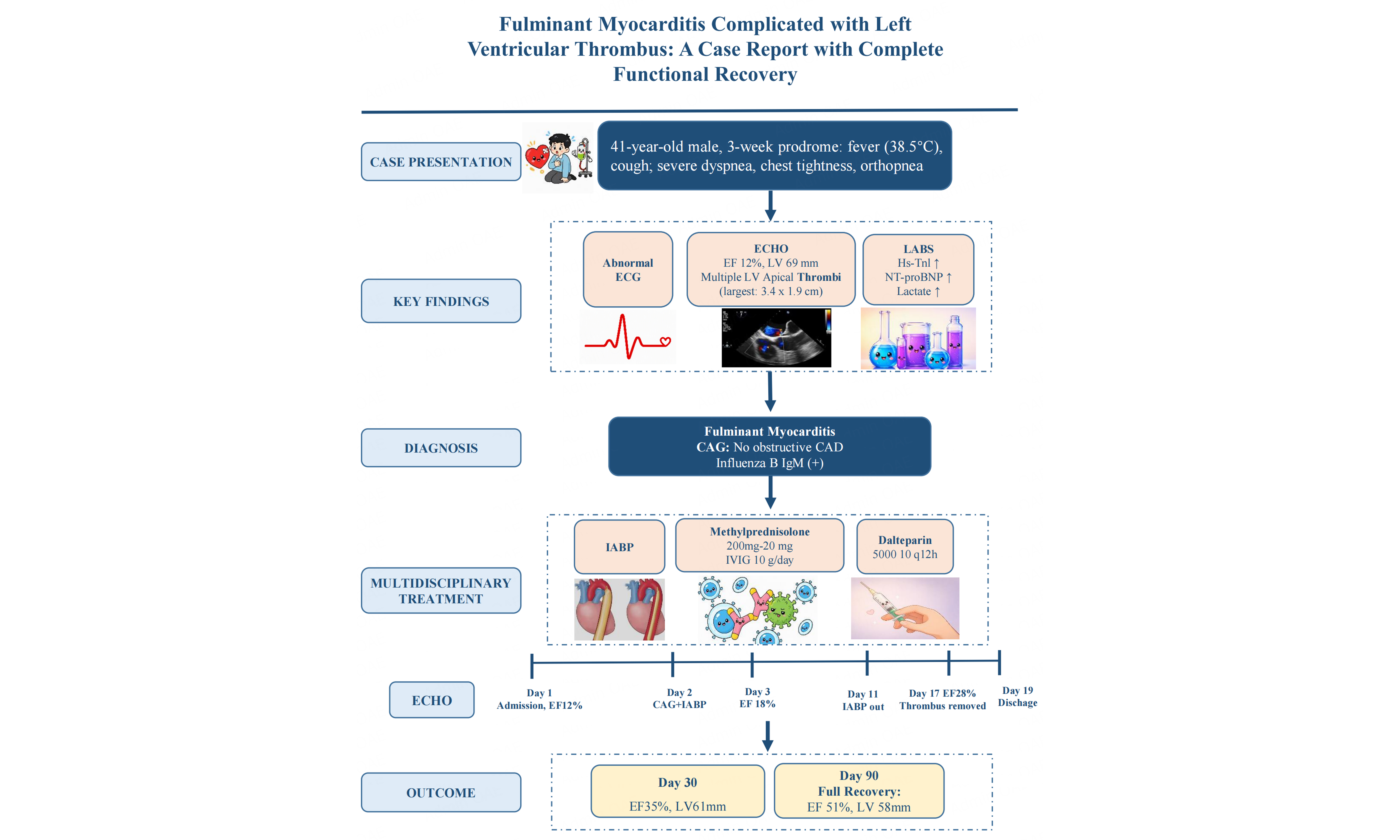
Fulminant myocarditis (FM) is a rapidly progressive, life-threatening inflammatory cardiomyopathy requiring early recognition, mechanical circulatory support, and immunomodulation. We report a 41-year-old man with a 20-day prodrome of cough and fever who presented with severe dyspnea and biventricular dysfunction (ejection fraction 12%) with multiple left ventricular apical thrombi. Coronary angiography excluded obstructive disease, and influenza B IgM was positive. Intra-aortic balloon pump (IABP), methylprednisolone (200 mg/day), intravenous immunoglobulin (10 g/day), and anticoagulation were initiated. IABP was weaned on day 11, and cardiac magnetic resonance on day 13 confirmed left ventricular excessive trabeculation with persistent thrombi. Follow-up echocardiography on day 17 demonstrated complete thrombus resolution. The patient was discharged on day 19, and at 3-month follow-up, ejection fraction normalized to 51%. This case underscores that even severe FM with multiple left ventricular thrombi remains potentially reversible with aggressive multidisciplinary management.
Keywords
INTRODUCTION
Fulminant myocarditis (FM) is a type of inflammatory cardiomyopathy characterized by sudden onset and severe hemodynamic disturbances. It is the most severe and distinct form of myocarditis, characterized by rapid onset, diverse clinical manifestations, variable course, and rapid progression[1-4]. If left untreated, patients may die within a few hours to a few days due to malignant arrhythmias, cardiogenic shock, or multi-organ failure[5]. FM complicated by a thrombus in the left ventricle is a rare but severe complication with a high risk of death. We present a case of FM occurring over three weeks in a middle-aged man, complicated by multiple left ventricular thrombus and severe biventricular dysfunction, who achieved complete functional recovery with multidisciplinary management including mechanical circulatory support (MCS), immunomodulatory therapy, and anticoagulation.
CASE PRESENTATION
History of presentation
A 41-year-old man was transferred with progressive dyspnea and chest tightness. Three weeks prior, he developed fever (38.5 °C), cough, and fatigue, diagnosed as respiratory tract infection. Two weeks before admission, exertional dyspnea worsened.
Past medical history
Local echocardiography revealed global cardiomegaly with severely reduced left ventricular contractility. He had a six-month history of untreated hypertension (167/113 mmHg). No prior cardiovascular disease or diabetes.
Physical examination
On admission, vital signs were: temperature 36.3 °C, heart rate 124 bpm, respiratory rate 22/min, blood pressure 159/113 mmHg, oxygen saturation 99% on room air. Bilateral basal lung crackles, tachycardia, and cool extremities were noted. These findings suggested peripheral hypoperfusion.
Diagnostic workup
Laboratory findings showed neutrophilia (79.5%), elevated IL-6 (12.50 pg/mL), D-dimer (5,994 ng/mL), high-sensitivity troponin I (4,344.0 pg/mL), NT-proBNP (9,810.0 pg/mL), and lactate (3.67 mmol/L)
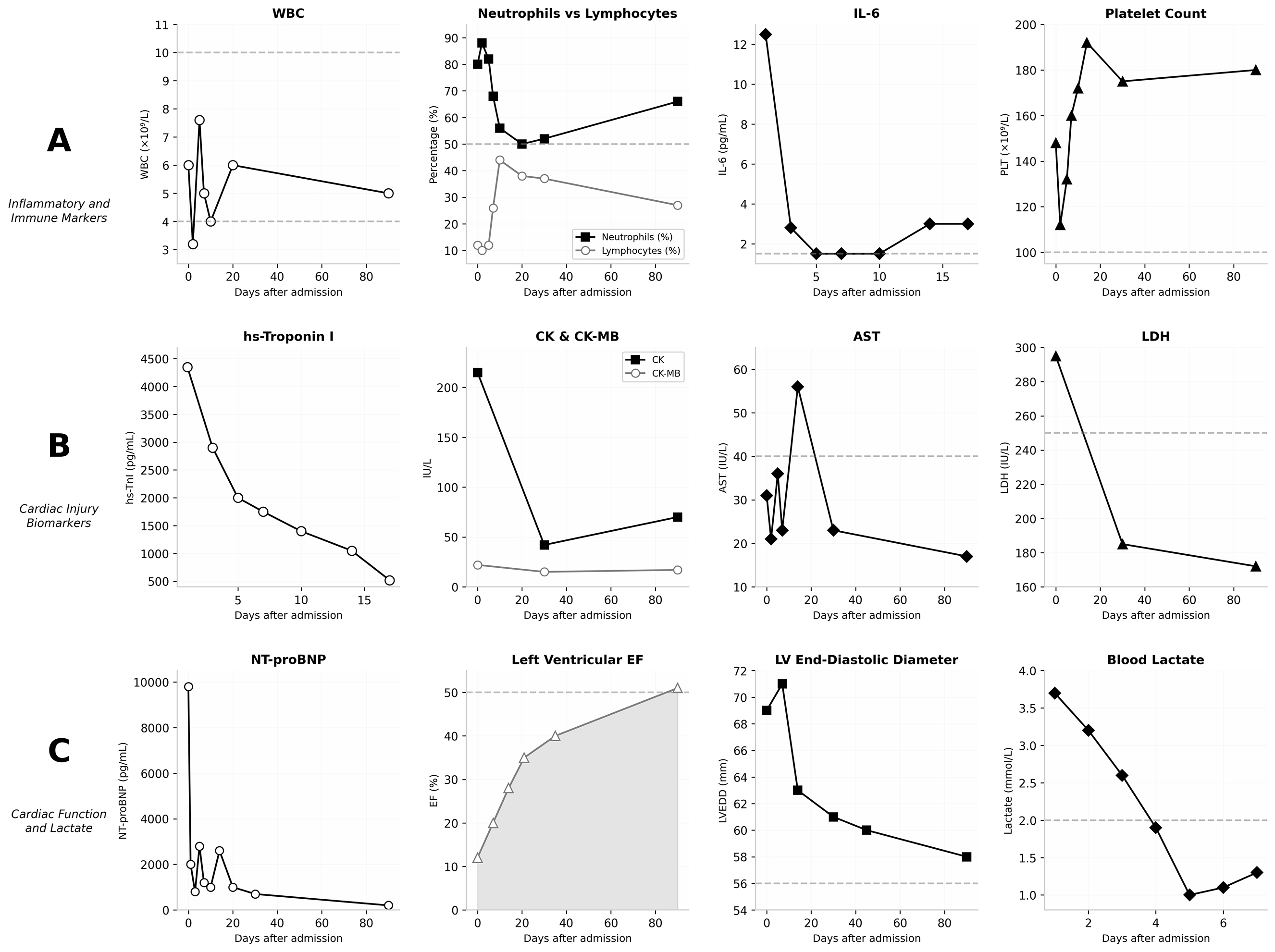
Figure 1. Clinical laboratory and cardiac parameters during hospitalization and follow-up. The figure is organized into three rows, displaying 12 panels in total. (A) Hematological and Inflammatory Markers; (B) Myocardial Injury and Hepatic Enzymes; (C) Cardiac Function and Metabolic Markers. In (C) (IL-6), the asterisks at days 5 and 8 indicate that the IL-6 concentration was below the lower limit of detection (i.e., not detectable), reflecting the rapid resolution of the systemic inflammatory response following immunomodulatory therapy. WBC: White blood cell; IL-6: interleukin-6; PLT: platelet; hs-TnI: high-sensitivity troponin I; CK: creatine kinase; CK-MB: creatine kinase-MB isoenzyme; AST: aspartate aminotransferase; LDH: lactate dehydrogenase; NT-proBNP: N-terminal pro-B-type natriuretic peptide; LVEF: left ventricular ejection fraction; LVEDD: left ventricular end-diastolic diameter.
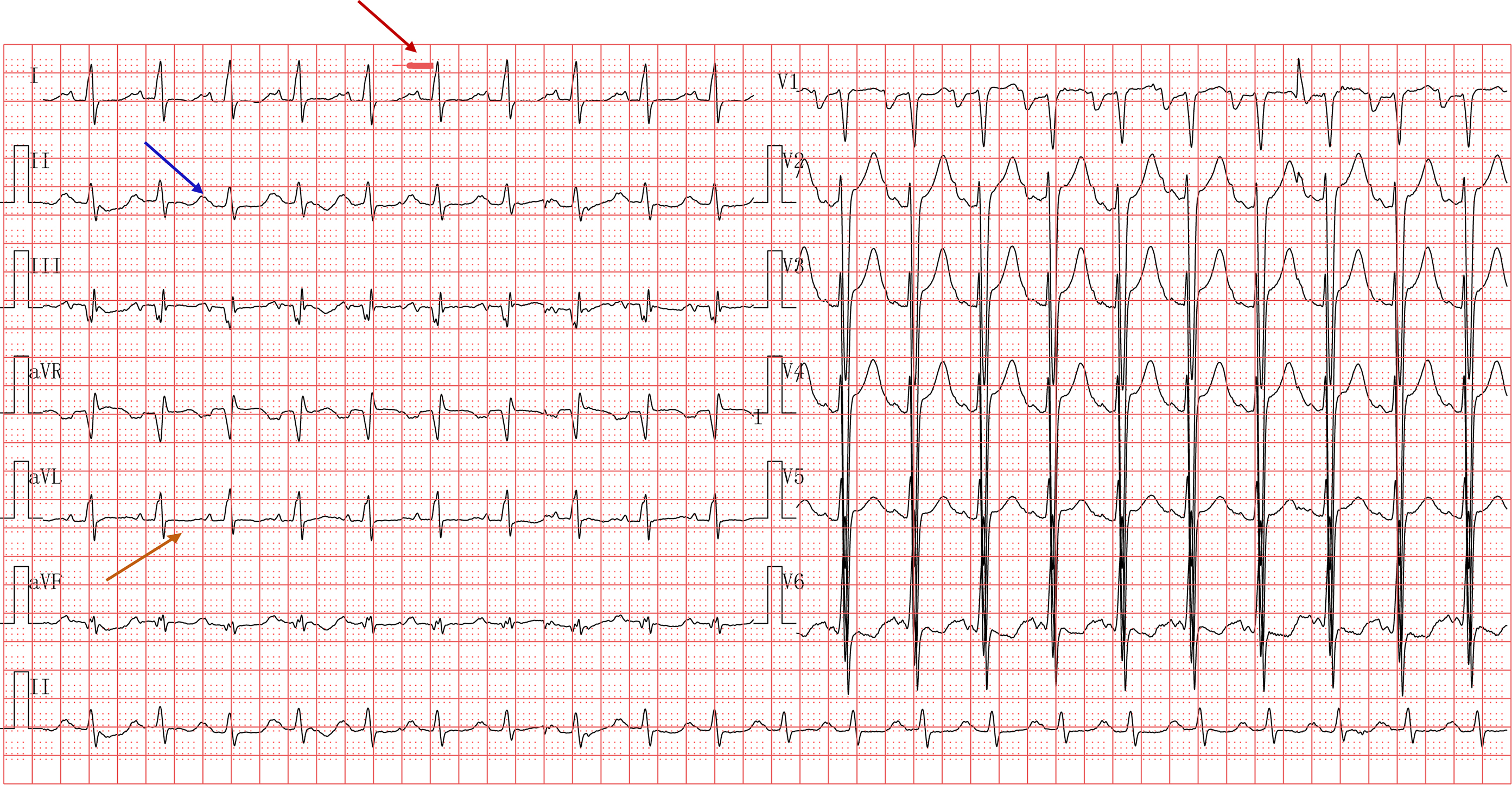
Figure 2. Electrocardiogram (paper speed 25 mm/s, sensitivity 10 mm/mV, 15 Hz) on the first day of hospitalization shows: sinus rhythm, clockwise rotation, prolonged PR interval (red arrow), left atrial abnormality (blue arrow) and T-wave changes (orange arrow).
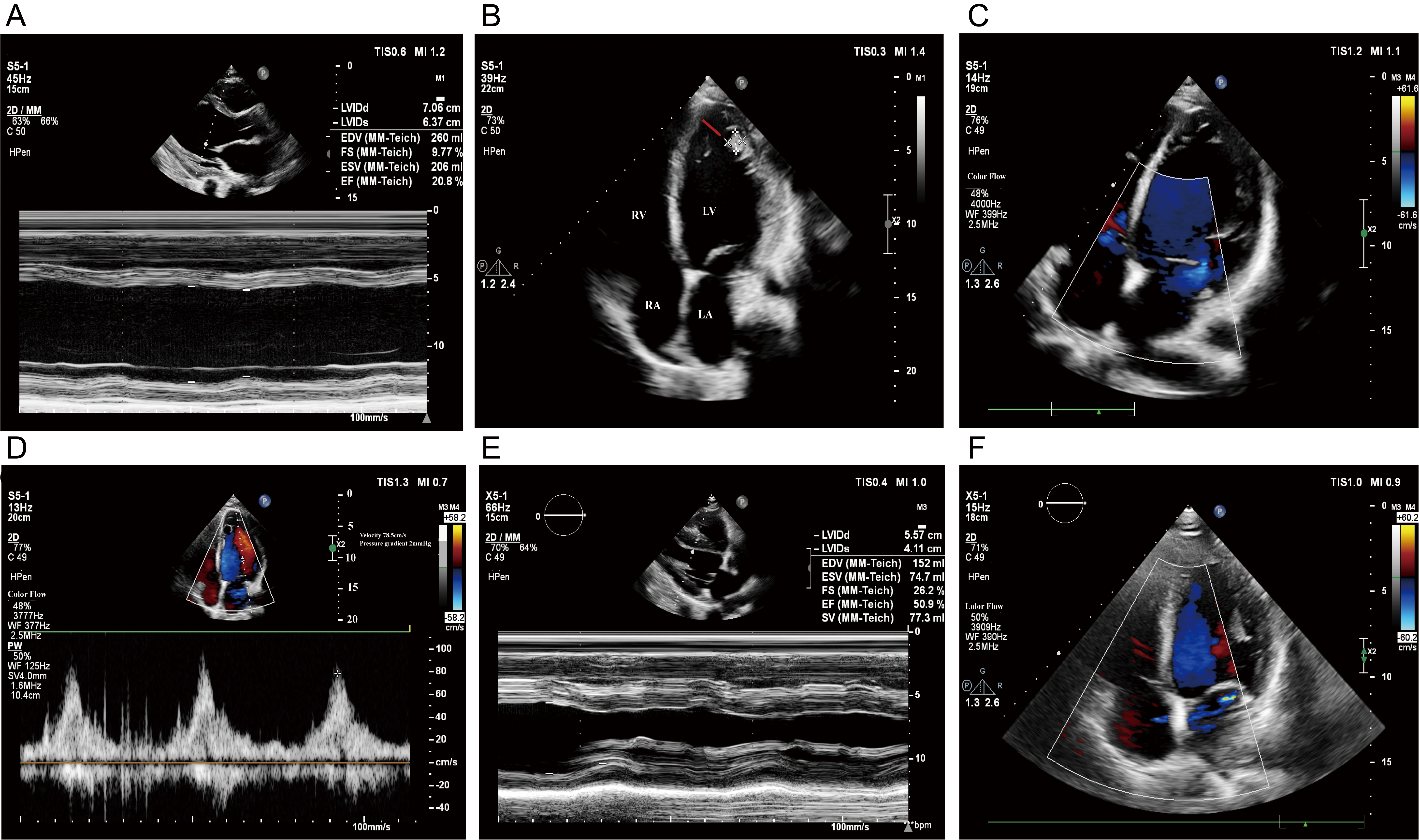
Figure 3. Serial echocardiographic images during follow-up. (A) Day 3: M-mode measurement of the left ventricle demonstrated reduced systolic function. (B) Day 3: Apical four-chamber view showing diffuse left ventricular enlargement and intraventricular thrombus formation. The thrombus is seen adjacent to the left ventricular wall (red arrow). (C) Day 17: Apical four-chamber view showing resolution of the left ventricular thrombus. (D) Day 30 follow-up: Apical four-chamber view showing mild reduction in left ventricular systolic and diastolic function. (E) Day 90 follow-up: M-mode measurement showing normalization of left ventricular systolic function. (F) Apical four-chamber view demonstrating restoration of normal chamber size. RA: Right atrium; RV: right ventricle; LA: left atrium; LV: left ventricle.
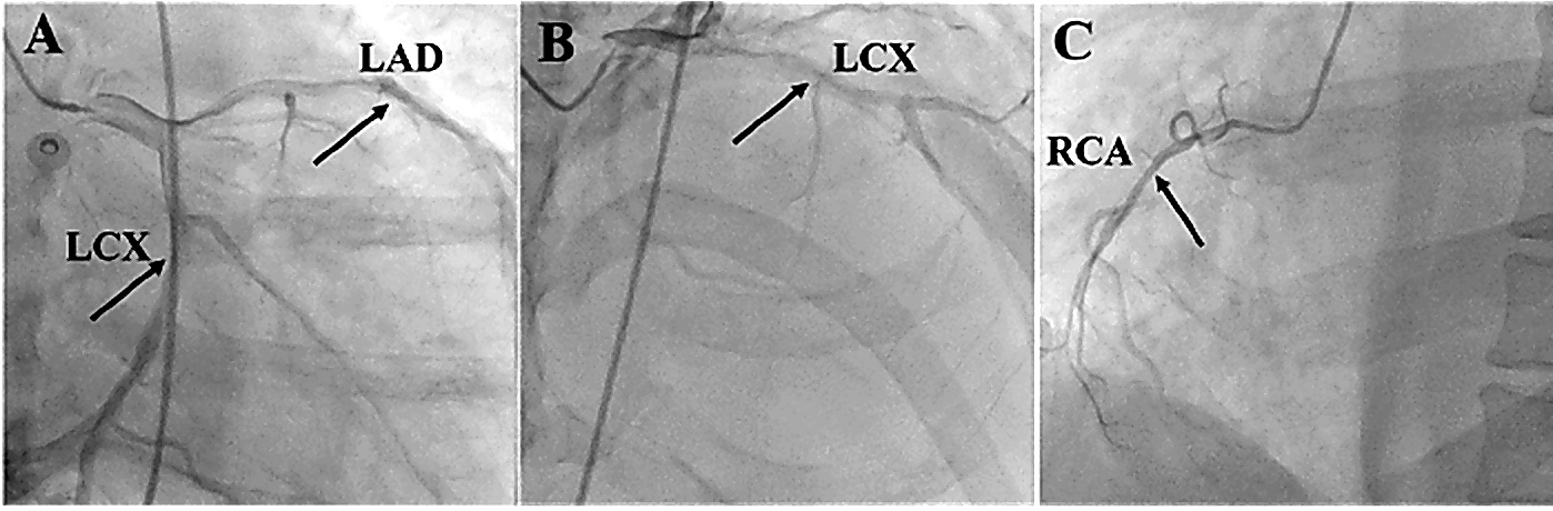
Figure 4. Result of coronary angiography: (A) There is no significant stenosis in the left main coronary artery, left anterior descending artery, or diagonal branches. (B) Mild to moderate stenosis in the mid-segment of the left circumflex artery with no significant stenosis in the obtuse marginal branch. (C) Mild stenosis in the mid-segment of the right coronary artery with no significant stenosis in the posterior left ventricular branch or posterior descending artery. Black arrows indicate the sites without obvious stenosis. LAD: Left anterior descending artery; LCX: left circumflex artery; RCA: right coronary artery.
Diagnosis
Based on the clinical presentation, laboratory findings, echocardiographic abnormalities, positive influenza B serology, and exclusion of obstructive coronary disease, the patient was diagnosed with FM secondary to influenza B virus infection, complicated by multiple left ventricular thrombi and cardiogenic shock. At admission, the patient was in compensated cardiogenic shock (Society for Cardiovascular Angiography and Interventions stage B-C)[6], as evidenced by a heart rate of 124 bpm, respiratory rate of 22/min, and cool extremities, meeting criteria for transition toward Stage C ("classic shock"). The diagnosis fulfilled the criteria for clinically suspected myocarditis[7] and met the core elements of FM[2,8]: preceding viral illness, acute onset within 2 to 4 weeks, rapidly progressive dyspnea, severe left ventricular systolic dysfunction (ejection fraction [EF] 12%), elevated cardiac biomarkers, and exclusion of acute myocardial infarction.
Differential diagnosis
The main diagnostic challenge in this case was to distinguish among three principal entities: FM, myocardial infarction with non-obstructive coronary arteries (MINOCA), and left ventricular excessive trabeculation. Discordant findings across clinical presentation, biomarkers, and imaging were pivotal in guiding our diagnostic reasoning.
(1) MINOCA: The prodromal influenza B viral illness was highly consistent with the typical clinical presentation of viral myocarditis. Coronary angiography revealed no significant stenosis, and the patient lacked typical ischemic chest pain, major cardiovascular risk factors (diabetes, hyperlipidemia, heavy smoking), or dynamic ECG changes. According to the 2018 Fourth Universal Definition of MI[9] and the 2019 AHA scientific statement[10], myocarditis has been explicitly reclassified as a "MINOCA mimicker" rather than MINOCA per se. Therefore, the final diagnosis for this patient is FM secondary to influenza B virus infection, complicated by left ventricular thrombus and cardiogenic shock—not MINOCA.
(2) Left ventricular excessive trabeculation: Cardiac Magnetic Resonance Imaging (MRI) showed a non-compacted/compacted ratio > 2.3, consistent with the diagnostic criteria for left ventricular excessive trabeculation. However, several factors argue against acute decompensation of excessive trabeculation as the primary event: the patient had no prior history of heart failure, the onset was abrupt with a febrile prodrome, and there was rapid and substantial improvement after immunomodulatory therapy—an outcome not typically expected in a purely structural cardiomyopathy. Furthermore, morphological criteria alone (NC/C > 2.3) are insufficient to establish a diagnosis of pathological left ventricular non compaction (LVNC). Notably, however, from a pathophysiological perspective, the subendocardial structural abnormalities associated with excessive trabeculation, blood stasis within deep recesses, and potential subclinical myocardial dysfunction could theoretically serve as a susceptible substrate for viral myocarditis—for instance, by altering local immune responses or promoting thrombus formation. In this case, the patient presented with multiple intraventricular thrombi, which, while not uncommon in FM, may have been exacerbated by the deep recesses and blood stasis associated with excessive trabeculation, thereby contributing to disease severity.
(3) Stress Cardiomyopathy (Takotsubo Syndrome): This is unlikely as there was no preceding significant emotional or physical stressor. The prodrome was an upper respiratory infection, and the echocardiogram showed diffuse, uncoordinated hypokinesis without the typical apical ballooning or segmental wall motion abnormalities characteristic of Takotsubo syndrome.
(4) Viral pneumonia/sepsis-induced myocardial dysfunction: These were considered but deemed unlikely because cardiac biomarkers were profoundly and persistently elevated—far beyond the mild, transient rise seen in sepsis—and because there was no clear infectious focus or predominant leukocytosis.
Management
According to the 2024 Chinese Society of Cardiology guidelines on the diagnosis and treatment of adult FM, FM should be managed with a "life support-based comprehensive treatment regimen", in which MCS constitutes the core component[2]. Our management included intra-aortic balloon pump (IABP) insertion, methylprednisolone (200 mg intravenous daily), intravenous immunoglobulin (IVIG) (10 g intravenous daily), and low-molecular-weight heparin (5000 IU twice daily). On day 2, symptoms markedly improved (blood pressure 136/74 mmHg, heart rate 74 bpm). Day 3 echocardiography showed ejection fraction improved to 18% with slight left ventricular cavity reduction (67 mm, Figure 3). Corticosteroid and IVIG doses were gradually tapered. The IABP was successfully removed on day 11. The patient was transferred to the general ward on day 12. Cardiac MRI on day 13 revealed left atrial and ventricular enlargement with reduced systolic function, increased left ventricular trabeculations with non-compacted to compacted myocardium ratio > 2.3, multiple thrombi, mild mitral regurgitation, and small pericardial effusion
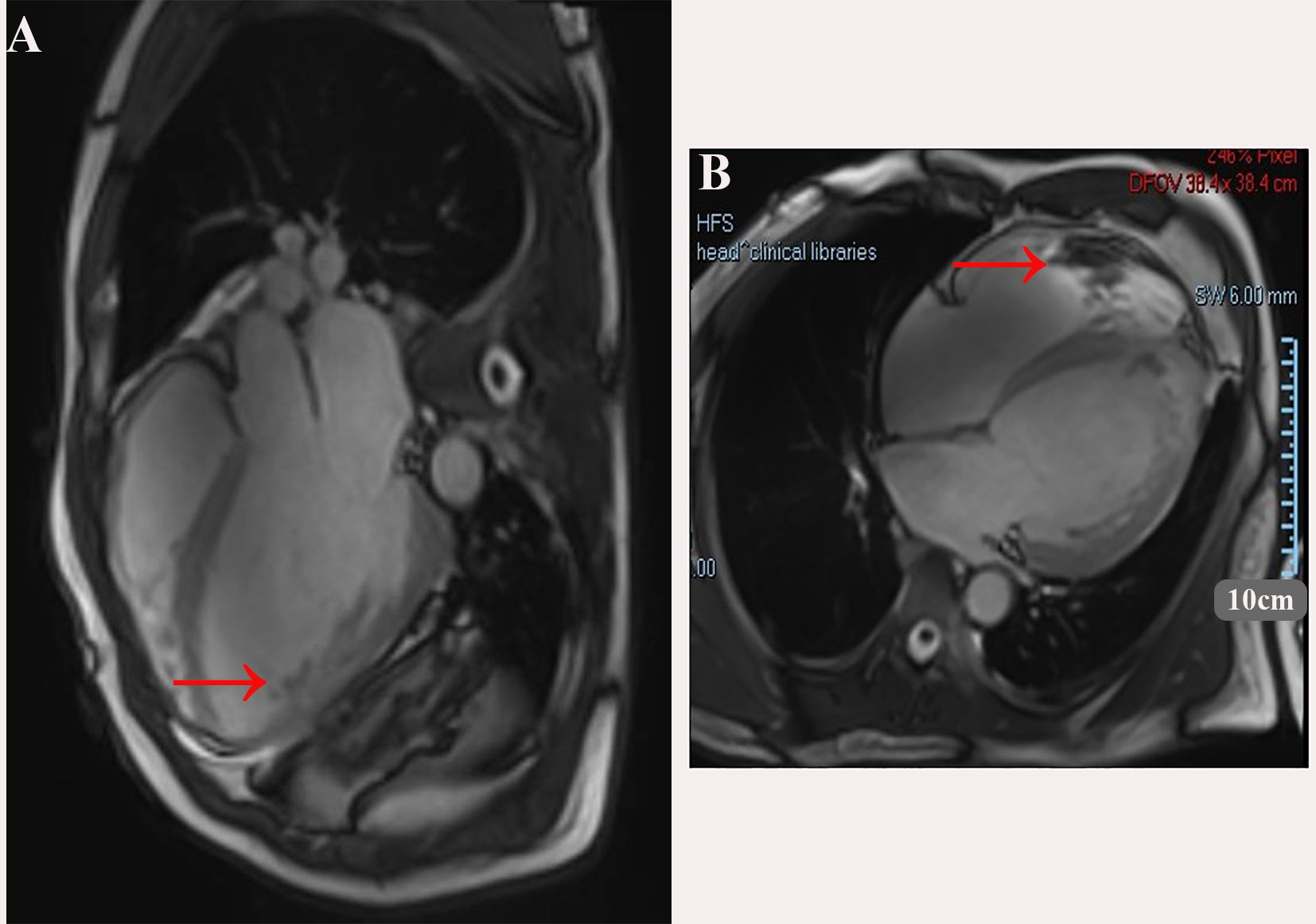
Figure 5. Showing the Cardiac magnetic resonance imaging, balanced steady-state free precession (bSSFP) cine sequence. (A) four-chamber view; (B) short-axis view. A large thrombus is seen adjacent to the apical/anterior wall of the left ventricle (red arrows). The left ventricle is markedly dilated.
Follow-up and outcomes
At one-month follow-up, echocardiography demonstrated significant improvement (ejection fraction 35%, left ventricular diameter 61 mm) and reconfirmed complete resolution of all left ventricular thrombus. Rivaroxaban was discontinued, and vericiguat 5 mg was added. At three-month follow-up, echocardiography showed normalization of left ventricular function (ejection fraction 51%, left ventricular diameter 58 mm, Figure 3). Timeline of the medical diagnosis and treatment process is shown in Figure 6.
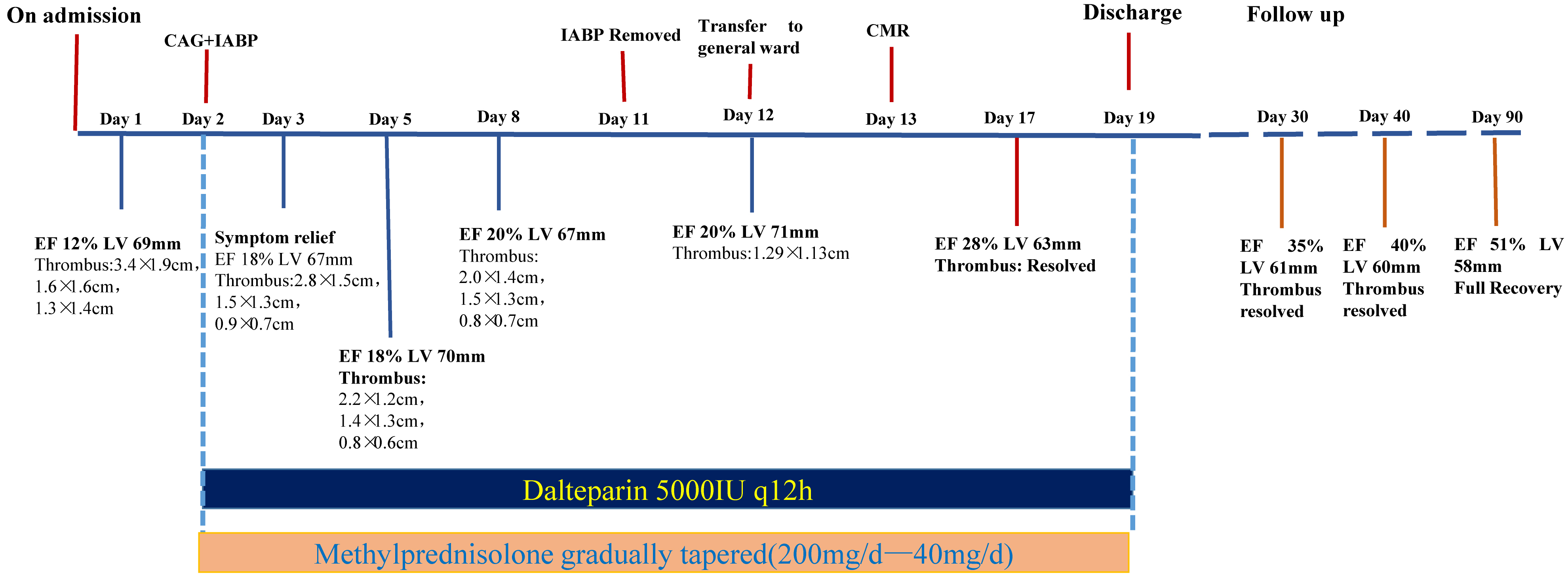
Figure 6. Timeline of the medical diagnosis and treatment process.
DISCUSSION
Myocarditis refers to inflammatory injury of the heart muscle induced by factors such as viral infection, autoimmune disease, or drugs/toxins, leading to impaired cardiac function including reduced myocardial contractility and diastolic function, as well as arrhythmias[1,3,5]. While ordinary acute myocarditis often presents with symptoms like chest tightness and palpitations, FM is characterized by a sudden onset and rapid progression. It can manifest directly as cardiogenic shock and malignant arrhythmias, potentially accompanied by multi-organ dysfunction such as respiratory, hepatic, and renal failure, and carries an extremely high early mortality rate[11,12]. Therefore, high vigilance is required for such critical cases of FM complicated by cardiogenic shock, emphasizing early recognition, assessment, and resuscitation. Corresponding measures should be promptly implemented if the condition progresses or responds poorly to initial treatment.
Currently, there is no specific diagnostic standard for FM. It is defined as a clinically diagnosed inflammatory heart disease with abrupt onset and hemodynamic compromise, rather than a histological or pathological diagnosis. Comprehensive analysis integrating clinical presentation, laboratory tests, and imaging findings is essential. The 2013 European Society of Cardiology (ESC) position paper[7] provides diagnostic criteria for clinically suspected myocarditis based on clinical presentation and non-invasive diagnostic techniques, while the 2024 Chinese Society of Cardiology guidelines[2] on adult FM emphasize very early identification, very early diagnosis, and very early treatment. In this case, the patient met the ESC criteria for clinically suspected myocarditis: a prodromal viral illness, acute onset within 2-4 weeks, rapidly progressive dyspnea, severe left ventricular systolic dysfunction (EF 12%), elevated cardiac biomarkers, and exclusion of acute myocardial infarction by coronary angiography. In this case, the patient's critical condition and the presence of multiple left ventricular thrombi precluded biopsy[13], and we have acknowledged this as a limitation.
In addition, the cardiac MRI findings—increased left ventricular trabeculations with a non-compacted-to-compacted myocardium ratio > 2.3, possible infero-apical infarction with local aneurysm formation, and multiple left ventricular thrombi—raise important differential diagnostic considerations. LVNC is a cardiomyopathy characterized by excessive trabeculations and a non-compacted-to-compacted ratio > 2.3[14]. The coexistence of LVNC with acute myocarditis has been described[15], and whether a pre-existing structural substrate contributed to the acute presentation remains uncertain. However, morphological criteria alone (NC/C > 2.3) are insufficient to establish a diagnosis of pathological LVNC, and a multiparametric approach is required. Nevertheless, from a pathophysiological perspective, the subendocardial structural abnormalities associated with excessive trabeculation, blood stasis within deep recesses, and potential subclinical myocardial dysfunction could theoretically serve as a susceptible substrate for viral myocarditis—for instance, by altering local immune responses or promoting thrombus formation. Therefore, the potential interaction between the LVNC phenotype and acute myocarditis cannot be entirely excluded. In this case, the patient presented with multiple intraventricular thrombi—which, while not uncommon in FM, may have been exacerbated by the deep recesses and blood stasis associated with excessive trabeculation, thereby contributing to disease severity. Continued anticoagulation therapy is warranted to prevent thrombus formation, and close longitudinal follow-up of the patient's myocardial disease status is necessary.
This severe condition demands urgent and specialized intervention. In China, a characteristic comprehensive management strategy has been developed for such critical cases, emphasizing proactive MCS coupled with early initiation of combined immunomodulatory therapy, such as glucocorticoids and IVIG[2,12,16]. The rationale for this approach is supported by the 2024 Chinese Society of Cardiology guidelines, which recommend a “life support-based comprehensive treatment regimen” with MCS and immunomodulation as the core[2]. Previous studies have demonstrated that immunomodulatory therapy can suppress myocardial inflammatory responses[17,18]. In addition, life-support therapy constitutes renal replacement, respiratory support, and circulatory support. Among the various circulatory support modalities. Among the various circulatory support modalities, the IABP and extracorporeal membrane oxygenation (ECMO) are commonly used in clinical practice[19,20]. In accordance with Chinese guidelines and previous research findings, this case employed a combined therapeutic approach consisting of anti-infection, antiviral, and immunomodulatory treatments, supplemented with circulatory support via IABP. However, in patients with severe left ventricle (LV) dysfunction and LV thrombus, the thromboembolic risk of IABP must be considered, and the choice of IABP versus ECMO depends on the degree of hemodynamic compromise and institutional experience. In this case, the patient's hemodynamics stabilized with IABP alone, and ECMO was not required. Subsequently, the patient's vital signs stabilized, follow-up examinations indicated gradual improvement in cardiac function, and no malignant arrhythmias occurred.
Furthermore, in this case, echocardiography revealed severe left ventricular heart failure accompanied by left ventricular dilation and thrombi. Left ventricular thrombus is a complication of myocarditis and severe left ventricular dysfunction. Embolization of ventricular thrombus can lead to systemic or pulmonary thromboembolism, causing serious consequences like cerebral or pulmonary embolism, which further worsens the condition and increases medical costs. The development of intraventricular thrombi in this patient was primarily attributable to the following factors: influenza B virus triggered a cytokine storm, with IL-6 serving as the central mediator causing endocardial injury and severe biventricular dysfunction (EF 12%); the resulting extremely low intracavitary blood flow velocity created profound apical blood stasis, with abnormal flow vortexes promoting thrombus formation; simultaneously, IL-6 drove a systemic hypercoagulable state via endothelial dysfunction, fibrinolysis inhibition, and platelet activation, as evidenced by a D-dimer level of 5,994 ng/mL. The convergence of these three elements explains the formation of multiple, large left ventricular apical thrombi—the patient's excessive trabeculation phenotype (NC/C > 2.3) may have further amplified thrombogenic risk by providing structural anchor points within deep recesses. Studies suggest that low molecular weight heparin is the most commonly used in-hospital treatment for left ventricular thrombi[21,22]. Other research suggests that surgical intervention, with left ventriculotomy being the standard method for thrombus removal, may be necessary if there is a risk of systemic embolism or left ventricular outflow tract obstruction[23]. However, no consensus exists on the optimal heparin regimen. In this case, the patient received low-molecular-weight heparin (5000 IU twice daily) during hospitalization, followed by rivaroxaban after discharge. Echocardiography on day 17 indicated thrombus disappearance. Recent case reports have demonstrated that anticoagulation alone can be effective for thrombus resolution in myocarditis[15]. The American Heart Association scientific statement on left ventricular thrombus provides guidance on anticoagulation strategies, though optimal regimens remain debated[24].
This case report has several important limitations. First, the diagnosis of FM was not confirmed by endomyocardial biopsy or by cardiac magnetic resonance meeting the updated Lake Louise criteria. Second, invasive hemodynamic data were not obtained, limiting objective documentation of cardiogenic shock. Third, the viral etiology remains speculative, as respiratory polymerase chain reaction (PCR) and myocardial viral PCR were not performed. Finally, as a single case report, the findings are not generalizable. Despite these limitations, this case demonstrates that significant recovery of cardiac function is possible even in severe myocarditis complicated by multiple left ventricular thrombi, and it underscores the importance of early recognition, multidisciplinary management, and close follow-up.
CONCLUSION
FM complicated by left ventricular thrombosis is uncommon; however, the reversibility of cardiac dysfunction should not be underestimated, provided that the condition is promptly recognized and addressed through a coordinated, multidisciplinary effort. Key therapeutic elements—early diagnosis, immediate anticoagulation, judicious application of MCS, and immunomodulatory treatment—are paramount. In the present case, the patient achieved complete functional recovery with left ventricular ejection fraction returning to 51% at the 3-month follow-up after discharge, suggesting that even severe cases with multiple left ventricular thrombi can achieve remarkable recovery. This case further serves as a reminder that anticoagulation must be carefully personalized, and that extended follow-up is indispensable.
DECLARATIONS
Authors’ contributions
Conceptualization, resources, data curation, formal analysis, writing - original draft: Peng L
Conceptualization, data curation, formal analysis, writing - original draft: Lessomo FYN
Acquisition, analysis, interpretation of data: Chen M, He T, Cheng W
Writing – review and editing: Xiong W
Conceptualization, funding acquisition, supervision, writing - review and editing, supervision: Wang H.
Availability of data and materials
All data and materials related to this work are available from the corresponding authors upon reasonable request.
AI and AI-assisted tools statement
Not applicable.
Financial support and sponsorship
This study was supported and funded by the Natural Science Foundation of Hubei Province of China (No. 2023AFC016) and Clinical Study MA-CVD-IV (20250236).
Conflicts of interest
All authors declared that there are no conflicts of interest.
Ethical approval and consent to participate
The Institutional Review Board (IRB) of Zhongnan Hospital of Wuhan University determined that, this retrospective case report involving a single patient was not classified as human subjects research; therefore, ethics approval was not required. Written informed consent was obtained from the patient for participation in this study.
Consent for publication
Written informed consent for publication of this case report and all accompanying images was obtained from the patient.
Copyright
© The Author(s) 2026.
REFERENCES
1. Ammirati E, Veronese G, Bottiroli M, et al. Update on acute myocarditis. Trends Cardiovasc Med. 2021;31:370-9.
2. Jiang J, Shu H, Wang DW, et al. Chinese Society of Cardiology guidelines on the diagnosis and treatment of adult fulminant myocarditis. Sci China Life Sci. 2024;67:913-39.
3. Li H, Chen X, Wang JJ, et al. Spatiotemporal transcriptomics elucidates the pathogenesis of fulminant viral myocarditis. Sig Transduct Target Ther. 2025;10:59.
4. Schulz-Menger J, Collini V, Gröschel J, et al. 2025 ESC Guidelines for the management of myocarditis and pericarditis. Eur Heart J. 2025;46:3952-4041.
5. Kanaoka K, Onoue K, Terasaki S, et al. Features and outcomes of histologically proven myocarditis with fulminant presentation. Circulation. 2022;146:1425-33.
6. Naidu SS, Baran DA, Jentzer JC, et al. SCAI SHOCK stage classification expert consensus update: a review and incorporation of validation studies: This statement was endorsed by the American College of Cardiology (ACC), American College of Emergency Physicians (ACEP), American Heart Association (AHA), European Society of Cardiology (ESC) Association for Acute Cardiovascular Care (ACVC), International Society for Heart and Lung Transplantation (ISHLT), Society of Critical Care Medicine (SCCM), and Society of Thoracic Surgeons (STS) in December 2021. J Am Coll Cardiol. 2022;79:933-46.
7. Caforio ALP, Pankuweit S, Arbustini E, et al. Current state of knowledge on aetiology, diagnosis, management, and therapy of myocarditis: a position statement of the European Society of Cardiology Working Group on Myocardial and Pericardial Diseases. Eur Heart J. 2013;34:2636-48.
8. Ammirati E, Cipriani M, Lilliu M, et al. Survival and left ventricular function changes in fulminant versus nonfulminant acute myocarditis. Circulation. 2017;136:529-45.
9. Thygesen K, Alpert JS, Jaffe AS, et al. Fourth universal definition of myocardial infarction (2018). J Am Coll Cardiol. 2018;72:2231-64.
10. Tamis-Holland JE, Jneid H, Reynolds HR, et al. Contemporary diagnosis and management of patients with myocardial infarction in the absence of obstructive coronary artery disease: a scientific statement from the American Heart Association. Circulation. 2019;139:e891-908.
11. Kanaoka K, Onoue K, Terasaki S, et al. Changes in cardiac function following fulminant myocarditis. Circ Heart Fail. 2024;17:e010840.
12. Kociol RD, Cooper LT, Fang JC, et al. Recognition and initial management of fulminant myocarditis: a scientific statement from the American Heart Association. Circulation. 2020;141:e69-92.
13. Huang F, Ammirati E, Ponnaiah M, et al. Fulminant myocarditis proven by early biopsy and outcomes. Eur Heart J. 2023;44:5110-24.
14. Di Fusco SA, Lucà F, Madeo A, et al. Left ventricular noncompaction: diagnostic approach, prognostic evaluation, and management strategies. Cardiol Rev. 2020;28:125-34.
15. Liang C, Liu H, Cao Z, et al. Successful treatment of massive biventricular thrombi associated with myocarditis: a case report. Front Cardiovasc Med. 2025;12:1530548.
16. Wen J, Li H, Zhou Y, et al. Immunoglobin attenuates fulminant myocarditis by inhibiting overactivated innate immune response. Br J Pharmacol. 2024;182:4984-5003.
17. Xuan Y, Gao X, Wang J, et al. Hydroxychloroquine cures autoimmune myocarditis by inhibiting the innate immune system via the C‐X‐C motif chemokine ligand 16 and C‐X‐C motif receptor 6 axis between macrophages and T cells. Br J Pharmacol. 2025;182:3649-68.
18. Shioji K, Kishimoto C, Sasayama S. Fc Receptor-Mediated inhibitory effect of immunoglobulin therapy on autoimmune giant cell myocarditis: concomitant suppression of the expression of dendritic cells. Circ Res. 2001;89:540-6.
19. Vishram-Nielsen JKK, Foroutan F, Rizwan S, et al. Patients with fulminant myocarditis supported with veno-arterial extracorporeal membrane oxygenation: a systematic review and meta-analysis of short-term mortality and impact of risk factors. Heart Fail Rev. 2022;28:347-57.
20. Van Den Brink F, Zivelonghi C, Vossenberg T, et al. VA-ECMO with IABP is associated with better outcome than VA-ECMO alone in the treatment of cardiogenic shock in ST-elevation myocardial infarction. J Invasive Cardiol. 2020;141:e69-92.
21. Yang XT, Chen YJ, Zeng H, et al. Successful treatment of large left ventricular thrombosis during extracorporeal membrane oxygenation (ECMO): a case report and review of the literature. Clin Case Rep. 2025;13:e70123.
22. Di Odoardo LAF, Bianco M, Gil IJN, et al. Left ventricular thrombus management after acute myocardial infarction in clinical practice: results from LEVITATION survey and narrative review. Cardiovasc Drugs Ther. 2022;38:483-92.
23. Tatewaki H, Hirata Y, Tanoue Y, Shiose A. Pediatric biventricular assist device for myocarditis and complicated left ventricular thrombus. Ann Thorac Surg. 2019;107:e307-8.
Cite This Article
 Open Access
How to Cite
Peng L, Lessomo FYN, Chen M, He T, Cheng W, Xiong W, Wang H. Fulminant myocarditis complicated with left ventricular thrombus: a case report with complete functional recovery. J Cardiovasc Aging. 2026;6:47. https://dx.doi.org/10.20517/jca.2026.12
Download Citation
Export Citation File:
Type of Import
Tips on Downloading Citation
Citation Manager File Format
Type of Import
Direct Import: When the Direct Import option is selected (the default state), a dialogue box will give you the option to Save or Open the downloaded citation data. Choosing Open will either launch your citation manager or give you a choice of applications with which to use the metadata. The Save option saves the file locally for later use.
Indirect Import: When the Indirect Import option is selected, the metadata is displayed and may be copied and pasted as needed.
About This Article
Special Topic
Copyright

Data & Comments
Data
0







Comments
Comments must be written in English. Spam, offensive content, impersonation, and private information will not be permitted. If any comment is reported and identified as inappropriate content by OAE staff, the comment will be removed without notice. If you have any queries or need any help, please contact us at support@oaepublish.com.