



Restoration of PINK1/Parkin-associated mitophagy via the NAD+/SIRT1 axis attenuates Kawasaki disease vasculitis
 0
0 CONVERGENT THREADS: IL-1, MITOCHONDRIA, AND THE NAD+/SIRT1 AXIS IN KAWASAKI DISEASE
Kawasaki disease (KD) is an acute, febrile, systemic vasculitis of unknown etiology that predominantly affects infants and young children, and it remains the leading cause of acquired heart disease in pediatric populations worldwide[1-3]. Approximately one quarter of untreated children develop coronary arteritis and/or coronary artery aneurysms, a figure that standard high-dose intravenous immunoglobulin G (IVIG) therapy reduces to roughly 3 to 5 percent. Yet up to 20 percent of patients are IVIG-resistant, and these children carry a substantially elevated risk of coronary artery lesions, with a pooled odds ratio of approximately 3.9 compared with IVIG-responsive patients in a recent meta-analysis[4-6]. The clinical imperative to find adjunctive or alternative therapies is therefore acute, particularly because IVIG is expensive, of limited availability in low-resource settings, and incompletely protective in a meaningful minority of recipients[7].
Over the past decade, the interleukin-1 (IL-1) axis has emerged as the dominant translational target in KD vasculitis. Genetic, transcriptomic, and murine data converge on NOD-like receptor family pyrin domain containing 3 (NLRP3) inflammasome activation and myeloid-derived IL-1β as drivers of coronary and aortic inflammation[8,9]. The Lactobacillus casei cell wall extract (LCWE) murine model reproduces the cardinal pathological features of human KD, including coronary arteritis, aortitis, myocarditis, and luminal myofibroblast proliferation, and it has served as the workhorse platform for mechanistic and preclinical therapeutic studies[10,11]. It should be noted, however, that histopathological and molecular analyses in this model frequently rely on the abdominal aorta, a large elastic vessel, whereas human KD predominantly affects medium-sized muscular arteries such as the coronary arteries; this vascular-bed discrepancy deserves explicit consideration when extrapolating molecular mechanisms from the model to the human disease. In this model, IL-1β is necessary and sufficient for coronary artery inflammation, and antibody-mediated or genetic blockade of the IL-1 pathway markedly attenuates lesion severity[12,13]. Consistent with the face validity of this platform, the beneficial effects of established therapies, including intravenous immunoglobulin, have been reproduced in the LCWE-induced model[7,11]. These preclinical observations have translated into clinical momentum: the KAWAKINRA phase II open-label trial demonstrated that the IL-1 receptor antagonist anakinra rapidly suppresses fever and inflammatory biomarkers in IVIG-resistant KD[14], with a follow-on immunological study identifying leucine-rich α2-glycoprotein (LRG1) as the biomarker most tightly linked to residual inflammatory activity and the need for dose escalation[15]. A separate phase I/IIa trial in acute KD with coronary artery aneurysms reported favorable safety and signals of coronary inflammation attenuation[16]. IL-1 blockade is now the subject of multiple international initiatives[7].
Parallel to the IL-1 narrative, a quieter but increasingly compelling body of work has implicated mitochondrial dysfunction and impaired autophagy/mitophagy in KD pathophysiology[9,17,18]. Damaged mitochondria that escape clearance release mitochondrial DNA and reactive oxygen species (ROS), both of which act as danger signals that potentiate NLRP3 inflammasome activation[19,20]. In the LCWE model, autophagy-mitophagy induction itself attenuates cardiovascular inflammation, and patients with KD show reduced mRNA expression of autophagy-related genes (LC3B, BECN1, ATG16L1) during the acute phase, with recovery after IVIG treatment[21]. The mechanistic link between mitophagy and inflammasome restraint is now well established in broader immunology: PTEN-induced kinase 1 (PINK1)/Parkin-mediated mitophagy suppresses NLRP3-driven inflammation by removing damaged mitochondria and limiting mitochondrial ROS, and deficiency in this pathway amplifies inflammasome activation across organ systems[22,23]. Sirtuin 1 (SIRT1), the best-characterized member of the nicotinamide adenine dinucleotide (NAD+)-dependent deacetylase family, sits at the intersection of these two narratives. SIRT1 deacetylates histones and non-histone proteins, including Forkhead box O (FOXO) transcription factors and peroxisome proliferator-activated receptor gamma coactivator 1-alpha (PGC-1α), to condense chromatin, suppress nuclear factor-κB-driven transcription, promote mitochondrial biogenesis, and enhance autophagy/mitophagy[24,25]. Through these activities, SIRT1 mitigates vascular remodeling, atherosclerosis, arterial stiffness, and aortic aneurysm formation, and its activity declines with age in association with endothelial dysfunction and heightened vascular inflammation[26,27]. Because SIRT1 is strictly NAD+-dependent, its activity is coupled to cellular NAD+ metabolism: chronic inflammation and oxidative stress activate NAD+-consuming enzymes such as PARP1 and CD38, depleting cellular NAD+ pools and consequently reducing SIRT1 activity, while SIRT1-driven upregulation of nicotinamide phosphoribosyltransferase (NAMPT) and PGC-1α-mediated mitochondrial biogenesis can regenerate NAD+ through the salvage pathway and oxidative phosphorylation[28]. It must be acknowledged that this positive-feedback loop, although mechanistically plausible and consistent with the metabolomic signature observed here, has not been directly interrogated in the LCWE model; rescue experiments such as vascular or myeloid NAMPT overexpression, or genetic deletion of the NAD+-consuming ectoenzyme CD38, would be required to close this mechanistic chain and should be regarded as indispensable rather than optional supplementary evidence in future studies. It is against this backdrop that Atici and colleagues report that the NAD+/SIRT1 axis is impaired in LCWE-induced KD vasculitis and that restoring it, genetically or pharmacologically, mitigates cardiovascular lesions by promoting mitophagy, blunting the vascular smooth muscle cell (VSMC) synthetic switch[29], and suppressing macrophage IL-1β production[30] [Figure 1]. The study advances the field on several fronts. First, the authors demonstrate through serum metabolomics that LCWE-injected mice exhibit a coordinated decline in nicotinamide, 1-methylnicotinamide, and nicotinamide N-oxide, indicating perturbation of the nicotinate and nicotinamide metabolism pathway, with a confirmed reduction in circulating NAD+[30]. SIRT1 protein and mRNA are correspondingly reduced in cardiac and abdominal aortic lesions. Second, using two complementary genetic gain-of-function approaches, Sirt1super mice (constitutive overexpression under the endogenous promoter) and Sirt1ER/ER mice (tamoxifen-inducible SIRT1-ER fusion), SIRT1 overexpression is confirmed to reduce heart vessel inflammation, abdominal aortic dilation, peritoneal IL-1β, and immune cell infiltration, and restore ejection fraction measured by echocardiography. Third, and most decisively, cell-type-specific deletion of Sirt1 in VSMCs (Myh11-Cre/ERT2) or myeloid cells (Csf1r-Cre) exacerbates LCWE-induced vasculitis, with myeloid deletion increasing IL-1β and TNFα production and favoring proinflammatory M1-like macrophage polarization[30]. Fourth, untargeted proteomics of abdominal aortas reveals that SIRT1 overexpression rescues pathways related to mitochondrial function (e.g., tricarboxylic acid cycle, fatty acid β-oxidation, mitochondrial protein degradation), autophagy/mitophagy, and VSMC contractile identity, while suppressing neutrophil degranulation and macrophage activation. Fifth, a therapeutic arm is included: daily SRT1720, resveratrol, β-nicotinamide mononucleotide (β-NMN), or nicotinamide riboside (NR), administered either prophylactically or beginning on day 3 post-LCWE injection, significantly reduces heart vessel inflammation and, for NR, abdominal aortic dilation[30]. In vitro, SRT1720 boosts autophagic flux in primary VSMCs (chloroquine accumulation of LC3-II and p62 in mitochondrial fractions) and reduces mitochondrial ROS, while EX527 abolishes these effects; these cell-based findings are distinct from, and complement, the separate in vivo chloroquine flux assay performed in aortic tissue of Sirt1super mice discussed below.
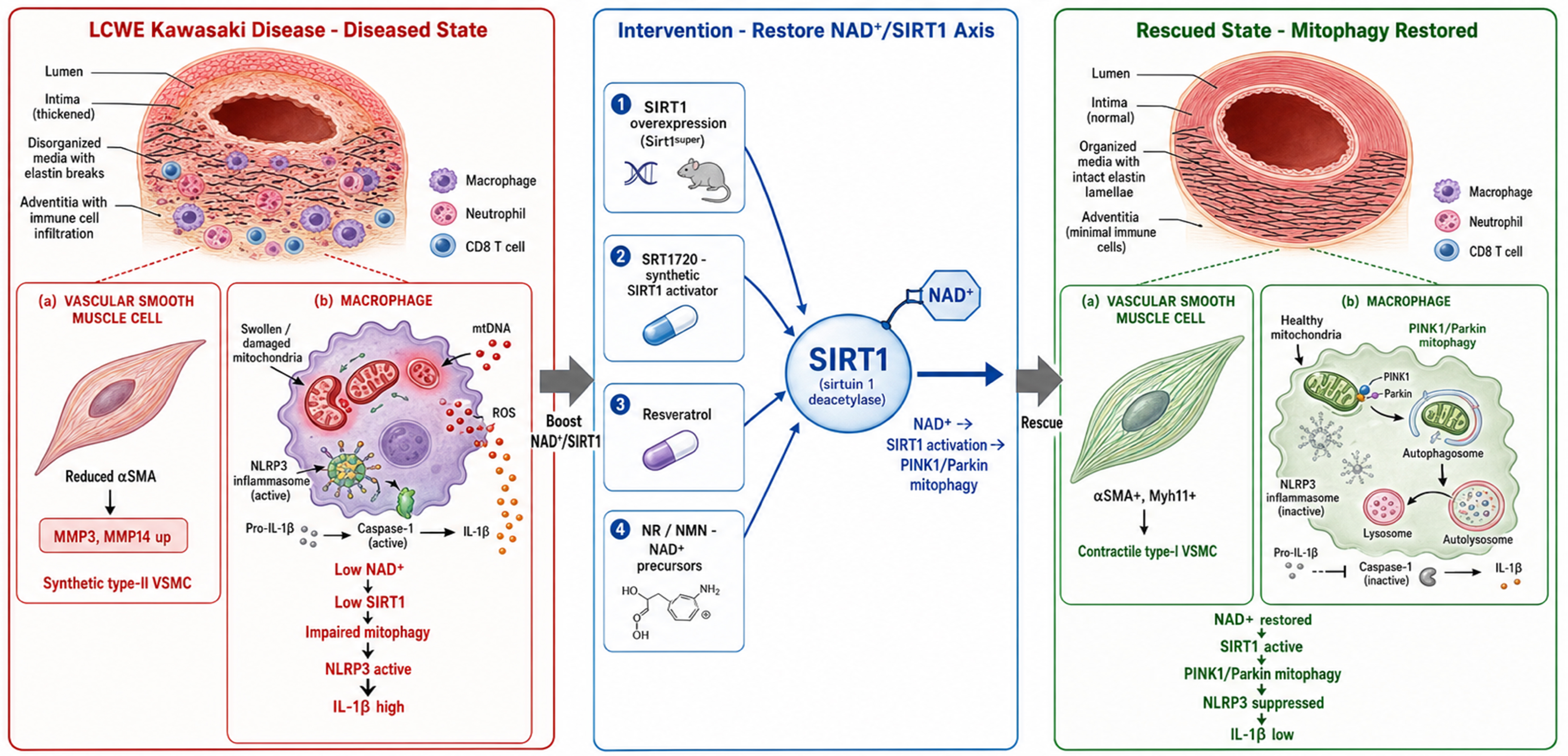
Figure 1. Restoration of PINK1/Parkin-associated mitophagy via the NAD+/SIRT1 axis attenuates KD vasculitis. In the LCWE murine model of KD, cardiovascular lesions are characterized by depletion of circulating NAD+ precursors (nicotinamide, 1-methylnicotinamide, nicotinamide N-oxide) and downregulation of the NAD+-dependent deacetylase SIRT1 in heart and abdominal aortic tissue (left panel). In VSMCs, low SIRT1 permits the pathogenic synthetic phenotypic switch from contractile type-I VSMCs (αSMA+, Myh11+) to synthetic type-II VSMCs (MMP3/14 up), contributing to luminal myofibroblast proliferation and coronary stenosis. In macrophages, impaired mitophagy leads to accumulation of damaged mitochondria that release mtDNA and ROS, activating the NLRP3 inflammasome and driving caspase-1-dependent maturation and secretion of IL-1β. Restoring the NAD+/SIRT1 axis through four complementary interventions (middle panel), constitutive SIRT1 overexpression (Sirt1super mice), tamoxifen-inducible SIRT1-ER activation (Sirt1ER/ER mice), pharmacological SIRT1 activation (SRT1720, resveratrol), or NAD+ precursor supplementation (NR, β-NMN), rescues the diseased phenotype (right panel). SIRT1 activation restores PINK1/Parkin-mediated mitophagic flux, clearing damaged mitochondria, suppressing NLRP3 inflammasome activity and IL-1β production, and preserving the contractile VSMC identity, thereby reducing coronary arteritis, abdominal aortic dilation, immune cell infiltration, and cardiac dysfunction. Cell-type-specific deletion of Sirt1 in VSMCs (Myh11-Cre/ERT2) or myeloid cells (Csf1r-Cre) exacerbates LCWE-induced vasculitis, confirming that both compartments are non-redundant nodes of SIRT1-dependent protection. The figure was created using LibreOffice and Inkscape. PINK1: PTEN-induced kinase 1; NAD+: nicotinamide adenine dinucleotide; SIRT1: Sirtuin 1; KD: Kawasaki disease; LCWE: Lactobacillus casei cell wall extract; VSMCs: vascular smooth muscle cells; αSMA+: alpha-smooth muscle actin-positive; MMP3/14: matrix metalloproteinase 3/14; mtDNA: mitochondrial DNA; ROS: reactive oxygen species; NLRP3: NOD-like receptor family pyrin domain containing 3; IL-1β: interleukin-1β; NR: nicotinamide riboside; β-NMN: β-nicotinamide mononucleotide.
A BROKEN AXIS, MAPPED AND RESCUED
The convergence of genetic overexpression, inducible overexpression, two independent cell-type-specific knockouts, and four pharmacological interventions constitutes a triangulation rarely achieved in a single preclinical vasculitis study. The integration of metabolomics, proteomics, echocardiography, flow cytometry, immunofluorescence, and in vitro flux assays provides a multi-layered mechanistic account rather than a single-line narrative. The cell-type-specific Cre drivers move the field beyond the question of whether SIRT1 matters to the more actionable question of where it matters, with the myeloid and VSMC compartments emerging as distinct and non-redundant nodes. The use of an in vivo chloroquine flux assay (a single chloroquine injection in LCWE-injected mice with aortic tissue harvested 4 h later, a design clearly distinct from the in vitro VSMC experiments summarized above) to demonstrate that SIRT1 overexpression restores, rather than merely increases, autophagic flux is methodologically sound and addresses a common pitfall in autophagy interpretation[31,32]. These strengths notwithstanding, several issues warrant scrutiny. Mechanistically, the proposed NAD+/SIRT1 positive feedback loop, in which SIRT1 upregulates NAMPT and PGC-1α to regenerate NAD+, is largely inferred from prior literature rather than directly tested in this model. A NAMPT-overexpression or CD38-knockout rescue experiment would have closed this loop. We concur with this assessment: until such rescue experiments are performed, the feedback loop should be viewed as a literature-derived framework rather than a validated circuit in KD vasculitis. The mitophagy readouts rely on surrogate markers (p62, LC3B puncta, phospho-ubiquitin Ser65) rather than mito-QC reporter mice such as mt-Keima or Mito-QC, which would have provided a more direct, quantitative measure of mitophagic flux in vivo. Static surrogate markers such as p62 and LC3B are technically simple and interpretable in paraffin-embedded vascular tissue, but they capture steady-state abundance rather than flux and can be confounded by altered lysosomal degradation; reporter systems such as mt-Keima and Mito-QC provide direct, single-cell resolution of mitophagic flux but require dedicated mouse lines, are sensitive to tissue autofluorescence and to the pH constraints of thick vascular-wall preparations, and demand specialized imaging. In our view, the absence of direct in vivo flux quantification does not invalidate the central conclusion of the study, which is supported concordantly by genetic, pharmacological, and proteomic data, but it does lower the resolution at which mitophagy can be claimed as the causal intermediary of vascular protection. Notably, SRT1720 did not alter phosphorylated Unc-51-like kinase 1 (ULK1) or AMP-activated protein kinase (AMPK) levels in VSMCs, leaving the upstream autophagy trigger unresolved and raising the possibility that SIRT1 promotes autophagy here through deacetylation of FOXO3a or direct mTOR inhibition rather than the canonical AMPK-ULK1 axis[33,34]. The proteomics comparison is restricted to the abdominal aorta, while the functional readouts span heart and aorta, leaving open whether the mitophagy rescue is uniform across vascular beds or concentrated where LCWE lesions are most severe. Methodologically, the exclusive use of 5-week-old male C57BL/6J mice is an acknowledged but underexplored limitation. KD shows a roughly 1.5:1 male predominance, and the LCWE model exhibits IL-1-mediated sex differences in vasculitis development and treatment response, so a sex-stratified analysis is not merely a robustness check but a mechanistic question in its own right[35]. The genetic-background confound is also non-trivial: 129S1 wild-type mice showed reduced baseline vasculitis and lower peritoneal IL-1β compared with C57BL/6J, complicating cross-strain comparisons of Sirt1ER/ER rescue magnitude.
THE TRANSLATIONAL CHASM: FROM SRT1720 TO A FEBRILE INFANT
Translationally, the gap between this murine work and human KD therapy remains wide. The LCWE model captures aortitis and coronary arteritis but not the full KD clinical syndrome, including the mucocutaneous features and the IVIG response biology that define the human disease. SRT1720 at 100 mg/kg intraperitoneally achieves exposures far above anything clinically attainable, and the broader SIRT1 activator field has struggled with isoform selectivity, pharmacokinetics, off-target effects, and inconsistent clinical efficacy, with resveratrol in particular acting on numerous sirtuin-independent pathways[36,37]. NAD+ boosters are more clinically advanced: chronic NR supplementation at 1,000 mg/day for six weeks was well tolerated and elevated blood NAD+ by approximately 60 percent in healthy middle-aged and older adults[38], and high-dose NR (up to 3,000 mg/day) was well tolerated in a Parkinson disease safety trial[39]. However, no pediatric dosing, pharmacokinetic, or KD-specific efficacy data exist, and the authors’ own data show an asymmetry in the therapeutic arm, with NR but not NMN reducing abdominal aortic dilation when dosed post-LCWE, a discrepancy they plausibly attribute to the need for extracellular NMN dephosphorylation to NR for cellular uptake[28]. Alternative or additional explanations for the divergent efficacy of NR and NMN deserve consideration, including differential reliance on distinct uptake mechanisms (direct transporter-mediated import vs. extracellular dephosphorylation by ectoenzymes such as CD73), variable expression of these enzymes across vascular beds and inflammatory states, and the narrow therapeutic window imposed by the day-3 post-LCWE dosing schedule, which may penalize a precursor that requires an extra enzymatic conversion step. Whichever mechanism prevails, the discrepancy argues that future pediatric trials should incorporate direct pharmacodynamic readouts of circulating and, where feasible, tissue NAD+ metabolites rather than assuming class equivalence among NAD+ precursors. Finally, the human validation rests on reanalysis of public transcriptomic datasets rather than direct immunohistochemical confirmation of SIRT1 or NAMPT protein in KD coronary lesions, which would have substantially strengthened the translational claim. In practical terms, circulating biomarkers offer the most realistic window on this axis in children: serial whole-blood and plasma NAD+ metabolomics (nicotinamide, 1-methylnicotinamide, nicotinamide N-oxide), SIRT1 and NAMPT expression in peripheral blood mononuclear cells, and inflammatory pharmacodynamic markers such as LRG1 could be measured in KD patients to gauge NAD+/SIRT1 axis activity and its restoration during therapy, thereby obviating the need for coronary lesion tissue.
A DELIBERATE PATH FORWARD
Looking forward, the study identifies a clear and testable translational agenda. Direct immunohistochemical quantification of SIRT1, NAMPT, and phospho-ubiquitin in human KD coronary artery and aortic lesions, ideally paired with non-KD febrile pediatric controls, would establish whether the NAD+/SIRT1 impairment observed in mice is present in the human disease it models. Sex-stratified LCWE studies, informed by the known IL-1-mediated sex differences, would determine whether SIRT1 rescue is equivalently effective in females[35]. If SIRT1-mediated protection were indeed sex-dependent, the clinical implications would be substantial: NAD+ precursor trials in KD would require sex-stratified enrollment and analysis, sex-specific expectations of treatment response, and pediatric trial designs powered to detect divergent efficacy, rather than assuming poolability of male and female responses on the basis of a male-only murine dataset. Mito-QC reporter mice crossed onto the LCWE model would replace surrogate flux markers with direct mitophagy quantification. A combination study of low-dose NR with sub-therapeutic anakinra would test whether NAD+ boosting and IL-1 blockade are additive or synergistic, a question with direct trial-design implications given the existing KAWAKINRA infrastructure[15]. Pediatric NR pharmacokinetics and a head-to-head therapeutic timing comparison of NR vs. NMN are necessary prerequisites before any KD-specific NAD+ booster trial. The NAD+/SIRT1 axis is an attractive therapeutic target, and Atici and co-workers have provided the most comprehensive preclinical case to date for pursuing it in KD vasculitis, but the path from a 5-week-old male mouse to a febrile infant in a pediatric cardiac intensive care unit remains long and must be walked deliberately.
DECLARATIONS
Authors’ contributions
Drafted the manuscript: Qin Y, Kansakar U
Critically revised the manuscript for important intellectual content: Pande S
Conceived the work, drafted and critically revised the manuscript, and approved the final version for publication: Santulli G
Availability of data and materials
Not applicable.
AI and AI-assisted tools statement
Not applicable.
Financial support and sponsorship
Santulli G was supported in part by the National Institutes of Health (NIH): National Heart, Lung, and Blood Institute (NHLBI: R01-HL146691, R01-HL164772, R01-HL159062), National Institute of Diabetes and Digestive and Kidney Diseases (NIDDK: R01-DK123259, R01-DK033823), and by the American Heart Association (AHA, 24IPA1268813). Kansakar U was supported in part by the NIH (T32-HL-172255) and by a postdoctoral fellowship of the AHA (AHA-23POST1026190). Pande S was supported in part by a postdoctoral fellowship of the AHA (AHA-26POST1563063).
Conflicts of interest
All authors declared that there are no conflicts of interest.
Ethical approval and consent to participate
Not applicable.
Consent for publication
Not applicable.
Copyright
© The Author(s) 2026.
REFERENCES
1. McCrindle BW, Rowley AH, Newburger JW, et al.; American Heart Association Rheumatic Fever, Endocarditis, and Kawasaki Disease Committee of the Council on Cardiovascular Disease in the Young; Council on Cardiovascular and Stroke Nursing; Council on Cardiovascular Surgery and Anesthesia; and Council on Epidemiology and Prevention. Diagnosis, treatment, and long-term management of Kawasaki disease: a scientific statement for health professionals from the American Heart Association. Circulation. 2017;135:e927-99.
2. Wang Q, Li W, Wan J, et al. Electrocardiographic alterations combined with hematological, biochemical, and metabolic profiles predict prognosis in Kawasaki disease. J Cardiovasc Dev Dis. 2026;13:228.
3. Eleftheriou D, Moraes YC, Purvis C, et al. Multi-centre, randomised, open-label, blinded endpoint assessed, trial of corticosteroids plus intravenous immunoglobulin (IVIG) and aspirin, versus IVIG and aspirin for prevention of coronary artery aneurysms (CAA) in Kawasaki disease (KD): the KD CAA prevention (KD-CAAP) trial protocol. Trials. 2023;24:60.
4. Zheng X, Li J, Yue P, et al. Is there an association between intravenous immunoglobulin resistance and coronary artery lesion in Kawasaki disease? - Current evidence based on a meta-analysis. PLoS One. 2021;16:e0248812.
5. Watanabe M, Fukazawa R, Kiriyama T, et al. Hemodynamic evaluation of coronary artery lesions after Kawasaki disease: comparison of fractional flow reserve during cardiac catheterization with myocardial flow reserve during 13N-ammonia PET. J Cardiovasc Dev Dis. 2024;11:229.
6. Amorim-Figueiredo R, Pereira Lemos A, Rito T, Conde M, Brito MJ, Pinto F. Multiresistant Kawasaki disease in a young infant with giant aneurysms growing fast. J Cardiovasc Dev Dis. 2024;11:149.
7. Burns JC, Koné-Paut I, Kuijpers T, Shimizu C, Tremoulet A, Arditi M. Review: Found in translation: international initiatives pursuing interleukin-1 blockade for treatment of acute Kawasaki disease. Arthritis Rheumatol. 2017;69:268-76.
8. Atici AE, Noval Rivas M, Arditi M. The central role of interleukin-1 signalling in the pathogenesis of Kawasaki disease vasculitis: path to translation. Can J Cardiol. 2024;40:2305-20.
9. Zhang R, Zhang J, Yang Y, Wang Y, Cao H. The autophagy-inflammasome axis as a molecular switch: from persistent inflammation to vascular remodeling in IVIG-resistant Kawasaki disease. Int J Mol Sci. 2026;27:6405.
10. Suganuma E, Sato S, Honda S, Nakazawa A. A novel mouse model of coronary stenosis mimicking Kawasaki disease induced by Lactobacillus casei cell wall extract. Exp Anim. 2020;69:233-41.
11. Noval Rivas M, Arditi M. Kawasaki disease: pathophysiology and insights from mouse models. Nat Rev Rheumatol. 2020;16:391-405.
12. Lee Y, Schulte DJ, Shimada K, et al. Interleukin-1β is crucial for the induction of coronary artery inflammation in a mouse model of Kawasaki disease. Circulation. 2012;125:1542-50.
13. Gorelik M, Lee Y, Abe M, et al. IL-1 receptor antagonist, anakinra, prevents myocardial dysfunction in a mouse model of Kawasaki disease vasculitis and myocarditis. Clin Exp Immunol. 2019;198:101-10.
14. Koné-Paut I, Tellier S, Belot A, et al. Phase II open label study of anakinra in intravenous immunoglobulin-resistant Kawasaki disease. Arthritis Rheumatol. 2021;73:151-61.
15. Kessel C, Koné-Paut I, Tellier S, et al. An immunological axis involving interleukin 1β and leucine-rich-α2-glycoprotein reflects therapeutic response of children with Kawasaki disease: implications from the KAWAKINRA trial. J Clin Immunol. 2022;42:1330-41.
16. Yang J, Jain S, Capparelli EV, et al. Anakinra treatment in patients with acute Kawasaki disease with coronary artery aneurysms: a phase I/IIa trial. J Pediatr. 2022;243:173-80.e8.
17. Xu Q, Wu Y, Ding Y. The autophagy-inflammation axis in Kawasaki disease: pathogenic mechanisms and translational opportunities. J Clin Med. 2026;15:3918.
18. Lin M, Xian H, Chen Z, et al. MCM8-mediated mitophagy protects vascular health in response to nitric oxide signaling in a mouse model of Kawasaki disease. Nat Cardiovasc Res. 2023;2:778-92.
19. Shimada K, Crother TR, Karlin J, et al. Oxidized mitochondrial DNA activates the NLRP3 inflammasome during apoptosis. Immunity. 2012;36:401-14.
20. Gupta S, Cassel SL, Sutterwala FS, Dagvadorj J. Regulation of the NLRP3 inflammasome by autophagy and mitophagy. Immunol Rev. 2025;329:e13410.
21. Marek-Iannucci S, Ozdemir AB, Moreira D, et al. Autophagy-mitophagy induction attenuates cardiovascular inflammation in a murine model of Kawasaki disease vasculitis. JCI Insight. 2021;6:e151981.
22. Mouton-Liger F, Rosazza T, Sepulveda-Diaz J, et al. Parkin deficiency modulates NLRP3 inflammasome activation by attenuating an A20-dependent negative feedback loop. Glia. 2018;66:1736-51.
23. Jankauskas SS, Mone P, Avvisato R, et al. miR-181c targets Parkin and SMAD7 in human cardiac fibroblasts: validation of differential microRNA expression in patients with diabetes and heart failure with preserved ejection fraction. Mech Ageing Dev. 2023;212:111818.
24. Rodgers JT, Lerin C, Haas W, Gygi SP, Spiegelman BM, Puigserver P. Nutrient control of glucose homeostasis through a complex of PGC-1alpha and SIRT1. Nature. 2005;434:113-8.
25. Jankauskas SS, Mone P, Santulli G. From resveratrol to ISIDE11: how to activate SIRT1 and improve endothelial function? New therapeutic insights for methylenetetrahydrofolate reductase deficiency. Cell Mol Life Sci. 2022;79:451.
26. Ding YN, Wang HY, Chen XF, Tang X, Chen HZ. Roles of Sirtuins in cardiovascular diseases: mechanisms and therapeutics. Circ Res. 2025;136:524-50.
27. Ministrini S, Puspitasari YM, Beer G, Liberale L, Montecucco F, Camici GG. Sirtuin 1 in endothelial dysfunction and cardiovascular aging. Front Physiol. 2021;12:733696.
28. Yoshino J, Baur JA, Imai SI. NAD+ intermediates: the biology and therapeutic potential of NMN and NR. Cell Metab. 2018;27:513-28.
29. Kansakar U, Jankauskas SS, Gambardella J, Santulli G. Targeting the phenotypic switch of vascular smooth muscle cells to tackle atherosclerosis. Atherosclerosis. 2021;324:117-20.
30. Atici AE, Jena PK, Carvalho TT, et al. Sirtuin 1 activation mitigates murine vasculitis severity by promoting mitophagy. Circ Res. 2026;139:e327991.
31. Gottlieb RA, Andres AM, Sin J, Taylor DP. Untangling autophagy measurements: all fluxed up. Circ Res. 2015;116:504-14.
32. Khawaja RR, Griego E, Lindenau K, et al. Chaperone-mediated autophagy regulates neuronal activity by sex-specific remodelling of the synaptic proteome. Nat Cell Biol. 2025;27:1688-707.
33. Gupta S, Afzal M, Agrawal N, et al. Harnessing the FOXO-SIRT1 axis: insights into cellular stress, metabolism, and aging. Biogerontology. 2025;26:65.
34. Wei X, Xiong X, Wang P, Zhang S, Peng D. SIRT1-mediated deacetylation of FOXO3 enhances mitophagy and drives hormone resistance in endometrial cancer. Mol Med. 2024;30:147.
35. Porritt RA, Markman JL, Maruyama D, et al. Interleukin-1 beta-mediated sex differences in Kawasaki disease vasculitis development and response to treatment. Arterioscler Thromb Vasc Biol. 2020;40:802-18.
36. Curry AM, White DS, Donu D, Cen Y. Human Sirtuin regulators: the “success” stories. Front Physiol. 2021;12:752117.
37. Bonkowski MS, Sinclair DA. Slowing ageing by design: the rise of NAD+ and sirtuin-activating compounds. Nat Rev Mol Cell Biol. 2016;17:679-90.
38. Martens CR, Denman BA, Mazzo MR, et al. Chronic nicotinamide riboside supplementation is well-tolerated and elevates NAD+ in healthy middle-aged and older adults. Nat Commun. 2018;9:1286.
Cite This Article
 Open Access
How to Cite
Qin Y, Kansakar U, Pande S, Santulli G. Restoration of PINK1/Parkin-associated mitophagy via the NAD+/SIRT1 axis attenuates Kawasaki disease vasculitis. J Cardiovasc Aging. 2026;6:49. https://dx.doi.org/10.20517/jca.2026.123
Download Citation
Export Citation File:
Type of Import
Tips on Downloading Citation
Citation Manager File Format
Type of Import
Direct Import: When the Direct Import option is selected (the default state), a dialogue box will give you the option to Save or Open the downloaded citation data. Choosing Open will either launch your citation manager or give you a choice of applications with which to use the metadata. The Save option saves the file locally for later use.
Indirect Import: When the Indirect Import option is selected, the metadata is displayed and may be copied and pasted as needed.
About This Article
Special Topic
Copyright

Data & Comments
Data
0







Comments
Comments must be written in English. Spam, offensive content, impersonation, and private information will not be permitted. If any comment is reported and identified as inappropriate content by OAE staff, the comment will be removed without notice. If you have any queries or need any help, please contact us at support@oaepublish.com.