
fig3
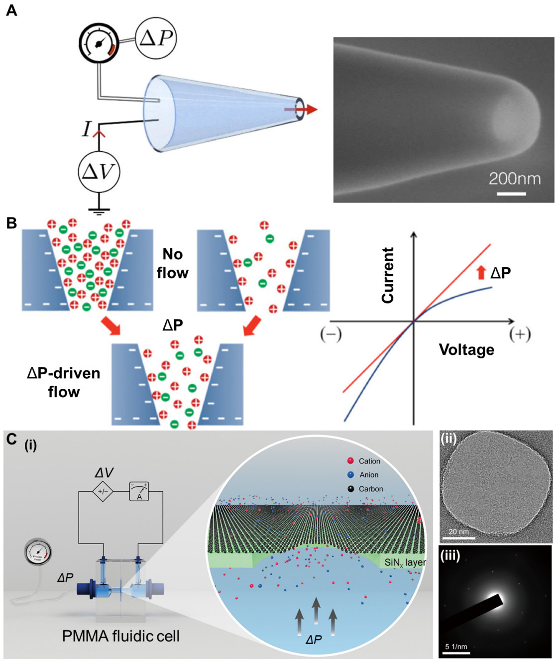
Figure 3. Illustration of bioinspired mechanosensitive ion channels based on glass channels and graphene channels. (A) The single conical glass nanopore and its SEM image. Reproduced with permission[85]. Copyright 2022, National Academy of Sciences. (B) Ion distribution around the pore of the negatively charged glass nanochannel membrane under both positive and negative potentials, with or without ΔP-driven flow. Reproduced with permission[11] Copyright 2011, American Chemical Society; (C) (i) Ion transport through graphene nanopores under the influence of pressure. (ii)TEM image of graphene membrane on 70nm supported SiNx. (iii) Electron diffraction pattern of the graphene membrane. Reproduced with permission[12]. Copyright 2022, The American Association for the Advancement of Science. ΔP: Pressure difference; ΔV: voltage difference; PMMA: poly(methyl methacrylate).

