
fig2
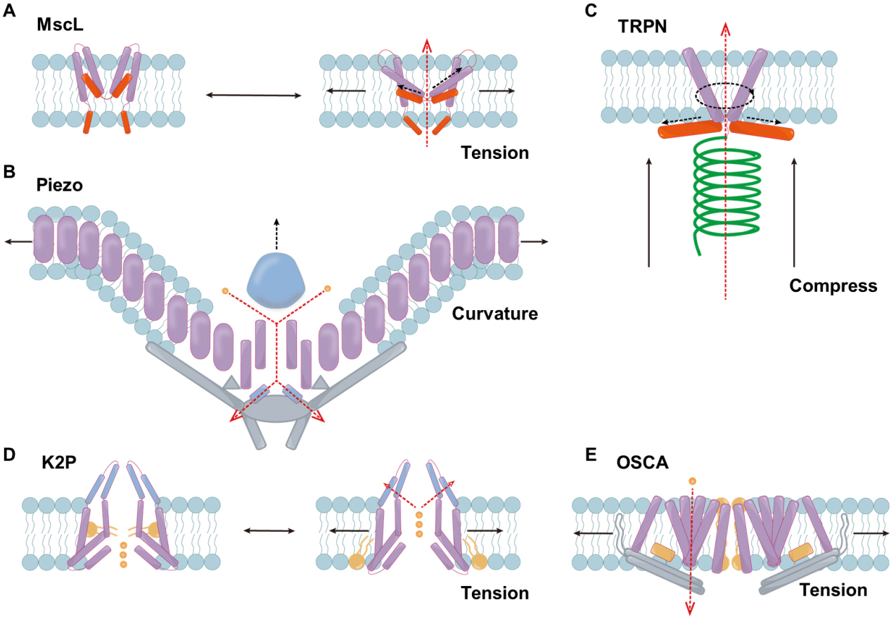
Figure 2. Illustration of the structure of mechanosensitive ion channels from different families. (A) Schematic diagram of the response mechanism of MscL; (B) The structure of the Piezo channel; (C) The structure of the TRPN channel; (D) The response mechanism of K2P; (E) The structure of the OSCA channel. Black solid line: direction of mechanical force. Dark blue: domain. Blue: membrane lipids. Purple: TM helices. Orange: lipid acyl. Red: amphipathic helices. Gray: bundled domain. Green: tether structure. Gold: ions. Black dotted line: change in protein domain. Red dotted line: direction of ion transport. MscL: Mechanosensitive channel of large conductance; TRPN: transient receptor potential channel, subfamily N; K2P: two-pore domain potassium; OSCA: osmosensitive calcium-permeable; ΔP: pressure difference.

