





fig1
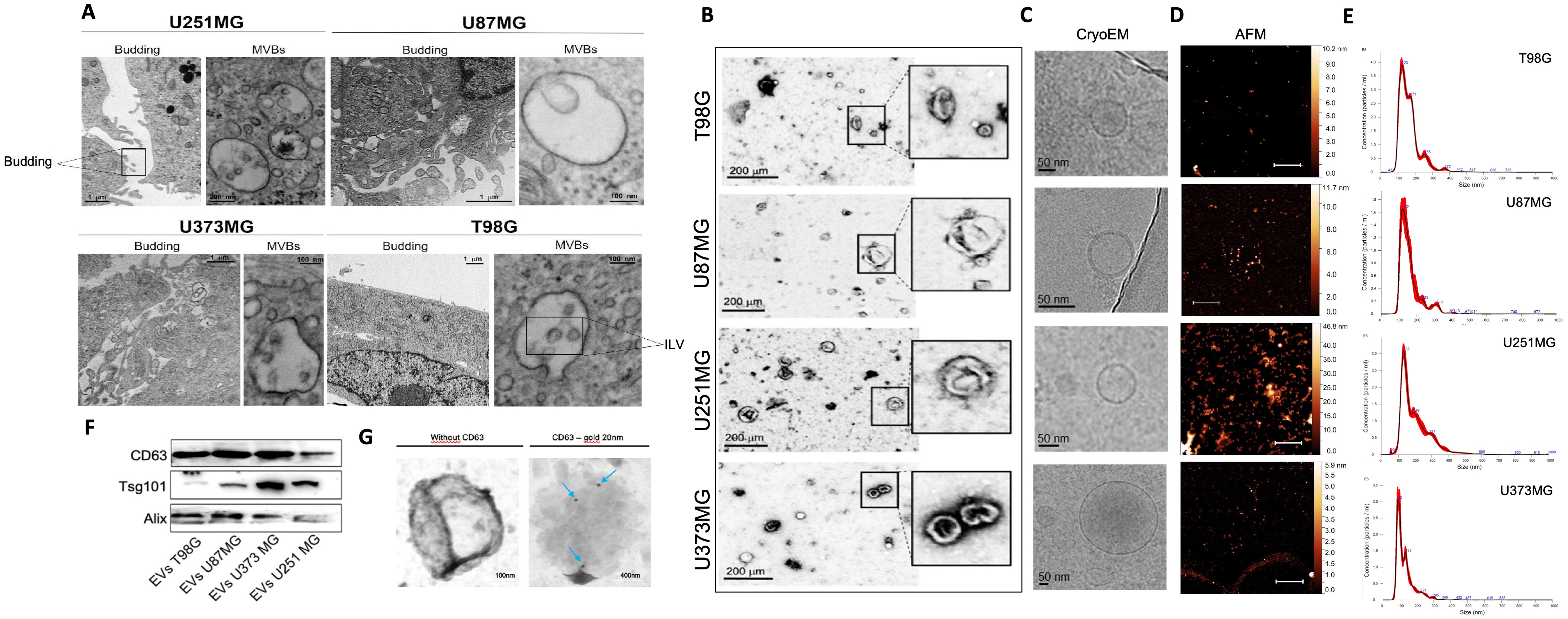
Figure 1. Ultrastructural characterization of GBM cells and GBM-derived EVs. (A) TEM micrographs showing budding vesicles and MVBs with ILVs in U251MG, U87MG, U373MG, and T98G cells; (B) TEM, (C) cryo-EM, and (D) AFM representative images of isolated EVs from T98G, U87MG, U251MG, and U373MG cells. AFM images scale bar: 20 µm; (E) Size distribution of EVs isolated from T98G, U87MG, U251MG, and U373MG cells measured by NTA; (F) Representative Western blot of CD63, TSG101, and ALIX in EV-enriched fractions isolated from T98G, U87MG, U251MG, and U373MG cells; (G) Representative TEM images of isolated EVs with immunogold enhancement with anti-CD63; blue arrows indicate Protein A-gold particles bound to the anti-CD63 antibody, labeling CD63-positive EVs. AFM: Atomic force microscopy; ALIX: ALG-2-interacting protein X; CD63: cluster of differentiation 63; cryo-EM: cryo-electron microscopy; EVs: extracellular vesicles; GBM: glioblastoma; ILVs: intraluminal vesicles; MVBs: multivesicular bodies; NTA: nanoparticle tracking analysis; TEM: transmission electron microscopy; TSG101: tumor susceptibility gene 101.




