





fig2
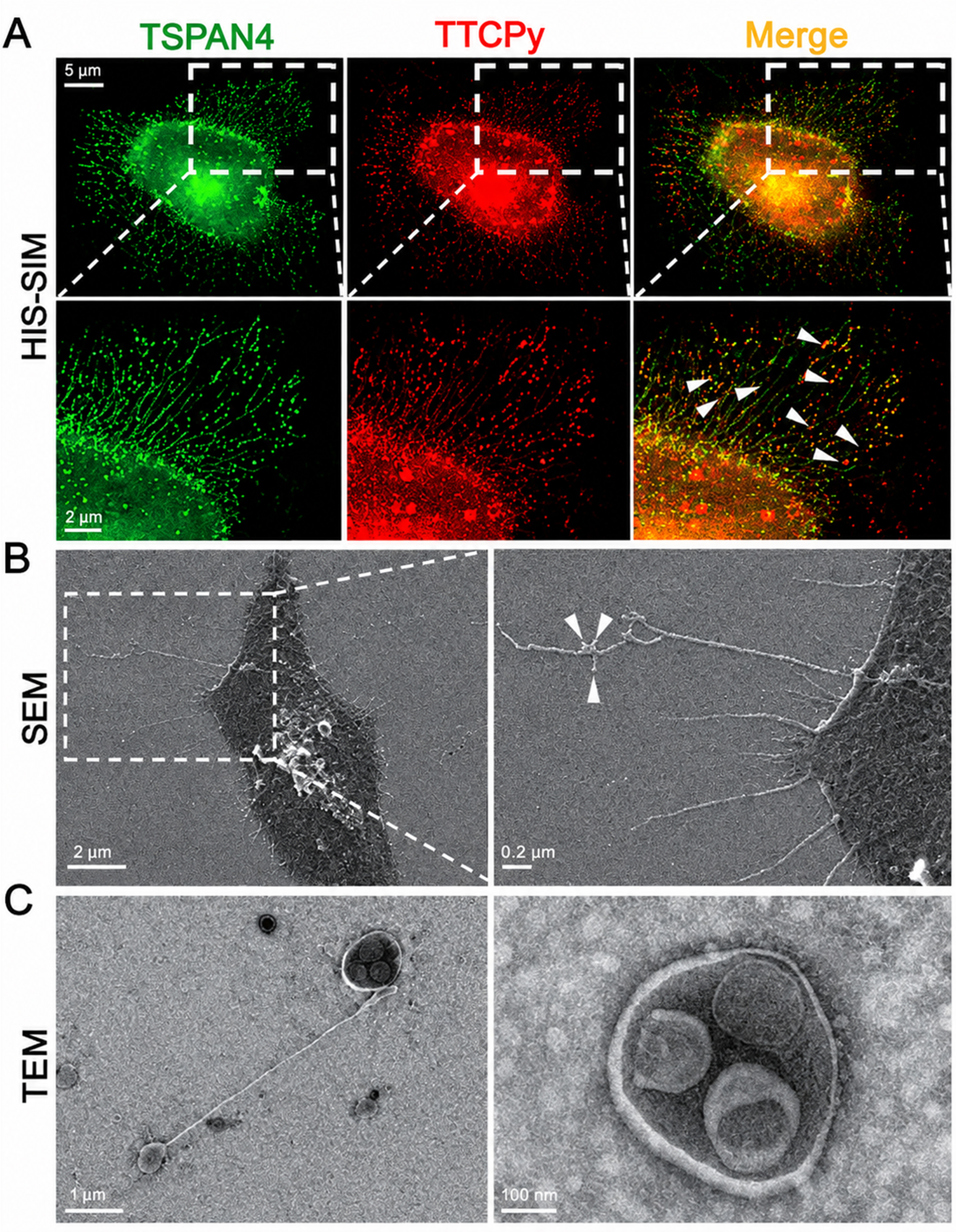
Figure 2. Morphological characterization of migrasomes. Representative (A) HIS-SIM (CSR Biotech, Guangzhou); (B) SEM (JSM-7900F, JEOL, Japan); and (C) TEM (JEM-1400, Japan) images showing migrasomes released from metastatic liver cancer cells (MHCC-97H). Arrows indicate the migrasomes[49]. HIS-SIM: High-sensitivity structured illumination microscopy; SEM: scanning electron microscopy; TEM: transmission electron microscopy; TSPAN4: tetraspanin 4; TTCPy: small-molecule fluorescent probe.




