





fig2
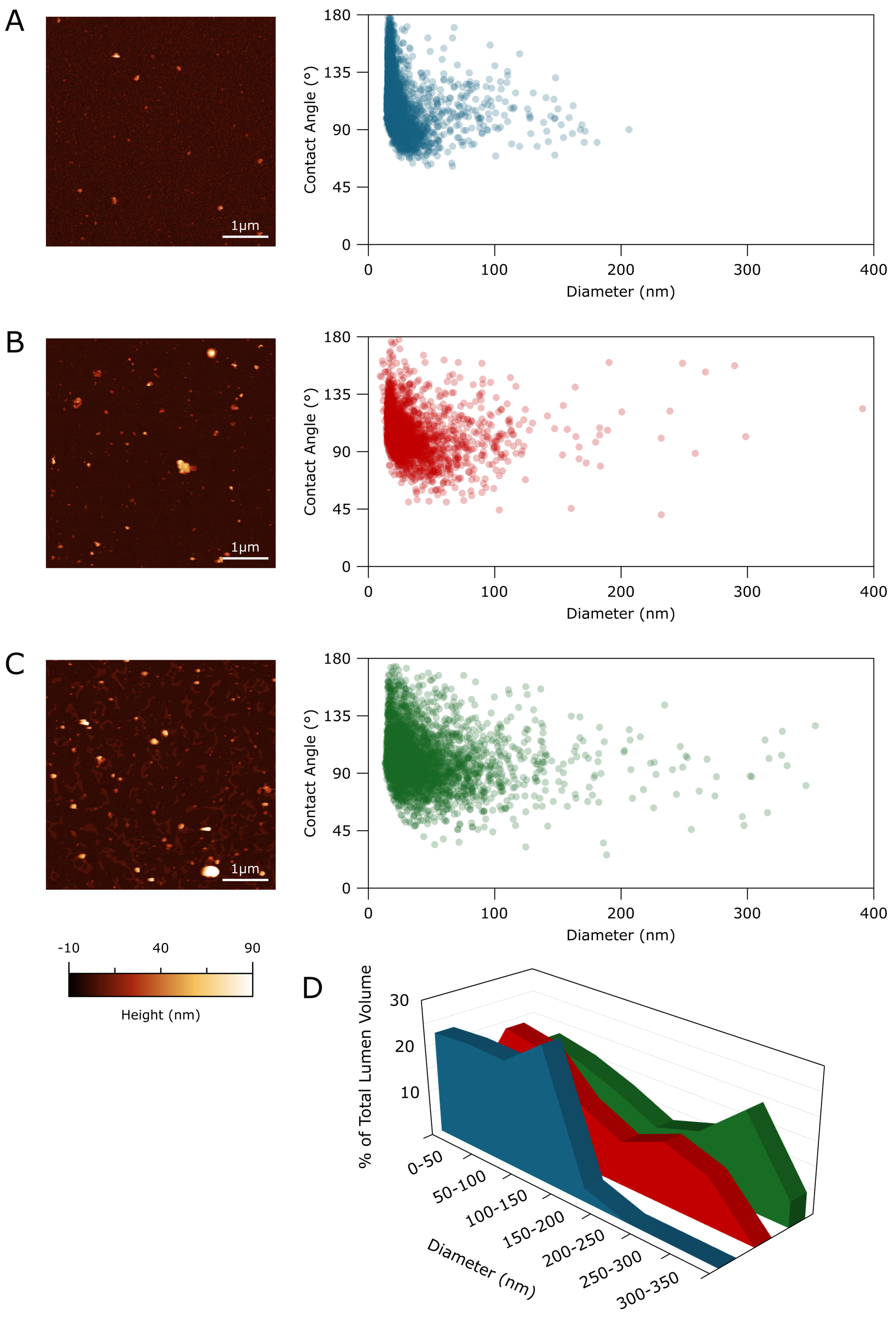
Figure 2. (A-C) Representative 5 µm × 5 µm AFM micrographs (left) and single-particle AFM morphometry plots (right) of sEVs isolated from (A) follicular fluids, (B) peritoneal fluids of control subjects, and (C) peritoneal fluids of endometriosis patients. Each dot in the morphometry plots corresponds to an individual particle plotted according to its free diameter in solution (nm) and surface CA, the latter reflecting its mechanical stiffness, and (D) Percentage of total particle lumen volume vs. vesicle diameter as estimated via AFM morphometry. Blue: follicular fluids, red: peritoneal fluids of control subjects, green: peritoneal fluids of endometriosis patients. The entirety of the lumen volume found in particles isolated from follicular fluids is enclosed by EVs with diameters below 200 nm, whereas in both peritoneal fluid samples, roughly half of the lumen volume is enclosed by vesicles with diameters in the 200-400 nm range. AFM: Atomic force microscopy; EVs: extracellular vesicles; sEVs: small extracellular vesicles.




