


fig1
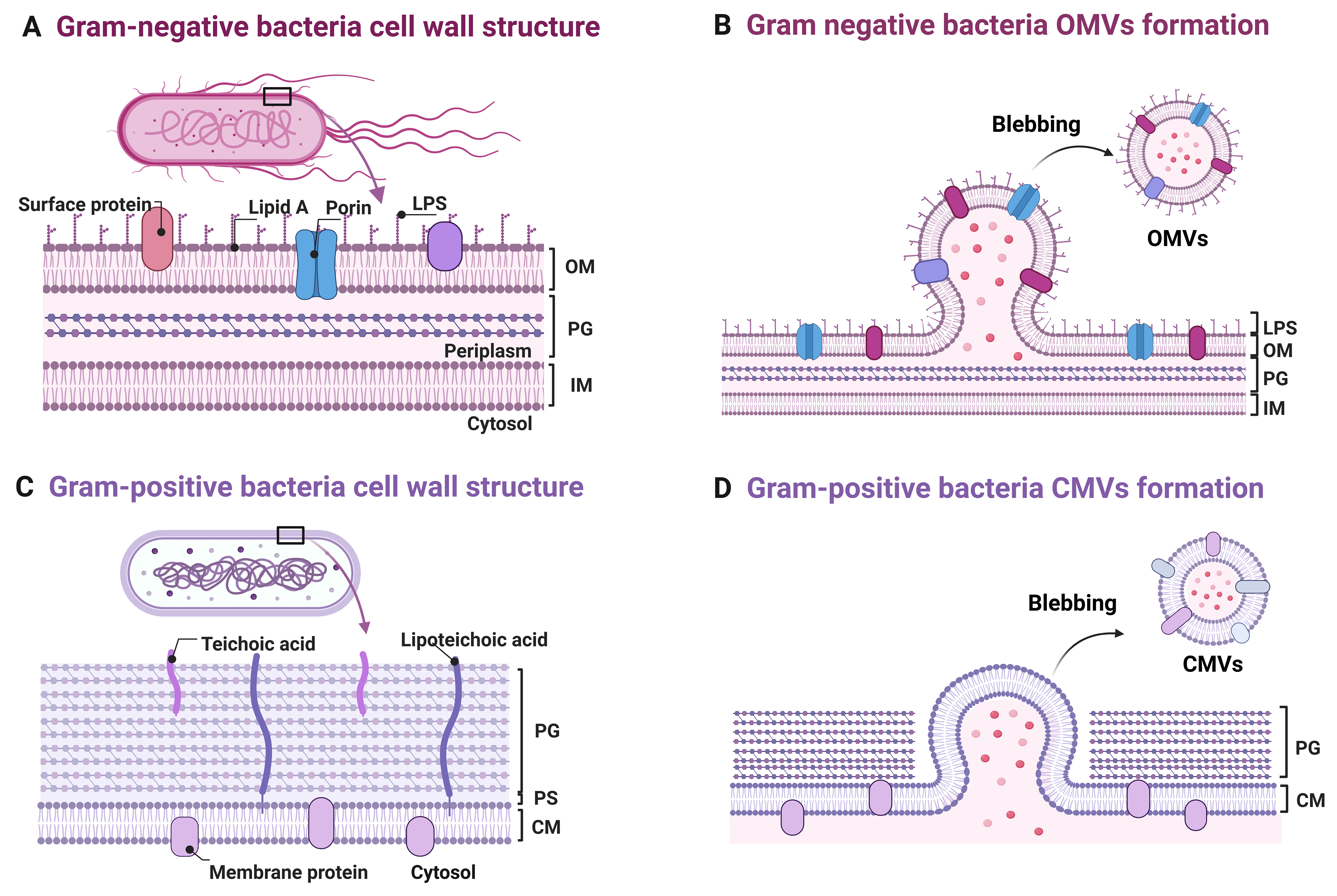
Figure 1. Schematic representation of bacterial membrane vesicle biogenesis and cell wall architecture. Multilayered cell wall structure comprising, from the outermost to innermost, LPS/OM, a PG layer, and an IM. The OM undergoes local evagination to form OMVs that encapsulate LPS, porins, and surface proteins; (B) Magnified view of OMV budding: Asymmetry in OM lipid composition and remodeling of lipid A drive membrane curvature and vesicle scission from the cell envelope; (C) Gram-positive cell envelope architecture consisting of a thick PG layer that directly encloses the CM, with teichoic acids and lipoteichoic acids interspersed throughout; (D) CM evagination leads to CMV formation, wherein membrane proteins and cytosolic cargo are encapsulated. These vesicles are released extracellularly via a yet-to-be-elucidated mechanism that likely traverse the thick PG layer. Note: Other vesicle subtypes (e.g., OIMVs, eOMVs, nanotubules) have been reported in bacteria but remain to be characterized in oral species; accordingly, they are not depicted in this schematic. Created in BioRender. Xumeng, D. (2026) https://BioRender.com/8w62rdx. LPS: Lipopolysaccharide; OM: outer membrane; PG: peptidoglycan; IM: inner membrane; OMVs: outer membrane vesicles; CM: cytoplasmic membrane; CMV: cytoplasmic membrane vesicle; OIMVs: outer-inner membrane vesicles; eOMVs: explosive outer membrane vesicles; PS: periplasmic space.




