





fig5
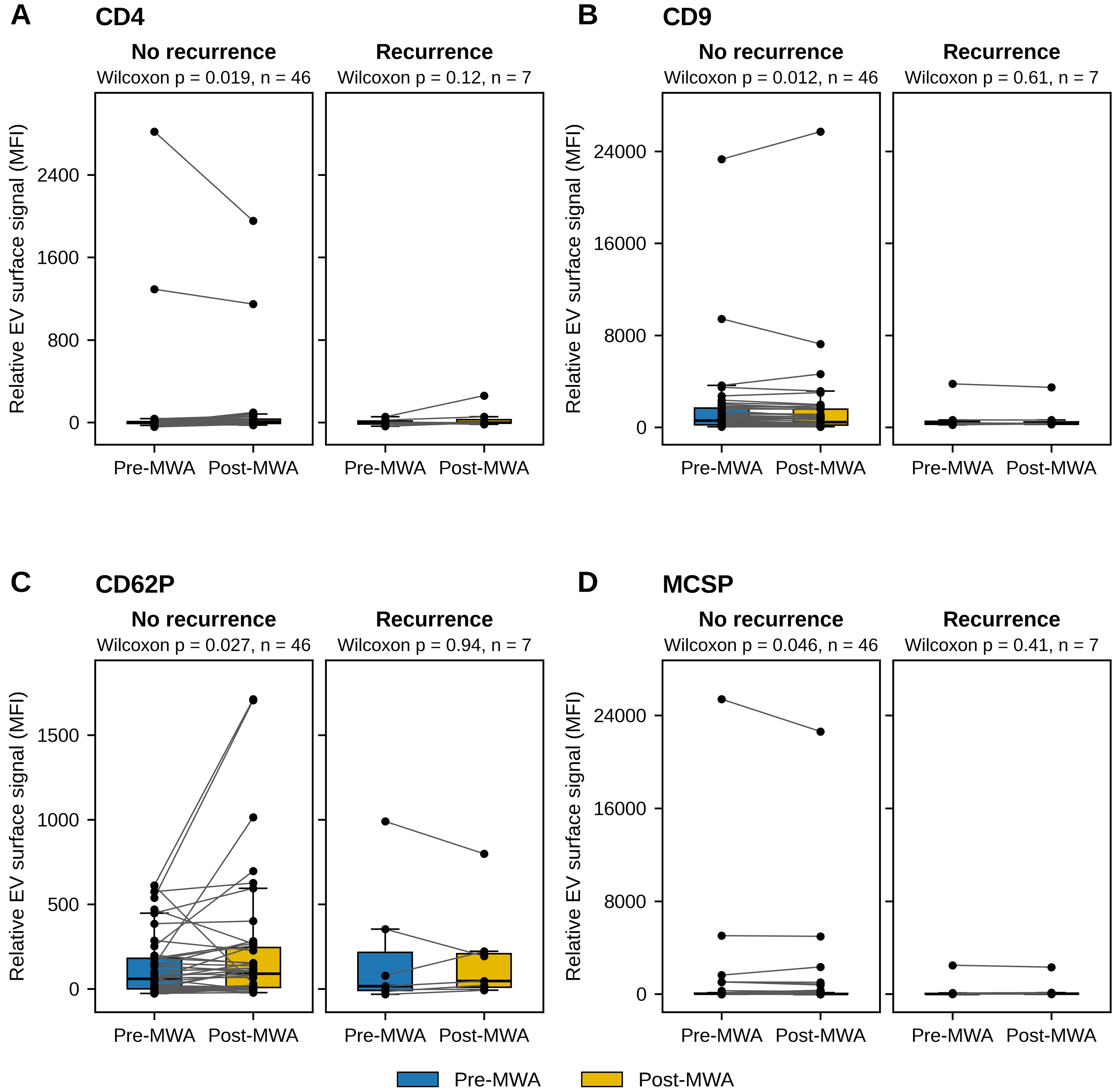
Figure 5. Plasma-derived EV marker levels pre- and post-MWA in the overall patient cohort, stratified by recurrence status (patients without documented recurrence, n = 46, and patients with documented recurrence, n = 7): (A) CD4; (B) CD9; (C) CD62P; (D) MCSP. Data are shown as box plots (median and IQR) with paired individual values. Paired Wilcoxon signed-rank test, nominal P-values. EV: Extracellular vesicle; IQR: interquartile range; MCSP: melanoma-associated chondroitin sulfate proteoglycan; MFI: median fluorescence intensity; MWA: microwave ablation.