fig2
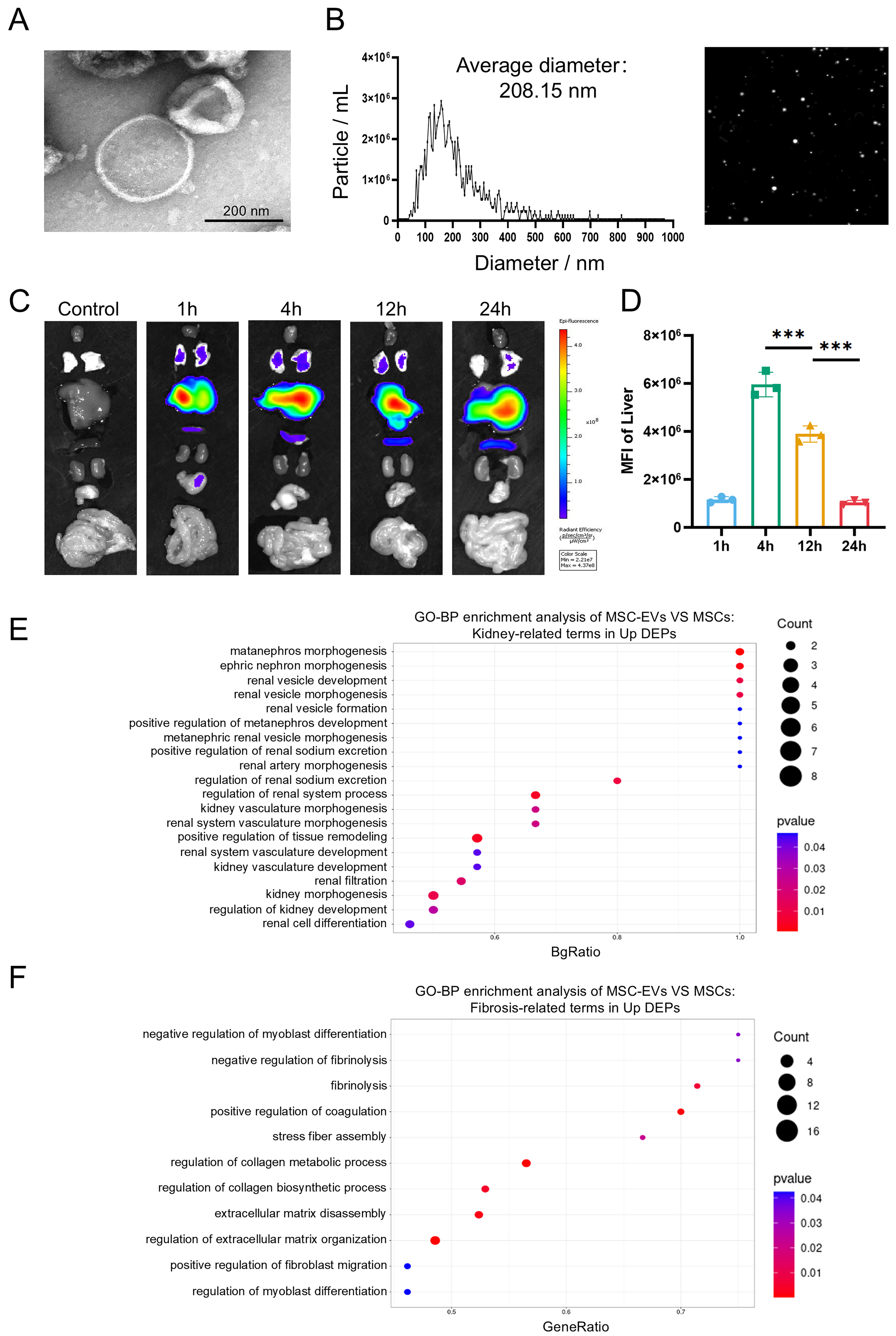
Figure 2. Identification and proteomic profiling of MSC-EVs. (A) Representative TEM image of MSC-EVs (scale bar = 200 nm); (B) Representative NTA image of MSC-EVs; (C) Organ distributions of MSC-EVs in mice detected by Cy5.5 fluorescence after intravenous injection of 30 μg EVs; (D) Statistical analysis of MFI values in the liver (n = 3); (E) GO enrichment analysis of significantly upregulated proteins in MSC-EVs. The Y-axis represents kidney-related GO terms, and the X-axis represents BgRatio. The color of the bubble represents enrichment significance and the size of the bubble represents number of upregulated proteins; (F) GO enrichment analysis of significantly upregulated proteins in MSC-EVs. The Y-axis represents fibrosis-related GO terms, and the X-axis represents GeneRatio. Data were analyzed by one-way ANOVA with Tukey’s post hoc test. Data are presented as mean ± SD. ***P < 0.001. MSCs: Mesenchymal stem cells; EVs: extracellular vesicles; TEM: transmission electron microscopy; NTA: nanoparticle tracking analysis; Cy5.5: cyanine 5.5; MFI: mean fluorescence intensity; GO: Gene Ontology; ANOVA: analysis of variance; SD: standard deviation.