



fig6
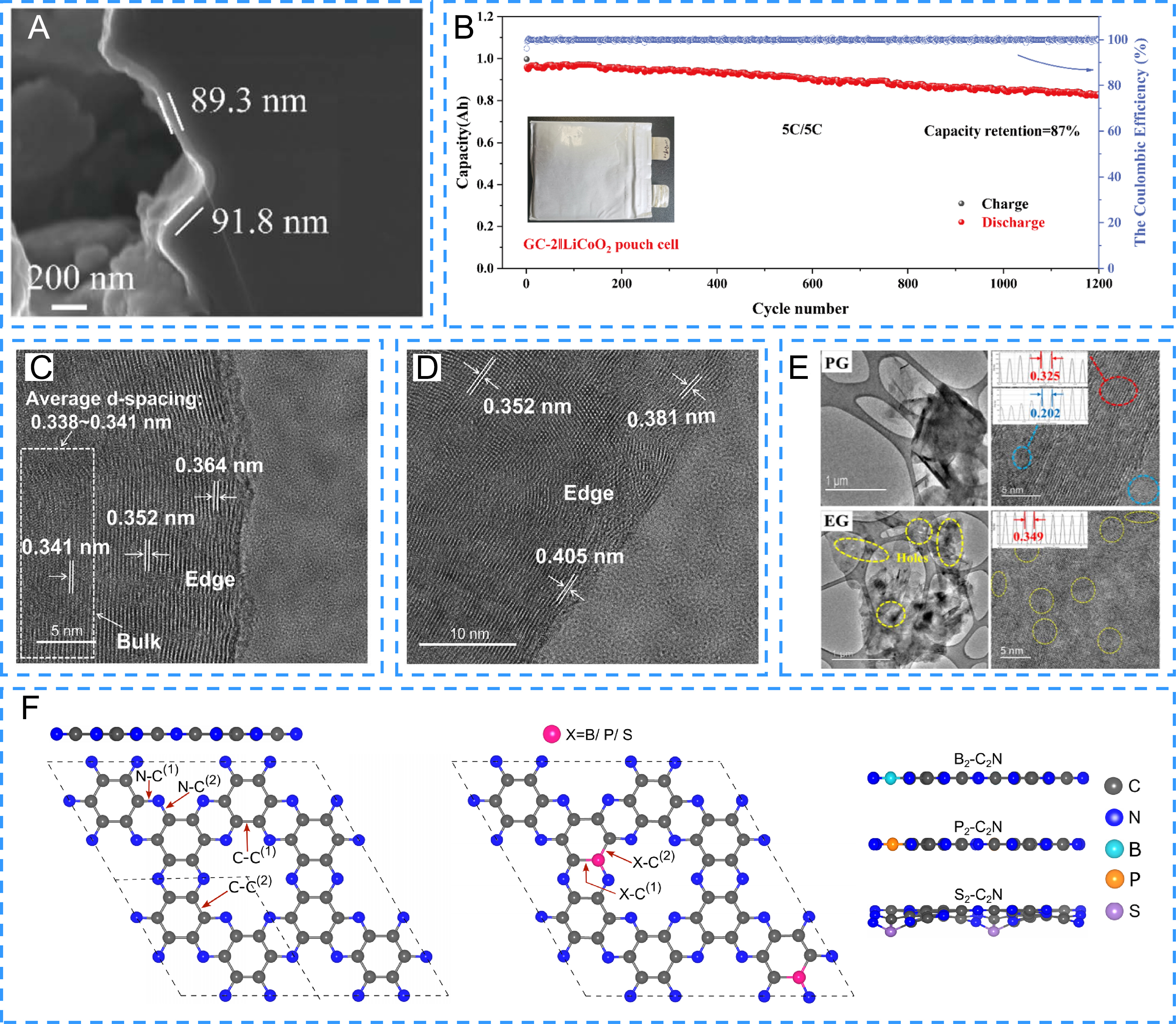
Figure 6. (A) Cross-sectional scanning electron microscopy image of the amorphous carbon coating on GC-2 graphite, showing local coating thicknesses of approximately 90 nm, (B) Cycling performance of GC-2 at 5 C, (A and B) Reproduced from Ref.[91], licensed under CC BY-NC 3.0. (C and D) High-resolution transmission electron microscopy images revealing locally expanded graphite interlayer spacings, particularly near the edges, in CO2-derived expanded graphite, (C and D) Reproduced from Ref.[94], licensed under CC BY 4.0. (E) HRTEM images of pristine PG and EG, (E) Reproduced from Ref.[95], licensed under CC BY-NC 3.0. (F) Top- and side-view atomic structures of pristine C2N and B-, P-, or S-substituted C2N monolayers, illustrating dopant-dependent structural distortion, (F) Reproduced from Ref.[97], licensed under CC BY 4.0.