



fig3
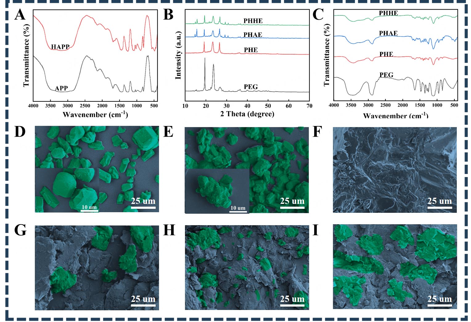
Figure 3. Structural and morphological characterization of the samples: (A) FTIR spectra of APP and HAPP; (B) FTIR spectra of PEG and the different CPCMs; (C) XRD patterns of PEG and the different CPCMs; SEM images of (D) APP, (E) HAPP, (F) PEG, (G) PHE, (H) PHAE, and (I) PHHE. The insets in (D) and (E) show enlarged views of APP and HAPP, respectively. The green regions were pseudo-colored to enhance particle visibility. HAPP: Hydroxylated ammonium polyphosphate; APP: ammonium polyphosphate; PEG: polyethylene glycol; PHAE: PEG/HDI/APP/EG; HDI: hexamethylene diisocyanate; EG: expanded graphite; CPCMs: composite phase change materials; XRD: X-ray diffraction; SEM: scanning electron microscopy; PHE: PEG/HDI/EG; PHHE: PEG/HDI/HAPP/EG; FTIR: fourier transform infrared spectroscopy.