

fig2
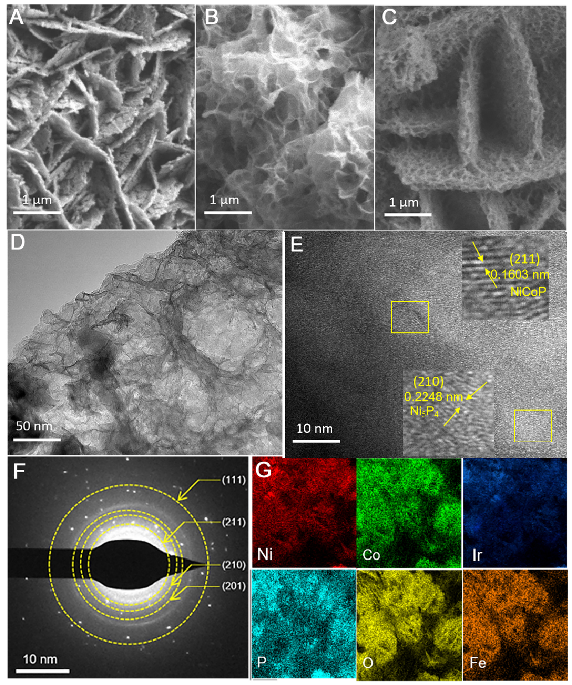
Figure 2. Morphology of the prepared samples. (A) SEM image of NiCoP/Ni5P4@NiFe-LDH-1; (B) SEM image of Ir-NiCoP/Ni5P4@NiFe-LDH-1; (C) SEM image of Ir-NiCoP/Ni5P4@NiFe-LDH-1-OV; (D) TEM image of Ir-NiCoP/Ni5P4@NiFe-LDH-1-OV; (E) HRTEM image; (F) SAED pattern of Ir-NiCoP/Ni5P4@NiFe-LDH-1-OV; (G) Elemental mapping of Ir-NiCoP/Ni5P4@NiFe-LDH-1-OV. SEM: Scanning electron microscopy; LDH: layered double hydroxide; TEM: transmission electron microscopy; HRTEM: high-resolution TEM; SAED: selected area electron diffraction.


