


fig1
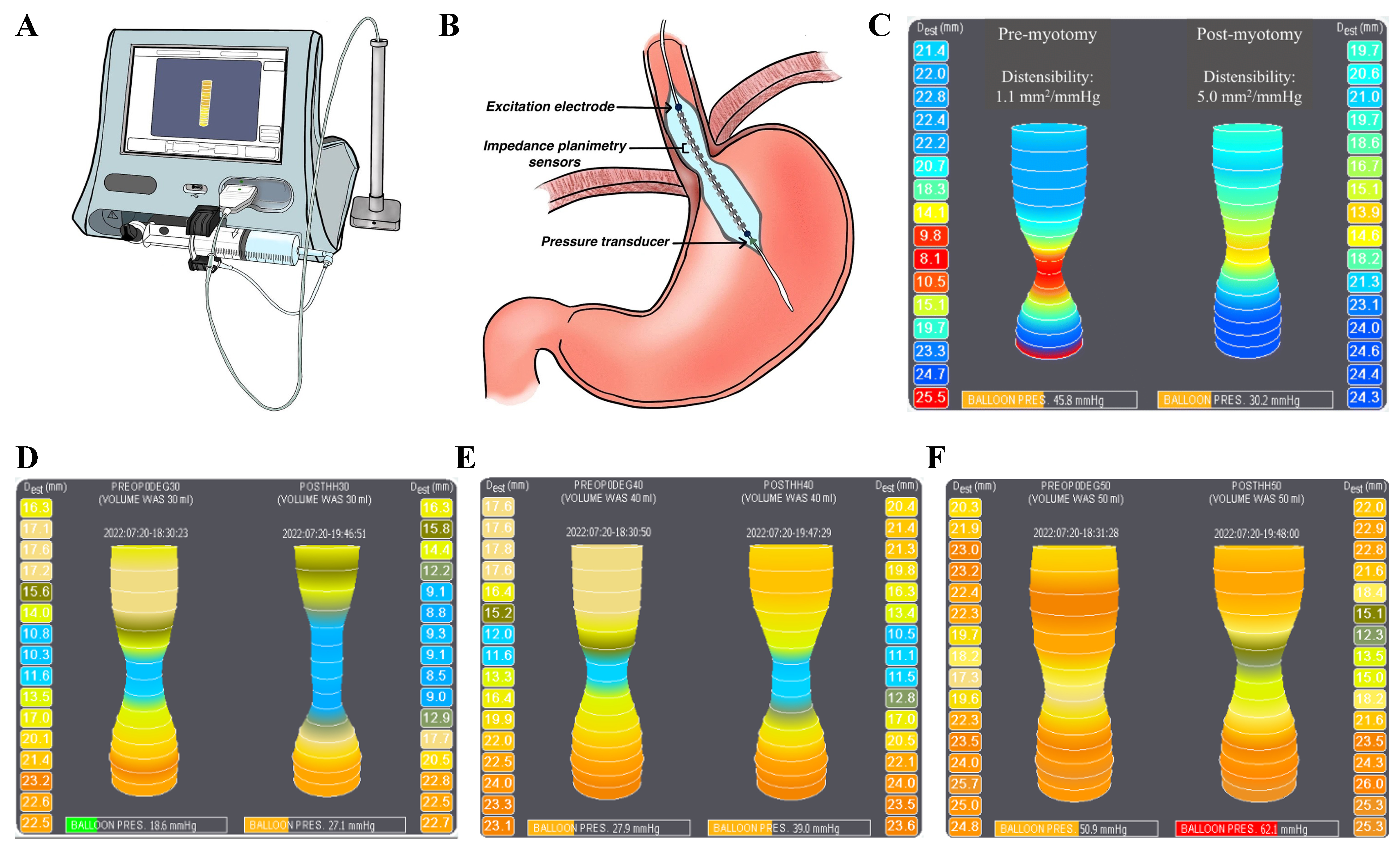
Figure 1. EndoFLIP system and intraoperative assessment of EGJ distensibility. The EndoFLIP console and catheter (A), EGJ catheter placement schematic (B), and representative achalasia tracing demonstrating an increase in DI from 1.1 to 5.0 mm2/mmHg after myotomy (C); Intraoperative topography at 30, 40, and 50 mL (D-F) shows volume-dependent changes in EGJ geometry and pressure, with postoperative findings consistent with symptom relief thresholds and safe distension profiles[14-18], supporting multi-volume functional assessment[19]. (A and B) adapted from Ref.[13]. (C-F) original. Figure assembled in BioRender. Turaga, A. (2026) https://BioRender.com/zihmyto. EndoFLIP: Endoluminal functional lumen imaging probe; EGJ: esophagogastric junction; DI: distensibility index.


