


fig3
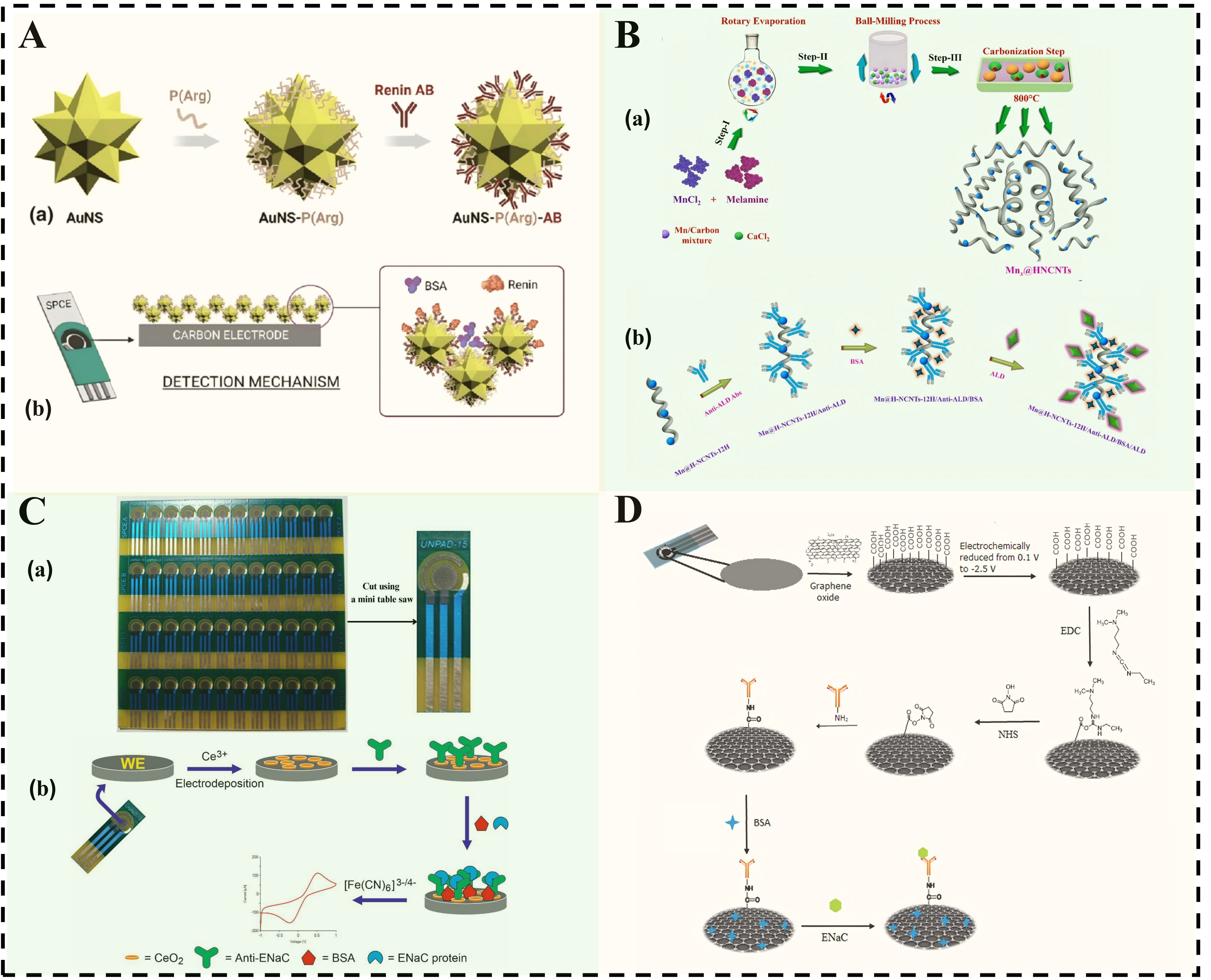
Figure 3. (A) Schematic diagram of renin detection sensing mechanism: (a) After the AuNS surface is coated with P (Arg), renin antibodies are immobilized on its surface; (b) When using a double-layer AuNSs-P (Arg) structure for detection, after immobilizing renin antibodies on the top layer, the active layer is then processed. Reproduced from Ref.[104] with permission from Copyright Clearance Center; (B) Synthesis and fabrication process of ALD immunosensor: (a) Schematic diagram showing the synthesis process of Mnx@H-NCNTs nanostructures; (b) Schematic diagram showing the construction of the BSA/anti-ALD/Mn@H-NCNTs-12H biosensor electrode used for ALD detection. Reproduced from Ref.[105] with permission from Copyright Clearance Center; (C) Schematic diagram of batch preparation of ENaC immunosensors: (a) PCB-SPCE printed circuit board-screen-printed carbon electrode sheets; (b) Schematic diagram of the electrochemical immunosensor used for the detection of ENaC protein. Reproduced from Ref.[109]; (D) Immunosensing scheme based on anti-ENaC/SPCE-rGO. Reproduced from Ref.[109]. AuNS: Gold nanostars; P(Arg): poly(arginine); HNCNTs: hollow nitrogen-doped carbon nanotubes; PCB: printed circuit board; BSA: bovine serum albumin; ENaC: epithelial sodium channel; NHS: N-hydroxysuccinimide; EDC: 1-ethyl-3-(3-dimethylaminopropyl) carbodiimide hydrochloride; ALD: aldosterone; H-NCNTs: hollow nitrogen-doped carbon nanotubes.


