


fig2
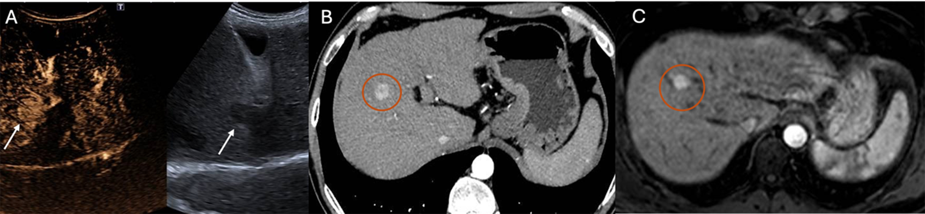
Figure 2. Detection of HCC using CEUS after contrast agent administration (SonoVue®) (A), arterial-phase CT (B), and arterial-phase MRI (C). Arrows in (A) indicate the HCC lesion, while circles in (B and C) highlight the tumor area identified on cross-sectional imaging. HCC: Hepatocellular carcinoma; CEUS: contrast-enhanced ultrasound; CT: computed tomography; MRI: magnetic resonance imaging.


