

A movable long-term implantable soft microfiber: driving a new paradigm for dynamic disease monitoring
 0
0 Abstract
Implantable bioelectronic devices are key to intervening in neurological diseases and realizing brain-computer interfaces. Ideal devices should possess mechanical properties that match biological tissues to reduce rejection reactions and support dynamic control after implantation to enhance monitoring flexibility. Recently, a controllable soft microfiber inspired by earthworms was reported. This microfiber is self-encapsulated into a one-dimensional fiber structure through a two-dimensional thin-film electrode, integrating multi-channel sensing capabilities for simultaneous monitoring of electroencephalogram and muscle mechanical signals. Unlike conventional passive implantable fibers that remain stationary after implantation, this fiber achieves magnetically controlled movement by embedding magnetic particles, enabling dynamic navigation within cortical or muscular tissues and maintaining long-term stability (over 43 weeks). However, its clinical translation still faces challenges such as precision in motor control, long-term signal stability, and complex manufacturing processes. Future research should focus on closed-loop control systems, material optimization, and multifunctional integration to promote the application of such dynamic intelligent implantable devices in neural rehabilitation and personalized medicine.
Keywords
INTRODUCTION
Implantable bioelectronic devices play a vital role in enabling deep intervention in neurological disorders and facilitating precise interpretation of brain–computer interfaces. By designing multimodal micro-bioelectronic systems, such devices can directly interface with biological tissues at minimal scales to enable bidirectional exchange of information or substances[1]. Ideally, these systems should exhibit mechanical properties that closely match those of the host tissue to mitigate immune responses[2]. Moreover, developing bioelectronic implants that are controllable and movable is essential for improving monitoring flexibility and interactive performance - particularly in long-term monitoring or dynamic stimulation scenarios, where fixed electrode placement may limit efficacy and hinder personalized treatment[3]. From early patch-based electroencephalographic (EEG) systems to the recent proliferation of wearable devices, the field of bioelectronics has consistently advanced toward miniaturization and multifunctional integration[4]. Conventional implantable bioelectronic fibers, while possessing basic recording or stimulating functions, are typically passive and remain stationary after implantation. This static characteristic limits their adaptability to dynamic physiological changes or disease progression, as well as their application in scenarios requiring targeted repositioning. A key challenge for next-generation systems lies in creating soft, multimodal micro-bioelectronic devices that are both mechanically compatible with biological tissues and dynamically controllable after implantation [Figure 1A]. Addressing this challenge, Xie et al. report in Nature a controllable soft microfiber capable of long-term dynamic monitoring of neural activity[5].
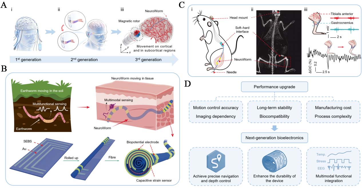
Figure 1. Development and Prospects of Bioelectronic Devices. (A) Evolution of bioelectronic devices from rigid patch electrodes to implantable electronic fibers; (B) Design and fabrication of soft fibers with multimodal monitoring capabilities inspired by earthworms; (C) (i) Schematic diagram and (ii) X-ray image of the soft fiber system implanted in the hind limb muscle of a rat, as well as (iii) simultaneous recording of electromyography from two muscle sites and acquisition of strain signals during leg extension; (D) Prospective upgrading pathways for next-generation bioelectronic devices. Modified with permission from Springer Nature[5].
DESIGN AND MECHANISM OF THE SOFT MICROFIBER
The design of this controllable soft microfiber is inspired by the morphology and locomotion mechanisms of earthworms[5]. By rolling a two-dimensional thin-film electrode into a one-dimensional fiber structure, a longitudinally distributed multi-channel electrode array is realized, enabling simultaneous monitoring of bioelectrical and biomechanical signals [Figure 1B]. A key step in the fabrication is the abrupt rolling self-encapsulation process, which forms “C”- or “L”-shaped conductive pathways by thermally evaporating gold onto a 400-nm-thick styrene-ethylene/butylene-styrene (SEBS) flexible substrate. This self-encapsulation yields flexible electronic fibers with a minimum diameter of 109 μm, a tensile strain of 93%, and a Young’s modulus of 3.1 MPa.
Traditional implantable bioelectronic fibers, such as those used for neural probes or stimulation electrodes, are typically made of rigid materials, lacking maneuverability after implantation. They rely on fixed insertion sites and cannot adapt to tissue movement or changes caused by disease. The flexible microfibers reported in this paper integrate magnetic responsiveness, enabling active movement and positioning after implantation. This provides excellent flexibility for long-term monitoring and intervention.
Researchers from Donghua University and the University of Chinese Academy of Sciences have used these fibers to fabricate magnetically controlled mobile multimodal sensors for detecting EEG signals and mechanical signals in muscle tissue. Unlike conventional passive implantable electrodes, the soft microfibers can be actively moved within the cerebral cortex or muscle tissue under the guidance of an external magnetic field, enabled by embedded magnetic microparticles at the fiber tip. By adjusting the position and rotation speed of the external magnet, the fibers can navigate around blood vessels and reach specific brain regions or muscle tissues, enabling dynamic targeted monitoring [Figure 1C]. The integrated L-shaped conductive pathway acts as a flexible electrode for collecting EEG or electromyographic (EMG) signals, while the C-shaped pathway functions as a capacitive strain sensor, allowing simultaneous monitoring of tissue deformation for multimodal physiological sensing.
Xie et al. also demonstrated the long-term implantability of these soft microfibers in brain and muscle tissues[5]. Owing to their tissue-matching mechanical properties, neither active movement nor deformation caused by muscle tissue leads to functional failure, unless the strain exceeds typical levels (~ 40%) found in most muscular and neural systems. The implanted fibers maintain stable contact with muscle tissue for over 43 weeks, exhibiting a high signal-to-noise ratio and low immune rejection. This controllable, movable, and long-term implantable flexible electronic fiber offers a new approach for dynamic monitoring of neurological diseases.
CHALLENGES AND LIMITATIONS
Although the soft microfibers have shown stable signal recording for over 43 weeks and induce minimal fibrous encapsulation (< 23 μm) in animal models, several challenges remain before clinical translation [Figure 1D]:
(1) Motion control accuracy and imaging dependence: The current open-loop magnetic control strategy has limitations in motion speed and positioning accuracy, particularly in deep tissues where magnetic field attenuation affects control consistency. Moreover, navigation in non-visual regions still requires imaging guidance, limiting autonomy.
(2) Long-term stability and biocompatibility: Although inflammatory responses around the fibers remain mild after 54 weeks, some samples exhibited conductor fracture, likely due to crack propagation in the gold electrodes under chronic deformation. Protein adsorption on the gold surface may also impair signal stability.
(3) Manufacturing cost and process complexity: Current fiber fabrication relies on manual crimping and precision masking, hindering scalable standardized production. The adoption of microfabrication techniques, such as lithography and roll-to-roll processing, holds promise for addressing this challenge, enabling the automation of winding and packaging steps. Furthermore, laser-assisted patterning and automatic alignment systems can replace manual operations, thereby achieving high-throughput production while maintaining consistent quality. These methods not only reduce variability in the manufacturing process but also facilitate the transition from laboratory-scale research to industrial production. The magnetic control system also requires further miniaturization and integration to support multi-channel signal acquisition for comprehensive disease monitoring and diagnosis.
OUTLOOK
To advance the translation of these controllable soft microfibers from the laboratory to the clinic, future efforts may focus on the following areas:
Closed-loop control systems: Integrating electromagnetic coil arrays with real-time imaging feedback could enable precise navigation and depth control in complex tissues. Material and structural optimization: Exploring highly conductive and stretchable materials, such as liquid metals, may enhance the durability of devices. Liquid metals, such as eutectic gallium-indium (EGaIn), possess both high conductivity and fluid deformability, enabling them to maintain electrical continuity under extreme mechanical strain without cracking or fatigue, which are common issues encountered by thin-film metals such as gold during long-term deformation. Multifunctional integration: Incorporating additional modalities such as optogenetics, drug release, or temperature sensing could broaden applications in neural regulation and therapy.
Furthermore, it is worth considering that this technology holds promise for expanding existing Brain-Computer Interface (BCI) platforms. While high-density electrode arrays, such as the Utah Array or the Michigan Array, provide excellent spatial resolution for cortical mapping, they are typically rigid and fixed, limiting their application in dynamic or moving tissues. In contrast, soft microfibers offer mechanical compliance and magnetically controllable capabilities, making them highly suitable for navigating around neural structures and maintaining stable contact over extended periods. In clinical scenarios such as epilepsy monitoring or closed-loop deep brain stimulation, these fibers can be used in conjunction with fixed high-density arrays to achieve large-area mapping and targeted adaptive intervention. This hybrid approach can enhance the precision and adaptability of next-generation BCIs.
Controllable soft microfibers represent significant progress in bioelectronics from static fixation toward dynamic intelligence. Their movable, soft, and multi-channel characteristics offer a new paradigm for long-term monitoring and precise neural interfacing. With continued advances in control strategies, materials science, and integration technologies, these dynamically controllable implantable fibers are poised to play a transformative role in brain–computer interfaces, neural rehabilitation, and personalized medicine.
DECLARATIONS
Authors’ contributions
Conceived the research: Liu, R.; Yue, Q.
Wrote the manuscript: Liu, R.
Supervised the research and revised the manuscript: Yue, Q.
Availability of data and materials
Not applicable.
AI and AI-assisted tools statement
Not applicable.
Financial support and sponsorship
This work is supported by the Natural Science Foundation of Sichuan Province (2025NSFTD0003, 2025ZDZX0083).
Conflicts of interest
Yue, Q. is an Editorial Board Member of the journal Micro Nano Science. Yue, Q. was not involved in any steps of the editorial process, including reviewers’ selection, manuscript handling, or decision-making. The other author declares that there are no conflicts of interest.
Ethical approval and consent to participate
This work does not involve any ethical issues.
Consent for publication
Not applicable.
Copyright
© The Author(s) 2026.
REFERENCES
1. Sahasrabudhe, A.; Rupprecht, L. E.; Orguc, S.; et al. Multifunctional microelectronic fibers enable wireless modulation of gut and brain neural circuits. Nat. Biotechnol. 2024, 42, 892-904.
2. Khatib, M.; Zhao, E. T.; Wei, S.; et al. High-density soft bioelectronic fibres for multimodal sensing and stimulation. Nature 2025, 645, 656-64.
3. Yang, J.; Zhang, Y.; Liu, Z.; et al. Magnetically actuated multimodal bioelectronic catheter for minimally invasive surgery and sensing. Nat. Mater. 2025, 24, 2019-31.
4. Wang, Z.; Shi, N.; Zhang, Y.; et al. Conformal in-ear bioelectronics for visual and auditory brain-computer interfaces. Nat. Commun. 2023, 14, 4213.
Cite This Article
How to Cite
Download Citation
Export Citation File:
Type of Import
Tips on Downloading Citation
Citation Manager File Format
Type of Import
Direct Import: When the Direct Import option is selected (the default state), a dialogue box will give you the option to Save or Open the downloaded citation data. Choosing Open will either launch your citation manager or give you a choice of applications with which to use the metadata. The Save option saves the file locally for later use.
Indirect Import: When the Indirect Import option is selected, the metadata is displayed and may be copied and pasted as needed.
About This Article
Copyright

Data & Comments
Data
0


Comments
Comments must be written in English. Spam, offensive content, impersonation, and private information will not be permitted. If any comment is reported and identified as inappropriate content by OAE staff, the comment will be removed without notice. If you have any queries or need any help, please contact us at support@oaepublish.com.