





Mitochondrial transfer in solid tumors: dual roles in tumor progression and therapeutic targeting
 0
0 Abstract
Solid tumors account for the majority of cancer cases, and their incidence continues to increase. A solid tumor is not an isolated mass but interacts dynamically with cells in the tumor microenvironment. Mitochondria, which are essential for energy production and the regulation of apoptosis, play a critical role in cellular function and may serve as therapeutic targets. A growing body of evidence demonstrates that mitochondria can be transferred between cells via extracellular vesicles, tunneling nanotubes, gap junctions, and cell fusion. This process across various cell types, including stromal cells, immune cells, and cancer cells, and is enhanced under stress conditions, such as hypoxia and chemotherapy. Under these conditions, mitochondrial transfer enables tumor cells to acquire functional mitochondria from neighboring cells, thereby enhancing their metabolic plasticity and survival capacity. Mitochondrial transfer thus represents a critical adaptive mechanism that supports tumor progression. Targeting this dynamic form of intercellular communication offers a promising therapeutic strategy to overcome tumor metabolic plasticity, immunosuppression, and resistance to therapy.
Keywords
INTRODUCTION
The global burden of cancer continues to increase. According to the latest data from the Global Burden of Disease Study 2023 (GBD 2023), there were 18.5 million new cancer cases worldwide in 2023, of which 10.4 million resulted in death[1]. Solid tumors are among the primary contributors responsible for cancer-related mortality, and are distinguished by aggressive tumor proliferation, as well as local invasion and distant metastases. Remarkable therapeutic gains have been realized for both in situ disease management and non-metastatic survival thanks to modern developments in multimodal regimens, with surgical removal of the primary neoplasm serving as a core contributor[2]. An intricate interconnected system known as the tumor microenvironment (TME) consists of neoplastic cells and an array of supporting components, among which are endothelial cells, immune cells, stromal cells, and extracellular matrix[3]. From the initial phases of tumor development, reciprocal interactions between malignant cells and their surrounding niche contribute to tumor survival, local invasive growth, and metastatic propagation[3].
Recently, accumulating evidence has shown that mitochondria not only play a critical role in host cells, but can also be transferred into recipient cells, where they regulate various cellular functions[4]. Mitochondrial transfer occurs across different tissues under both physiological and pathological conditions[4]. Released mitochondria may originate from both dying cells and viable cells[5]. Stress conditions, such as oxygen-glucose depletion, drug-induced oxidative stress and inflammation, can promote the release of damaged or fragmented extracellular mitochondria, thereby facilitating intercellular mitochondrial transfer[6]. More recently, accumulating evidence has shown that mitochondrial transfer can occur between different cell types. To date, four primary mechanisms underlying mitochondrial transfer have been identified: extracellular vesicles (EVs)[7], tunneling nanotubes (TNTs)[8], gap junctions (GJs)[9] and cell fusion[10]. Physiological mitochondrial rescue is a transient homeostatic protective mechanism in normal tissues triggered by mild stress such as temporary hypoxia. Mitochondria move bidirectionally between adjacent normal cells to supplement impaired energy supply, and this transfer will stop automatically after cellular metabolic balance recovers; it only serves tissue repair without malignant risks. It has been suggested that the transfer of intact mitochondria may serve as a preventive mechanism in a variety of conditions requiring metabolic rescue, including acute myocardial infarction and stroke-induced ischemic injury[11]. The acquisition of functional mitochondria or their components may help restore or enhance mitochondrial function[4]. In contrast, tumor-promoting mitochondrial transfer is sustained pathological communication activated by long-term harsh tumor microenvironmental stress, including nutrient deprivation, persistent hypoxia, and chemotherapy damage. This transfer is predominantly unidirectional toward metabolically defective tumor cells. Recipient tumor cells acquire intact functional mitochondria to achieve metabolic remodeling, stronger invasive capacity and cross-drug resistance, ultimately accelerating tumor growth, metastasis and treatment failure[12].
The transferred mitochondria may also impact other cellular structures. In the context of previous findingson mitochondrial transfer, functional mitochondria acquired by recipient tumor cells may integrate into thehost mitochondrial-associated endoplasmic reticulum (ER) membranes (MAMs) network, restore proper ER- mitochondrial Ca2+ homeostasis, and mitigate chronic ER stress, promoting chemoresistance and survival. Conversely, if transferred mitochondria fail to establish appropriate contact sites, they might exacerbate Ca2+ dysregulation and sensitize cells to apoptosis[13]. In solid tumors, the balance between mitochondrial biogenesis and mitophagy is often dysregulated, contributing to metabolic flexibility and therapy resistance[14]. Additionally, mitochondrial-derived vesicles (MDVs) can deliver oxidized mitochondrial components directly to lysosomes for degradation, representing an alternative quality control pathway[14]. In tumors with high basal autophagy (a common feature of many solid tumors), transferred mitochondria could be rapidly targeted for mitophagy and degraded, limiting their functional impact.
This review aims to provide a comprehensive synthesis of the complex biological functions of mitochondrial transfer in solid tumors, with a particular focus on its bidirectional and context-dependent mechanisms. Evidence for both pro-tumorigenic and potential anti-tumor effects is critically examined. Building on this foundation, the review systematically evaluates emerging intervention strategies-such as inhibiting detrimental transfer or enhancing beneficial transfer-and discusses the prospects and challenges for their clinical translation.
MECHANISMS OF MITOCHONDRIAL TRANSFER IN CANCER CELLS
Up to now, four primary mechanisms underlying mitochondrial transfer have been identified: EVs, TNTs, GJs and cell fusion [Figure 1].
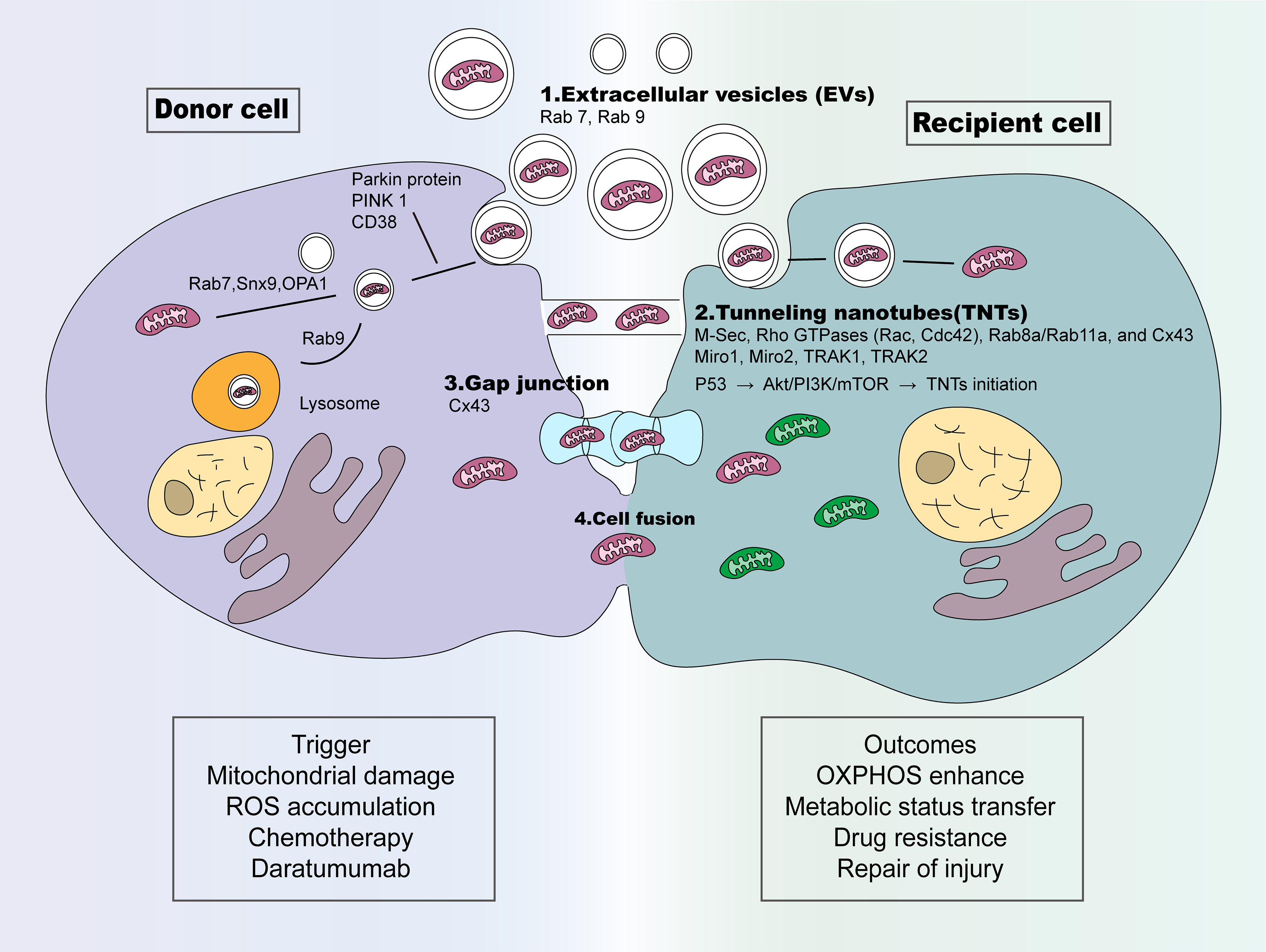
Figure 1. The ways of mitochondrial transfer in cancer cells. Four primary mechanisms underlying mitochondrial transfer have been identified: (1) EVs: Mitochondria-containing EVs form under the regulation of Snx9, Rab9 and OPA1, while Rab7 mediates EVs-lysosome fusion for cargo degradation. Alternatively, PINK1/Parkin signaling drives extracellular release of mitochondrial cargo-laden vesicles; (2) TNTs: Multiple signaling molecules coordinate TNT formation, including M-Sec, Rho GTPases (Rac, Cdc42), Rab8a/Rab11a, and Cx43 gap junction proteins. Miro1/2 together with TRAK1/2 form adaptor complexes that mediate mitochondrial transfer within microtubule-containing TNTs. TNT development is further modulated by stress and pro-survival cascades such as p53, PI3K/Akt, MAPK/ERK, and mTOR pathways; (3) GJ: Gap junctions are assembled by two hemichannels, each composed of six oligomerized connexin subunits, enabling direct molecular exchange between adjacent cells. Cx43 is the best-characterized connexin subtype supporting mitochondrial transfer; (4) Cell fusion: Increased cell fusion events between different cells enhance mitochondrial transfer. CD38: Cluster of differentiation 38; Cx43: connexin 43; M-Sec: Mammalian Sec14-like protein 2; Miro1: mitochondrial Rho GTPase 1; Miro2: mitochondrial Rho GTPase 2; OPA1: Optic atrophy 1; PINK1: phosphatase and tensin homolog-induced kinase 1; Rab11a: Ras-related protein 11a; Rab7: Ras-related protein 7; Rab8a: Ras-related protein 8a; Rab9: Ras-related protein 9; Rac: Rac family small GTPase; Snx9: Sorting nexin 9; TRAK1: trafficking kinesin protein 1; TRAK2: trafficking kinesin protein 2; EVs: extracellular vesicles; TNTs: tunneling nanotubes; GJ: gap junction; Cdc42: cell division control protein 42 homolog; p53: tumor protein p53; PI3K: phosphoinositide 3-kinase; Akt: protein kinase B; MAPK: mitogen-activated protein kinase; ERK: extracellular signal-regulated kinase; mTOR: mechanistic target of rapamycin.
EVs
EVs are membranous structures enclosed by lipid bilayers that are produced and released by cells[15,16], with sizes ranging from 30 to 1,000 nm[15]. Two dominant classes of EVs have been identified, namely microvesicles (MVs) and exosomes. The biogenesis of MVs depends on outward membrane budding, while exosome production originates from the cellular endocytic pathway[17]. A wide range of biomolecules are packaged within EVs, including proteins, lipids, and metabolites. These vesicles also harbor diverse genetic materials such as mRNA, non-coding RNA, nuclear DNA, and mitochondrial DNA, with sporadic innate organelle components observed in specific EV subsets. The transfer of intracellular cargo to recipient cells reshapes cellular biological phenotypes through the regulation of gene and protein expression profiles, as well as nutrient metabolic status, thereby disrupting cell homeostasis[18]. Cargo composition, coupled with the dynamic release and internalization patterns of EVs, collectively dictates their functional performance within the TME[18]. Accumulating experimental evidence obtained via transmission electron microscopy, immunofluorescence, and western blotting confirms the presence of mitochondrial components in EVs[19]. Mitochondria-derived vesicles (MDVs) typically range from 80 to 150 nm in diameter and are characterized by a double membrane structure lacking cristae[19]. The formation of MDVs requires sorting nexin 9 (Snx9) and Ras-related protein 9 (Rab9). Through the coordinated action of Snx9 and optic atrophy 1 (OPA1), mitochondria and their components are selectively incorporated into MDVs for degradation[20]. Ras-related protein 7 (Rab7) mediates the trafficking of MDVs to lysosomes[21]. In addition to being targeted to lysosomes, MDVs are capable of extracellular release, and this secretory process is regulated by two key molecules: the E3 ubiquitin ligase Parkin and PTEN-induced kinase 1 (PINK1)[22]. Notably, depletion of arrestin domain-containing protein 1 (ARRDC1) in mouse embryonic fibroblast cells markedly increases the abundance of mitochondrial proteins encapsulated in EVs, supporting the regulatory role of ARRDC1 in mediating mitochondrial protein incorporation into vesicle cargos[23]. CD38 catalyzes the production of the calcium-mobilizing messenger cyclic ADP-ribose (cADPR) within mitochondrial membranes[24]. Stimulation of astrocytes with cADPR activates CD38 signaling, resulting in increased levels of extracellular mitochondrial in conditioned media, along with enhanced mitochondrial function in a calcium-dependent manner[11]. Activation of the CD38/inositol 1,4,5-trisphosphate receptor (IP3R)/Ca2+ pathway generates “super donor” mesenchymal stem cells (MSCs) which produce EVs containing mitochondrial (EV-Mito) at threefold higher levels than those normal MSCs[25]. Inhibition of CD38 reduces extracellular mitochondrial levels and their astrocyte-to-neuron transfer[11]. Furthermore, pharmacological inhibition of MV formation using GW4869 resulted in a decrease in the extracellular release of mitochondria[26].
TNTs
TNTs serve as intercellular channels enabling communication between spatially separated cells in the same tissue, even spanning distances up to hundreds of micrometers[27]. Two primary mechanisms for the formation of TNTs have been proposed[27,28]. The first mechanism is largely influenced by cell movement and takes place when cells initially in close proximity subsequently separate[27]. The second mechanism involves the extension and fusion of actin filament-containing membrane protrusions. These protrusions originate from the donor cell and fuse with the target cell’s membrane, without requiring cell motility or prior contact[27]. As a direct form of cell-to-cell communication, TNTs have been reported in several pathological conditions, including cancer, in both in vitro and in vivo models[29]. Two types of TNTs based on the cytoskeletal components involved in their formation[30]. One subset comprises thin filaments composed solely of F-actin, whose formation can be induced by the mammalian protein M-sec. A thicker subset, measuring 0.7 μm, known as microtubule-bearing TNTs, contains both F-actin and microtubules[30]. It may facilitate kinesin- and dynein-dependent transport of mitochondria between interconnected cells[8]. The movement of mitochondria along microtubules is enabled by a protein complex containing the calcium-sensitive adaptor protein mitochondrial Rho GTPase 1 (Miro1, encoded by RHOT1). Miro1, along with associated factors such as Miro2, trafficking kinesin-binding protein 1 (TRAK1), TRAK2, and myosin XIX (Myo19), is essential for regulating mitochondrial transport[31]. Miro family proteins modulate mitochondrial transfer along TNTs, acting as molecular adaptors to connect mitochondria with cytoskeletal regulatory proteins[32]. They are also crucial for actin rearrangement via Guanosine diphosphate (GDP)-guanosine triphosphate (GTP) cycling.[27]. The downstream effectors of Miro are essential to this process because they regulate actin polymerization via Arp2/3 nucleation and include the Wiskott-Aldrich syndrome protein (WASP) and WASP family verprolin-homologous 2 (WAVE2). Inhibition of the Arp2/3 complex resulted in decreased formation of tumor microtubes in pancreatic cancer cells, highlighting the significant role of actin polymerization in their development[33]. Rac and Cdc42, two pivotal members of the Rho GTPase family responsible for actin cytoskeleton polymerization, are essential for TNT generation. Localization of these proteins within TNTs of T cells further confirms their regulatory role, and targeted Cdc42 inhibition is sufficient to hinder TNT formation[34]. Another study demonstrated that Rab8a/Rab11a regulate intercellular communications between neural cells via TNTs[35]. Knockdown of Rab8a/Rab11a promotes apoptosis in Schwann cells and inhibits their migration by suppressing TNT formation[35]. The integral GJ protein, connexin 43 (Cx43), which is present in TNT-like structures, plays a key role in communication between TNT-connected cells[36]. Knockdown of Cx43 has been reported to significantly affect TNT formation and reduce mitochondrial transfer between MSCs and epithelial cells[37]. The biogenesis of TNTs involves various stress-signaling pathways, such as p53 and mitogen-activated protein kinase (MAPK), alongside pro-survival mechanisms, including epidermal growth factor receptor (EGFR), protein kinase B (Akt), Rho-associated coiled-coil kinase (ROCK), p21-activated kinase (PAK), MAPK/extracellular signal-regulated kinase (ERK), phosphoinositide 3-kinase (PI3K), or mechanistic target of rapamycin (mTOR)[6]. Signaling pathway activation ultimately targets effector proteins that mediate membrane recycling processes and cytoskeletal dynamic remodeling[6]. tumor microtubes (TMs) constitute a type of tubular structure different from TNTs. TNTs are thin, transient intercellular channels composed exclusively of actin filaments and are ubiquitously present in all mammalian cell types. By contrast, TMs are thick, extraordinarily stable membrane protrusions characterized by the coexistence of F-actin and uninterrupted microtubule bundles[38].
GJs
GJs constitute direct intercellular routes that enable molecular exchange between neighboring cells. They are established through the connection of hemichannels from each cell, which are hollow tube-like structures resulting from the oligomerization of six connexin subunits in the plasma membrane[39]. GJs also represent an important mechanism for mitochondrial transfer, although fewer studies have examined GJs compared with TNTs. This is partly because TNTs are more readily observed in in vitro co-culture systems[40]. Connexin family proteins play crucial roles in the formation of GJs. Among these, Cx43 is the most extensively studied. Latest studies reveal that the reactive oxygen species (ROS)-mediated oxidative stress response during emergency granulopoiesis initiates PI3K-Akt activation, thereby opening Cx43 channels and supporting intercellular mitochondrial transfer from bone marrow mesenchymal stem cells (BMSCs) to hematopoietic stem cells[41]. Mitochondrial transfer occurs exclusively when GJs operate normally at areas of close contact between cells[42]. One study indicates that during the process of GJ internalization, one cell engulfs small portions of a neighboring cell, facilitating mitochondrial transfer[43]. Emerging evidence indicates that GJs strengthen the adhesive interaction between TNTs and recipient cells, while simultaneously enhancing EV internalization efficiency[44]. Following treatment with the GJ enhancer retinoic acid, mitochondrial transfer was increased, which means that GJs participated in the transfer of mitochondria from BMSCs to VSC 4.1 motor neurons[9]. In glioblastoma, GJs formed by Cx43 may facilitate TNT formation and enhance mitochondrial transfer[45]. This may occur by enabling tumor microtubules to establish contact with adjacent cells, thereby promoting increased signal exchanges and ultimately advancing tumor progression[45].
Cell fusion
Cell fusion involves the fusion of the plasma membranes between two separate cells. Such intercellular fusion facilitates the exchange of cytosolic substances and organelles, whereas the nuclei of the two cells remain structurally distinct[46]. Owing to the low prevalence of cell fusion events in normal higher eukaryotic physiology, this process is not classified as a primary route for mitochondrial intercellular transfer[40]. Some studies suggest that the events described above may represent partial or transient cell fusion, whereas permanent cell fusion results in hybrid cells that share cytoplasm and often exhibit abnormal karyotypes. This leads to a more complex interplay of mitochondrial dynamics, including transfer, fusion, and fission events[10]. Increased cell fusion events between MSCs and differentiated cells enhance mitochondrial transfer and promote tissue regeneration, as evidenced by the detection of heterokaryons in regenerated tissues[47]. Primary glioblastoma cells can acquire mitochondria through phagocytosis of tumor-activated stromal cells, utilizing cytosolic components of these cells for their own benefit[48]. Similarly, mitochondrial transfer mediated by cell fusion has been observed in multiple systems. For instance, mitochondria from human stem cells transferred to mature cardiomyocytes persist over time in mouse cardiac progenitor-like cells generated via cell fusion[49]. In another example, mitochondrial transfer between BMSCs and myeloma cells is bidirectional and occurs through partial cell fusion, and this process can be enhanced significantly with chemotherapeutic drugs. This process leads to enhanced survival and elevated adenosine triphosphate (ATP) levels, as well as reduced mitochondrial superoxide levels[50]. BMSC-mediated mitochondrial transfer to Müller cells occurs via cell fusion. Such intercellular mitochondrial supplementation restores mitochondrial activity, reduces oxidative damage, and suppresses gliotic responses[51].
PATTERNS OF MITOCHONDRIAL TRANSFER IN CANCER
Mitochondrial transfer among tumor cells predominantly enhances their resistance to stress conditions. Recent research has highlighted the dynamic exchange of mitochondria within the TME, occurring among different cell types [Table 1 and Figure 2][4].
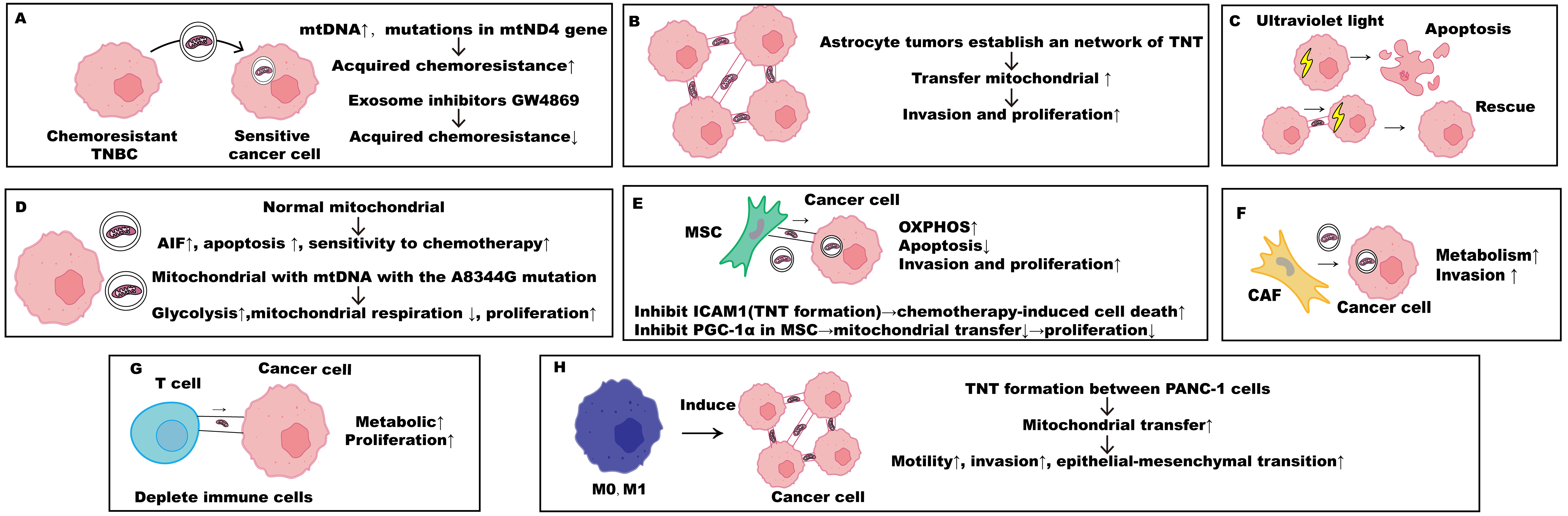
Figure 2. Patterns of mitochondrial transfer in cancer. (A-C) Mitochondrial transfer among cancer cells. (A) Chemoresistant TNBC cells transfer mitochondria to sensitive cancer cells by EVs; (B) Astrocyte tumors establish an interconnected network of TNTs and tumor microtubules and can transport mitochondria, which leads to increased tumor invasion and proliferation; (C) UV-induced stressed rat pheochromocytoma cells formed TNTs with normal cells and obtained transferred mitochondria that participate in the rescue effect; (D-F) Mitochondrial transfer between cancer and stromal cells. (D) MCF-7 breast cancer cells can internalize complete normal mitochondria and manifest as inhibition of cell proliferation; (E) Mitochondria extracted from MSCs to cancer cells result in an enhancement of OXPHOS activity that promotes the proliferative and invasive characteristics of recipient cancer cells. Inhibition of ICAM-1 blocks TNT formation, which promotes chemotherapy-induced cell death; Inhibition of PGC-1α in MSC suppresses mitochondrial transfer and impairs tumor cell proliferation; (F) CAFs transfer mitochondria to prostate cancer cells, further enhancing their metabolic and motile features; (G and H) Mitochondrial transfer between cancer and immune cells. (G) Mitochondrial transfer from T cells to cancer cells, facilitated by nanotubes, enhances the metabolic capabilities of the cancer cells while simultaneously depleting the immune cells; (H) M0 and M1 macrophages induced TNT network formation in PANC-1 cells, mitochondria delivered through TNTs. AIF: Apoptosis-inducing factor; CAF: cancer-associated fibroblast; ICAM-1: intercellular adhesion molecule-1; M0: M0-macrophage; M1: M1-macrophage; MSC: mesenchymal stem cell; mtDNA: mitochondrial deoxyribonucleic acid; OXPHOS: oxidative phosphorylation; PGC-1α: peroxisome-proliferator-activated receptor-gamma coactivator-1α; TNBC: triple-negative breast cancer; TNT: tunneling nanotube; EVs: extracellular vesicles; TNTs: tunneling nanotubes; UV: ultraviolet light.
The mechanism and effects of mitochondrial transfer
| Donor cells | Recipient cells | Transfer approach | Mechanism (signaling pathways, molecules, and structural basis) | Effect on recipient cells | Ref. |
| CAFs | Prostate cancer cells | TNT | Lactate shuttle from CAFs → intracellular NAD+/NADH ratio ↓ → activation of SIRT1 deacetylase → PGC-1α activation → mitochondrial biogenesis; Concurrent direct transfer of whole mitochondria via TNTs | Mitochondrial mass and oxidative activity ↑; TCA cycle dysregulation; oncometabolite accumulation; altered ETC complex expression; superoxide generation ↑; proliferation, invasion & malignancy ↑ | [60] |
| Immune Cells (CD8 T cells, NK cells, others) | Melanoma cells, breast cancer cells, colon adenocarcinoma | TNT | Mitochondrial hijacking; Exogenous mitochondrial fuse with endogenous networks → mtDNA leakage into cytosol → cGAS/STING pathway activation → Type I interferon-mediated immune evasion | Immune function (antigen presentation, NK/CD8 activation) ↓; Lymph node metastasis ↑; cGAS/STING/IFN pathway activation promotes immune evasion | [70] |
| Melanoma cells | Melanoma cells | TNT | Oxidative stress (ROS accumulation) → M-sec-dependent TNT formation → Miro-1-dependent mitochondrial transfer → Cargo (IR780) hitchhikes on mitochondrial for cotransport along TNTs | TNT network formation ↑; Deep tumor penetration of cargo bypassing physical barriers; Enhanced photodynamic killing upon NIR irradiation; Controllable drug delivery via TNT regulation; High biocompatibility for precision cancer therapy | [83,84] |
| Cancer cells (including melanoma, breast cancer, skin squamous cell carcinoma) | Tumor-infiltrating lymphocytes (TILs, primarily CD8+ T cells) | TNT, EV | Mitochondrial transfer via TNTs and small EVs; Co-transfer of mitophagy-inhibitory molecules USP30 (deubiquitinating enzyme inhibiting Parkin-mediated mitophagy) attached to cancer cell mitochondria → Transferred mitochondria resist ROS-induced mitophagy → Homoplastic replacement of endogenous T cell mtDNA; USP30 inhibitor CMPD-39 or siUSP30 partially prevents replacement | Impaired antitumor immunity: Basal respiration and OXPHOS, ATP ↓; ROS production ↑; Membrane potential ↓; Senescence (β-galactosidase, p16, p53) ↑; Central memory formation (CCR7+CD45RAlow, KLRG1low) ↓; Effector activation (PD-1, CD69) ↓; Apoptosis ↑ | [71] |
| Cancer-associated neurons | Breast cancer cells | TNT | Metabolic reprogramming of neurons when co-cultured with cancer cells ↑ → neuronal mitochondrial mass → Direct transfer of functional mitochondria via TNTs | Metabolic capacity, stemness, and resistance to metastatic stressors (oxidative/shear stress) ↑; Selective enrichment of mitochondrial-receiving cancer cells at metastatic sites | [65] |
| Astrocytes | Glioblastoma cells | TNT | GAP43-dependent intercellular connections (first identification of GAP43 in mitochondrial transfer; GAP43 knockdown significantly reduces transfer); TM-mediated transfer (network-forming intercellular connections) | Basal and maximal respiration ↑; mitochondrial biology genes (ETC components) ↑; ATP5A expression ↑; ATP levels; cell cycle progression to G2/M ↑; self-renewal ↑; tumorigenicity ↑ | [64] |
| BMSC | CD8+ T cells | TNT | TNT-mediated connections (nanotube structures visualized by field-emission SEM; mitochondria confirmed inside nanotubes by confocal); Talin 2-dependent on both donor and recipient cells (optimal transfer requires Talin 2) | Mitochondrial content ↑; mitochondrial respiration ↑; spare respiratory capacity ↑; tumor infiltration ↑; expansion in vivo ↑; exhaustion markers ↓; antitumor efficacy ↑; enhanced CAR-T function | [8] |
| NKT cells | Breast cancer cells | TNT | TNT formation (actin-based nanotubes visualized by SEM); Protein farnesylation/geranylgeranylation-dependent assembly (inhibited by L-778123 targeting FTase/GGTase1); Miro-1-dependent mitochondrial transferring along cytoskeleton; Exocyst complex (Sec3/Sec5) localization to TNT assembly sites | Cancer cell oxidative metabolism ↑; Antitumor immune cell viability and function ↓; Immune evasion; TNT inhibition + anti-PD-1 improves outcomes | [67] |
| Macrophage | Breast cancer cells | TNT | Macrophage-derived inflammatory mediators → enhanced TNT formation; TNT-mediated release of “microplasts” (independently migrating viable cytoplasmic fragments); Actin cytoskeleton-dependent (inhibited by cytochalasin-B); Dynamic TNT retraction and microplast resorption | Formation of TNT-based intercellular networks; mitochondrial transfer to microplasts; expanded intercellular communication range; organelle redistribution across cell population | [69] |
| Glioma cells | Glioma cells | TNT, TM | TM formation; Ultra-long membrane protrusions; Gap junction-mediated communication; 1p/19q status-dependent | Formation of interconnected malignant network; Resistance to cytotoxic therapies | [54] |
| Chemo-resistant breast cancer cellsMDA-MB-231 | Chemo-sensitive breast cancer cells | EV | Primarily EV/exosome-mediated (blocked by GW4869); Transfer of mutant mtND4 mtDNA; TNTs as secondary route | Acquired chemoresistance; mutant mtDNA copy number ↑; Propagation of resistant phenotype | [56] |
| Glioblastoma stem-like cells | Glioblastoma stem-like cells | TNT | TNT formation in 2D and 3D organoids; TM in 3D organoids; Heterogeneous stress responses to radiation; Open-ended functional TNTs | Formation of interconnected networks; heterogeneous radiation responses; potential contribution to therapy resistance | [45] |
| Macrophages | Breast cancer cells | NA | Cell contact-dependent transfer; Fragmented mitochondrial network in pro-tumorigenic macrophages → higher transfer rates; Donor mitochondrial dysfunction promotes transfer | Transfer of dysfunctional mitochondria → ROS accumulation → ERK activation; proliferation independent of bioenergetics ↑ | [68] |
| MSCs | Gastric cancer cells | EV | OXA administration → GC cells actively secrete ECM → matrix stiffness in tumor microenvironment ↑; Mechanical signals (ECM stiffness) promote MSCs to transfer mitochondria to GC cells via MVs; RhoA/ROCK1 pathway involved (inhibition alleviates OXA resistance) | Repair of mitochondrial function; mitophagy ↓; chemotherapy resistance | [7] |
| Isolated from HeLa-DsRed2-mito cells | Hepatocellular carcinoma cells, fibroblasts | Endocytosis | Endocytosis-mediated mitochondrial engulfment (blocked by macropinocytosis inhibitors EIPA and cytochalasin D); Requires intact mitochondrial outer membrane proteins; Heparan sulfate proteoglycan-dependent recognition (blocked by heparin/pentosan polysulfate) | Co-localization with endogenous mitochondrial network; Functional integration into existing network | [86] |
| Astrocytes | Glioma cells | Endocytosis | Serum-glucose starvation → NAD+ accumulation and release → extracellular NAD+ catalyzed by CD38 (NAD+ glycohydrolase) → generation of intracellular cADPR → Ca2+ release from ER stores → cytoskeleton remodeling and plasma membrane invagination → endocytosis-mediated internalization; TNT formation; Miro-1-dependent mitochondrial transferring along cytoskeleton; Exocyst complex (Sec3/Sec5) localization to TNT assembly sites | TCA cycle gene/protein expression ↑; aerobic respiration ↑; glycolysis (Warburg effect reversal) ↓; cytochrome c release ↑; cleaved caspase 9 ↑; proliferation ↓; radiosensitivity ↑; tumor growth in vivo ↓ | [62] |
| Pancreatic cancer cells | Pancreatic cancer cells | TNT | Macrophage-derived soluble factors induce TNT formation in cancer cells; TNT-mediated mitochondrial transfer facilitation | Motility ↑; Invasion ↑; EMT ↑; Facilitation of mitochondrial transfer | [52] |
Mitochondrial transfer among cancer cells
The main effect of mitochondrial transfer among tumor cells is to enhance their resistance to stress conditions. A study showed that conditioned media from M0 and M1 macrophages derived from human acute monocytic leukemia (THP-1) cells promote TNT formation in human pancreatic ductal adenocarcinoma (PANC-1) cancer cells. Mitochondria and lysosomes were observed to be transported through these TNTs. TNT formation was closely associated with increased cell motility and the induction of epithelial-mesenchymal transition (EMT) in PANC-1 pancreatic cancer cells[52]. Similarly, ultraviolet light-stressed PC12 rat pheochromocytoma cells form TNTs with unstressed PC12 cells and acquire mitochondria that contribute to a rescue effect[53]. TNT-mediated transfer of functional mitochondria can reverse cellular stress during early stages of apoptosis[53]. These findings suggest that cancer cells may enhance their proliferative capacity by exchanging mitochondria to overcome stress conditions. Chemo-resistant triple-negative breast cancer (TNBC) cells can transfer mitochondria to sensitive TNBC cells via EVs, thereby promoting chemoresistance[54]. This process is associated with elevated mtDNA levels, including mutations in the mtND4 gene that contribute to tumorigenesis[54]. Inhibition of mitochondrial transfer using exosome inhibitors such as GW4869 reduces the acquisition of chemoresistance in TNBC[54]. Astrocytic tumors establish interconnected networks of TNTs and tumor microtubules that facilitate mitochondrial transport[55]. Tumor microtubules act as key structural elements supporting glioma cell invasion and proliferation, thereby promoting efficient brain colonization[55]. Human breast adenocarcinoma (MCF-7) cells can internalize intact mitochondria from 143B osteosarcoma cybrids through simple co-culture. Such alterations restrain proliferative activity, initiate apoptosis-inducing factor (AIF)-mediated caspase-independent apoptosis, and markedly improve the responsiveness of tumor cells to chemotherapy treatment[56]. Furthermore, replacement with dysfunctional mitochondria harboring the A8344G mtDNA mutation (Mito8344) shifts cellular metabolism toward a glycolytic phenotype, driven by enhanced glycolysis and impaired mitochondrial respiration[56].
Mitochondrial transfer between cancer and stromal cells
Like other stromal cells, MSCs exhibit altruistic behavior in tumor formation, including the transfer of mitochondria to tumor cells. Tumor cells can establish TNT connections with endothelial cells and MSCs, which supports the bidirectional exchange of cytoplasmic cargoes encompassing miRNAs and mitochondria[57]. The transfer of limited quantities of MSC-derived mitochondria to cancer cells enhances oxidative phosphorylation, thereby promoting the proliferative and invasive capacities of recipient cells during co-incubation with exogenous mitochondria[58]. However, mitochondrial transfer from MSCs to osteosarcoma cells appears to occur primarily when the recipient cells are entirely depleted of mitochondria following pretreatment with ethidium bromide or rhodamine. Suppression of peroxisome proliferator-activated receptor gamma coactivator 1-alpha (PGC-1α) in BMSCs decreases the mitochondrial transfer, thereby limiting the proliferative potential of the tumor[59]. The transfer of mitochondria by MSCs to tumor cells also promotes drug resistance in tumor cells. For example, following oxaliplatin treatment, gastric cancer cells restructure the extracellular matrix, enhancing its stiffness, and stimulate the Ras homolog family member A (RhoA)/Rho-associated coiled-coil containing protein kinase 1 (ROCK1) pathway in MSCs, which facilitates mitochondrial transfer from MSCs to gastric cancer cells. Pharmacological treatment with the ROCK inhibitor Y-27632 decreases EVs‐dependent mitochondrial transfer and reinstates the tumor‐suppressive efficacy of oxaliplatin[7].
Another type of stromal cell, cancer-associated fibroblasts (CAFs), also represents an important source of mitochondria for tumor cells. CAFs deliver mitochondria to prostate cancer cells, thereby enhancing their metabolic and migratory capacities[60]. In particular, CAFs enhance mitochondrial function in prostate cancer cells through activation of the lactate/sirtuin 1 (SIRT1)/PGC1-α axis and other mitochondrial-related signaling. This leads to the accumulation of tricarboxylic acid cycle intermediates, such as citrate, succinate, and fumarate, as well as the emergence of a mitochondrial reactive oxygen species (mtROS)-dependent signature, including the oxidation of Src and pyruvate kinase M2 (PKM2)[60]. Similarly, mitochondrial transfer from CAFs increases migration in aggressive breast cancer[61].
Numerous studies have reported that astrocytes in the nervous system can promote glioma progression through mitochondrial transfer. In glioblastoma cells, it was found that tumor-activated stromal cells can transfer mitochondria to tumor cells through EVs[48]. Cancer cells lacking mtDNA (ρ0 cells) exhibit a significant delay in tumor formation compared to their parental counterparts; however, this growth impairment can be reversed through the acquisition of functional mitochondria from parental cells via mitochondrial transfer[48]. In addition, mitochondria derived from human astrocytes can be transferred to starved human glioma cells through co-incubation[62,63]. This process restores aerobic respiration and enhances sensitivity to radiotherapy[62]. Growth-associated protein 43 (GAP-43) has been implicated in facilitating mitochondrial transfer via tumor-astrocyte tumor microtubes[64]. Acquisition of astrocytic mitochondria facilitates the elevation of mitochondrial respiratory activity, while simultaneously boosting metabolic programs that promote tumor cell proliferation and malignant progression[64]. Tumor-associated neurons can transfer functional mitochondria to cancer cells in a contact-dependent manner through TNTs. Neuron-derived mitochondria integrate into the metabolic network of cancer cells to potentiate oxidative phosphorylation and ATP production, while reinforcing cellular redox homeostasis by increasing reduced glutathione levels. This mitochondrial reprogramming endows cancer cells with enhanced metabolic plasticity, stemness, and proliferative capability. Furthermore, the acquired mitochondria strengthen cancer cell resistance to oxidative stress and shear stress, facilitating the survival and colonization of circulating tumor cells in the bloodstream and distant tissue microenvironments. Lineage tracing evidence demonstrates that cancer cells acquiring neuronal mitochondria are significantly enriched in lung and brain metastatic foci, thereby conferring prominent metastatic advantages[65].
Mitochondrial transfer between cancer and immune cells
Mitochondrial uptake from immune cells enables tumor cells to proliferate aggressively and outcompete adjacent normal cells. Intercellular nanotubes serve as critical channels for mitochondrial shuttling from BMSCs to CD8+ T cells. This transfer process is governed by Talin2 (TLN2), a cytoskeletal protein responsible for actin assembly through the regulation of integrin-membrane protrusion interactions[8]. The transfer of mitochondria from BMSCs improves the metabolic capacity and antitumor immune response of CD8+ T cells against solid tumors. More significantly, the benefits resulting from mitochondrial transfer are not only immediate but also sustained over a longer duration[8]. Cancer cells acquire mitochondria from adjacent T cells, and this process can be tracked and quantified by single-cell RNA sequencing (RNA-seq) technology[66]. A classic study has shown that TNTs facilitate the transfer of mitochondria between breast cancer cells and T lymphocytes[67]. Mitochondrial tracing and quantitative metabolic analyses reveal that mitochondrial transfer from T cells to breast cancer cells boosts the metabolic fitness of cancer cells while concurrently weakening the immune cells[67]. Furthermore, the combined use of a farnesyltransferase inhibitor and a geranylgeranyl transferase I inhibitor effectively suppresses TNT formation and mitochondrial transfer. Meanwhile, treatment with a programmed cell death protein 1 (PD-1) immune checkpoint inhibitor has been shown to enhance antitumor effects in an aggressive immunocompetent breast cancer model[67]. Human breast cancer cells can acquire mitochondria from T cells and macrophages[66,68]. Macrophages have been shown to stimulate the formation of TNTs and microplasts (independently migrating viable cytoplasmic fragments) in cancer cells, leading to the development of extensive intercellular networks. Mitochondria can be packaged into microplasts and transported between cells via TNTs[69]. Cancer cells can seize mitochondria from multiple types of immune cells. Mitochondrial depletion in immune cells compromises their antigen presentation capability and costimulatory molecular machinery, accompanied by diminishing vitality of immune cells and cytotoxic potency of natural killer cells and CD8+ T cells. Upon internalization by cancer cells, exogenous mitochondria integrate with endogenous mitochondrial networks, leading to cytosolic mtDNA leakage. This process further activates the cyclic GMP-AMP synthase (cGAS)-stimulator of interferon genes (STING) signaling axis and induces type I interferonmediated immune evasion programs. Pharmacological blockade of mitochondrial transfer machineries, such as cGAS, STING, or type I interferons, effectively inhibits tumor lymph node metastasis. These observations demonstrate that tumor cells hijack immune cell mitochondria to abrogate antitumor immunosurveillance, while utilizing the acquired mitochondrial functions to meet the immunological demands of lymph node colonization and metastatic progression[70]. Tumor cells employ TNTs and EVs as critical transport channels to deliver mtDNA-mutated mitochondria to tumor-infiltrating lymphocytes in the tumor microenvironment. The persistent replacement of endogenous T-cell mitochondria by these aberrant exogenous organelles induces mitochondrial homogenization, a key mechanism underlying cancer immune evasion. Notably, these cancer-derived mitochondria carry the autophagy suppressor USP30, enabling them to resist intracellular ROS-triggered mitophagy and stably persist in T cells. The retained mutated mitochondria further induce T cell mitochondrial dysfunction (reduced membrane potential and defective oxidative phosphorylation) and excessive ROS accumulation, thereby disrupting T cell metabolism, accelerating cellular senescence, inhibiting proliferation, and impairing T cell cytotoxic effector function and memory formation, which ultimately blunts anti-tumor immune responses[71].
In summary, research on mitochondrial transfer in tumor cells should consider the TME and the dynamic interactions between tumor cells and surrounding cell populations. This is crucial for understanding how mitochondrial transfer contributes to tumor progression and for developing strategies to prevent tumor cells from exploiting this process[72].
MITOCHONDRIAL TRANSFER AS A STRATEGY FOR SOLID TUMOR THERAPY
In recent years, the organelle-specific delivery of bioactive molecules has been utilized in cancer therapy to enhance specificity, improve treatment efficacy, minimize side effects, and reduce chemoresistance. Approaches targeting mitochondria enable direct manipulation of the mitochondrial membranes and components, as well as mitochondrial metabolism and mitochondria-associated apoptotic or regulatory signaling pathways[73].
With increasing recognition of intercellular mitochondrial transfer, its potential applications are expanding across various medical disciplines [Table 2]. While spontaneous mitochondrial transfer within the TME typically promotes tumor invasion, the exogenous transplantation of healthy mitochondria into cells can exert opposing effects. Tumor cells employ TNTs and extracellular EVs as critical transport channels to deliver mtDNA-mutated mitochondria to tumor-infiltrating lymphocytes in the tumor microenvironment. The persistent replacement of endogenous T-cell mitochondria by these aberrant exogenous organelles induces mitochondrial homogenization, which serves as a key mechanism underlying cancer immune evasion. The therapeutic potential of targeting intercellular mitochondrial transfer lies in its promise as a novel anti-cancer strategy and its capacity to enhance cell-mediated immunotherapies. Future therapeutic approaches should aim to promote the cell-to-cell organelle transfer when it confers beneficial effects, or inhibit it when it supports tumor progression. Such strategies are particularly relevant for containing neoplastic cells and reducing their associated chemoresistance[74].
The strategies for targeted mitochondrial delivery and mechanisms
| Mechanism | Ref. | |
| Agents inhibiting mitochondrial transfer | ||
| Taxanes | Inhibit formation of TNT | [29] |
| Vinca alkaloids | Inhibit formation of TNT | [29] |
| Latrunculin | Actin-disrupting agents, inhibit formation of TNT | [75] |
| Cytochalasin B and D | Actin-disrupting agents, inhibit formation of TNT | [75] |
| L-778,123 | Inhibitor of farnesyltransferase and geranylgeranyl transferase I, inhibits formation of TNT | [67] |
| Belamaf | Microtubule-disrupting agent | [77] |
| 18-α-glycyrrhetinic acid | Inhibitors targeting gap junctions | [79] |
| Dynasore | Inhibits vesicular endocytosis | [79] |
| Mitochondrial transplantation | ||
| MitoCeption | Mitochondria entering recipient cells through internalization induced by centrifugation and heat shock during co-culture | [58] |
| Transplantation of mitochondria into prostate cancer cells and ovarian cancer cells | Increased apoptosis in these cells as well as enhancing the sensitivity of mice to chemotherapy | [92] |
| Transplantation of mitochondria into melanoma-bearing mice | General chromosome silencing and mitochondrial activity increasing | [93] |
| Mitochondrial transplantation in hepatocellular carcinoma | Decreased glycolysis, promoted dephosphorylation of p-Bad, downregulated the expression of Bcl-2, increased Bax | [94] |
| Mitochondria isolated from healthy mouse livers transferred into melanoma model mice | Reduced glycolysis and produced an oxidative intracellular environment, inhibiting tumor growth and prolonging the survival duration of the animals | [95] |
| Transplanted normal human GES-1 mitochondria to human gastric AGS | Reduced the expression of mesenchymal markers α-SMA, MMP-9, snail, vimentin and N-cadherin, whereas the epithelial markers E-cadherin and claudin-1 were not changed. The proteins implicated in the cell cycle, such as cyclin B1 and D1, were decreased | [96] |
| Promote spontaneous transfer of mitochondria | ||
| Promote the formation of TNT between tumor cells and facilitate mitochondrial transfer through siPINK1 | PINK1-mediated mitophagy inhibition enhances mitochondrial intercellular transport by increasing mitochondrial motility, oxidative phosphorylation, and TNT formation. Engineered mitochondria carrying photosensitizer IR780 achieve deep tumor penetration via TNTs, and subsequent NIR irradiation triggers potent photodynamic killing while disrupting the transfer network, achieving 43% complete tumor regression in melanoma models | [83,84] |
| Adjunct to immunotherapy | ||
| Transferred prepared intact mitochondria into CAR-T cells | Improved metabolic adaptability and enhanced proliferative capacity of CAR-T cells, increased cytokine production ability | [99] |
Inhibition of the mitochondrial transfer pathway
Most of the aforementioned studies indicate that spontaneous mitochondrial transfer predominantly facilitates tumor development. TNTs play a crucial role in mediating the transport of mitochondria between tumor and stromal cells; therefore, inhibiting TNT formation may effectively reduce mitochondrial transfer. Possible approaches for blocking mitochondrial transfer include: (i) perturbing actin filament dynamics to suppress TNT assembly; (ii) targeting critical structural and functional components supporting TNT formation; (iii) weakening cell adhesion and shortening cell-cell contact time to minimize intercellular mitochondrial transfer; and (iv) suppressing signaling pathways that stimulate horizontal mitochondrial transfer[42]. Accordingly, the development of pharmacological agents aimed at disrupting the mitochondrial transfer between tumor cells and TME may represent a promising strategy for tumor inhibition. Taxanes and vinca alkaloids, frequently used as routine chemotherapy, have been shown to disrupt microtubule polymerization, thereby inhibiting mitochondrial transfer[29]. Given that actin is an essential structural element of TNTs, the dynamic rearrangement and polymerization of the actin cytoskeleton are fundamentally required for TNT biogenesis. In line with this mechanism, small-molecule agents targeting and disrupting actin function, including latrunculin and cytochalasin isoforms B and D, substantially suppress TNT formation and assembly[75]. As a key protein modulating TNT generation and a specific marker of these structures, M-sec possesses therapeutic potential. Targeting M-sec could effectively inhibit the intercellular shuttling of mitochondria[76]. Downregulating the expression or impairing the functional activity of M-sec significantly impedes TNTs assembly, consequently diminishing mitochondrial transportation between malignant cells[76]. At concentrations that exert no cytotoxic effects, L-778123, a dual inhibitor of farnesyltransferase and geranylgeranyl transferase I, markedly blocks TNTs assembly and subsequent mitochondrial exchange[67]. Multiple myeloma (MM) cells can resist belantamab mafodotin (belamaf), a monoclonal antibody linked to the microtubule-disrupting agent monomethyl auristatin-F (MMAF), through enhancing mitochondrial transfer from stromal cells to allow them to evade apoptosis[77]. Doxycycline has emerged as a novel repurposed mitochondria-targeted reagent, which can interfere with mitochondrial transfer processes and thus benefit cancer therapy[78]. Experimental findings reveal that administration of doxycycline enhances intercellular mitochondrial transfer among tumor cells. Such restoration of normal mitochondrial function significantly improves cancer cell responsiveness to anticancer regimens[78]. Optimization of mitochondrial function and cellular metabolism mediated by these drugs can normalize aberrant mitochondrial behaviors in cancer cells, ultimately boosting tumor susceptibility to clinical anticancer interventions[78]. Additional druggable mechanisms are GJs, which can be targeted with 18-α-glycyrrhetinic acid, and dynamin, which can be inhibited by dynasore, to suppress vesicular endocytosis[79]. Despite the signaling mechanisms regulating mitochondrial transfer, the identification of key molecular components, such as Miro1, Cx43, and CD38, has laid the foundation for potential therapeutic interventions[80].
Promotion of spontaneous transfer of mitochondria
In mitochondrial diseases or ischemic conditions, spontaneous mitochondrial transfer can be beneficial. For example, pretreatment of human MSCs with iron oxide nanoparticles has been shown to promote the overexpression of Cx43 and significantly enhance the efficiency of mitochondrial transfer from human MSCs to damaged alveolar epithelial cells[81]. Similarly, overexpression of Miro1 in MSCs potentiates the metabolic protective effects of intercellular mitochondrial transfer against neuronal oxidative damage, attributable to improved transfer efficiency[82]. However, in solid tumors, mitochondrial transfer may instead contribute to tumor initiation and progression. Therefore, strategies that engineer mitochondria to remain harmless to normal cells while selectively targeting tumor cells. Peng et al. demonstrated that inhibition of PINK1-mediated mitophagy enhances intercellular mitochondrial transfer by increasing mitochondrial motility, oxidative phosphorylation, and TNT formation. Furthermore, engineered mitochondria carrying photosensitizer IR780 can achieve deep tumor penetration via TNTs. Subsequent near-infrared irradiation induces potent photodynamic killing while disrupting mitochondrial transfer networks, resulting in up to 43% complete tumor regression in melanoma models[83,84]. These findings suggest that using mitochondria as delivery vehicles for therapeutic agents, combined with promoting mitochondrial transfer, represents a novel and promising strategy for cancer treatment.
Artificial mitochondrial transplantation
Artificial mitochondrial or mitochondrial transplantation techniques have been developed and are expected to have broader applications in the future. Healthy human blood contains substantial numbers of mitochondria, suggesting a potential role in maintaining physiological homeostasis; this observation has served as the theoretical foundation for the concept of artificial mitochondrial transplantation[85]. These isolated mitochondria can be rapidly integrated into recipient cells, where they co-localize with endogenous mitochondria[86]. Currently, the initial step in mitochondrial transplantation involves isolating functional mitochondria from various cell types. Employing stem cells with high mitochondrial transfer capacity represents a viable alternative strategy for transplantation. MSC-derived mitochondria have been extensively utilized in mitochondrial therapies[87]. Another potential approach involves the use of induced pluripotent stem cells (iPSCs) to generate functional mitochondria suitable for transplantation. iPSCs can be derived from patients’ somatic cells and subsequently directed to differentiate into various cell types, including those affected by mitochondrial diseases[88]. Functional mitochondria can be produced by generating iPSCs from patients with mitochondrial disorders and subsequently transferring them to the affected tissues[88]. As the benefits of modifying cells to enhance effective mitochondrial transfer become increasingly evident and the therapeutic effects are progressively clarified, the use of engineered cells for mitochondrial supplementation may emerge as a promising strategy in the future[89]. Scientists have developed a technique known as MitoCeption, in which mitochondria are internalized by recipient cells through centrifugation-induced mechanisms and heat shock-mediated mechanisms during co-culture[58]. In this approach, labeled mitochondria are combined with similarly labeled recipient cells via centrifugation and subsequently co-cultured under standard conditions. During this co-culture period, recipient cells take up the isolated mitochondria, facilitating their incorporation into the cellular structure[58]. Another crucial step is mitochondrial infusion. Systemic administration via injection is a common method for delivering mitochondria and can be performed through routes such as the jugular or femoral veins[90]. Isolated mitochondria are inherently unstable; therefore, efficient delivery of exogenous mitochondria to somatic cells or tissues is essential. Recent advances in mitochondrial therapy have introduced an erythrocyte membrane vesicle-based encapsulation platform. This innovative transplantation approach substantially enhances the delivery efficiency of exogenous mitochondria into cells and tissues of mice and non-human primates[91]. Studies have shown that delivering mitochondria to Various types of solid tumor cells alongside low-dose chemotherapy markedly increases apoptosis in these cells and improves chemosensitivity in mouse models[92]. In melanoma-bearing mice, mitochondrial transplantation was found to inhibit tumor growth and lung metastasis. Transcriptomic analyses revealed a strong association between global chromosome silencing and mitochondrial activity in suppressing melanoma following mitochondrial transplantation in metastatic models[93]. Interestingly, mitochondria derived from female animals exhibited greater anti-tumor efficacy compared to those from males. This difference may be attributed to more robust communication between mitochondria and the nucleus mediated by female mitochondria[93]. Mitochondrial transplantation with intact healthy mitochondria exerts robust anti-tumor effects in hepatocellular carcinoma. This intervention suppresses tumor glycolysis, facilitates Bad dephosphorylation, modulates the B-cell lymphoma 2 (Bcl-2)/Bcl-2-associated X (Bax) balance by repressing Bcl-2 and increasing Bax expression, and eventually induces caspase-mediated apoptotic cell death in hepatocellular carcinoma[94]. Healthy mouse liver mitochondria were isolated and injected through tail veins into melanoma-bearing model mice, which suffered from lung metastasis. The results demonstrated that mitochondrial transplantation substantially suppressed tumor growth and extended the survival time of these mice[95]. The anti-tumor effects of mitochondria are linked to their ability to reprogram tumor cell metabolism by suppressing glycolysis and promoting an oxidative intracellular environment, thereby creating conditions that hinder tumor cell proliferation[95]. Delivery of healthy mitochondria isolated from the human normal gastric epithelial cell line GES-1 into the gastric adenocarcinoma cell line AGS effectively impairs tumor cell migration and invasion. Notably, this intervention does not alter cell viability or induce cellular apoptosis. These findings indicate that mitochondrial transplantation can effectively reduce the malignancy of gastric cancer cells[96].
Mitochondrial transplantation as an adjunct to immunotherapy
Chimeric antigen receptor (CAR) T-cell therapy is a revolutionary advancement in cellular immunotherapy, offering a personalized therapeutic strategy for cancer treatment[97]. Premature exhaustion of CAR-T cells serves as a critical bottleneck for therapeutic outcomes, which is closely associated with defective mitochondrial function and aberrant mitochondrial dynamic remodeling[98]. In addition, exogenous mitochondria have been successfully transferred into CAR-T cells in preclinical studies. This mitochondrial supplementation effectively improves cellular metabolic adaptability, enhances proliferative potential, promotes cytokine release, and significantly reinforces the anti-tumor function of CAR-T cells[99].
CONCERNS REGARDING MITOCHONDRIAL TRANSPLANTATION AS A TARGETED THERAPY
Despite the prominent therapeutic prospects of exogenous mitochondrial transplantation for tumor intervention, the clinical transformation and large-scale clinical application of this emerging therapeutic strategy still face multiple unavoidable bottlenecks and unresolved limitations. A variety of technical safety, delivery efficiency, and translational standardization barriers remain to be systematically addressed before mitochondrial transfer-targeted therapy can be safely and stably applied to clinical cancer treatment.
First, the biosafety risks associated with mitochondrial transfer intervention strategies have not been fully clarified and resolved. Small-molecule inhibitors targeting TNT formation mainly act on cytoskeletal proteins; such agents produce broad cytotoxicity against dividing normal cells and may trigger myelosuppression and organ toxicity. Moreover, these blockers indiscriminately disrupt physiological TNT-mediated mitochondrial rescue in parenchymal cells, neurons, and immune cells, disturbing tissue homeostasis, wound healing, and endogenous immune surveillance[12]. Notably, incomplete regulation of intercellular mitochondrial transfer may exert selective pressure on tumor subpopulations, enriching highly aggressive, drug-tolerant clones and ultimately accelerating tumor relapse. Allogeneic mitochondrial transplantation introduces additional safety and ethical concerns, as donor mitochondrial DNA may permanently integrate into recipient cells and trigger unforeseen long-term biological consequences.
Second, widespread delivery efficiency bottlenecks limit in vivo therapeutic performance. Isolated free mitochondria face rapid degradation and calcium-induced damage in circulating blood, while EV-based mitochondrial carriers also suffer from short systemic half-life and rapid clearance[100]. Dense fibrotic tumor stroma creates physical barriers that hinder deep intratumoral penetration of EVs, peptides, and small-molecule inhibitors, resulting in uneven drug distribution within tumor lesions. In addition, intratumoral heterogeneity leads to drastically variable TNT abundance across distinct tumor subclones and patient specimens. Although surface modification with “don’t-eat-me” markers (CD47, CD55, CD59, CD200) can partially reduce phagocytic clearance of EVs[101], inconsistent mitochondrial loading efficiency and variable vesicle quality from different isolation methods further weaken therapeutic reproducibility[102].
Thirdly, unresolved translational and standardization limitations further hinder the clinical progression of mitochondrial transfer-targeted therapy. At this stage, there is a lack of clinically validated specific imaging biomarkers and targeted detection probes that can accurately quantify TNT abundance and mitochondrial transfer activity in human tumor tissues. The absence of effective in situ detection techniques makes it impossible to precisely screen patients suitable for mitochondrial transplantation therapy, failing to achieve individualized and precise tumor treatment. Furthermore, the core clinical treatment specifications for mitochondrial targeted therapy have not yet been established. The optimal clinical administration dosage, safe and effective treatment intervention window, and standardized treatment course remain undefined. In addition, the synergistic application strategies of mitochondrial transfer inhibition combined with traditional clinical tumor treatment methods, including chemotherapy, radiotherapy, and tumor immunotherapy, lack systematic exploration and verification. At present, most relevant studies are limited to in vitro cell experiments and preliminary animal models, and there is a scarcity of standardized in vivo combination therapy trials to confirm the long-term stable anti-tumor efficacy and clinical safety of combined regimens, which greatly delays the clinical transformation and clinical application process of this innovative tumor therapy.
CONCLUSIONS
The progress in mitochondrial transfer research has not only deepened our understanding of intercellular communication and interaction mechanisms but has also driven the development of novel therapeutic strategies. Despite the innovative insights brought by current technical advances, the mechanistic basis of intercellular mitochondrial transfer remains incompletely defined. Specifically, the tissue- and cell-type-specific diversity of mitochondrial transfer patterns has yet to be systematically clarified. Elucidating the exact molecular pathways through which mitochondrial transfer contributes to tumor progression, including cell proliferation, invasion, metastasis, chemotherapy resistance, and immune evasion, may reveal multiple targets for therapeutic intervention. Continuing research in this field will play a crucial role in developing more personalized and precise cancer treatments. Mitochondrial transplantation therapy for tumors remains at an experimental stage. Although promising therapeutic effects have been demonstrated in animal models, its clinical translation has not yet been achieved. Bridging the gap between laboratory research and clinical implementation presents several challenges, including safety concerns, achieving efficient delivery of mitochondria to tumor cells, and overcoming preservation-related limitations. Resolving these limitations will facilitate the clinical translation of mitochondrial transfer research and yield meaningful therapeutic gains for cancer treatment. Concurrently, novel intervention strategies targeting tumor mitochondrial transfer are under continuous investigation. Early successes of this approach bear considerable potential, indicating that targeting metabolic interactions among tumor cells could represent a novel approach to suppress tumor growth and metastasis.
Accumulating evidence suggests that artificial transfer of mitochondria into tumor cells can exert either pro-tumorigenic or anti-tumor effects, depending on the context. Determining how to make reliably direct artificially transplanted mitochondria toward tumor-inhibitory outcomes under specific conditions remains a critical challenge that warrants further investigation.
DECLARATIONS
Acknowledgments
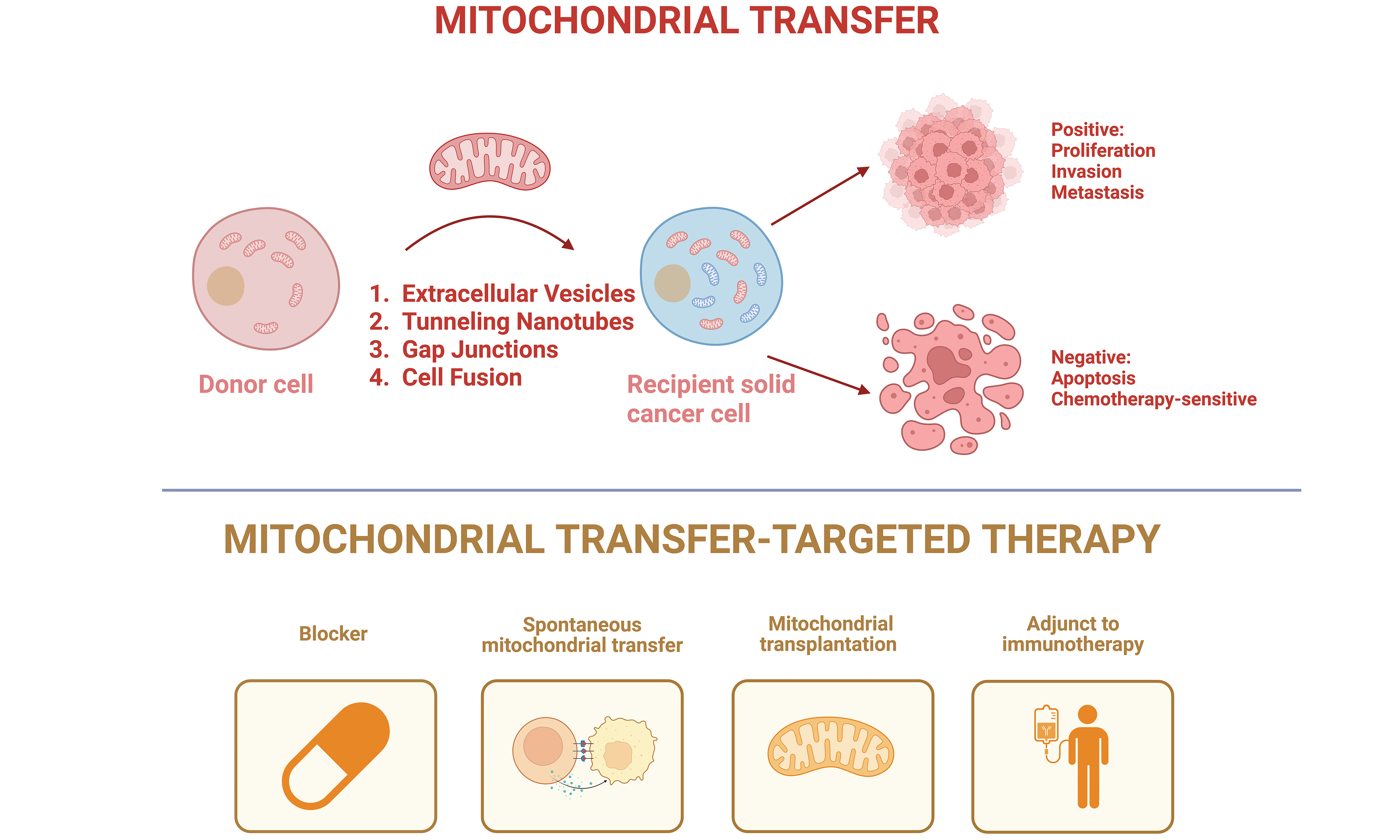
The Graphical Abstract was created with BioRender.com [Created in BioRender. Huang, L. (2026). https://BioRender.com/ewosq29].
Authors’ contributions
Contributed equally to drafting the manuscript, overseeing multiple revisions, and managing the overall writing process: Huang L, Li Z, Qin C
Contributed to the manuscript and designed Figure 1: Zhao B
Designed Figure 2 and also participated in writing the manuscript: Li T, Zhao Y
Contributed to the manuscript and designed Table 1: Shi H, Zhang X, Xie Y, Yan Y
Supervised all stages of the project, from idea selection to final editing: Wang W
All authors reviewed and approved the final version of the manuscript.
Availability of data and materials
Not applicable.
AI and AI-assisted tools statement
During the preparation of this manuscript, the AI tool DeepSeek (version V3, released 2024-12-26) was used solely for minor language revision. The tool did not influence the study design, data collection, analysis, interpretation, or the scientific content of the work. All authors take full responsibility for the accuracy, integrity, and final content of the manuscript.
Financial support and sponsorship
Wang W received support from the National Natural Science Foundation of China (No. 82573412), Beijing Natural Science Foundation (No. 7232127), National High Level Hospital Clinical Research Funding (2025-PUMCH-C-018), Capital’s Funds for Health Improvement and Research (2024-2-4017), National Multidisciplinary Cooperative Diagnosis and Treatment Capacity Building Project for Major Diseases (ZK12101). Qin C received support from the National Natural Science Foundation of China (No. 82503800), Postdoctoral Fellowship Program of CPSF (Grant Number GZC20240146), National High Level Hospital Clinical Research Funding (2025-PUMCH-A-075), Fundamental Research Funds for the Central Universities, Peking Union Medical College (3332025001). The grants supported this study financially and had no role in the study design, data collection, data analysis and interpretation, or manuscript writing.
Conflicts of interest
All authors declared that there are no conflicts of interest.
Ethical approval and consent to participate
Not applicable.
Consent for publication
Not applicable.
Copyright
© The Author(s) 2026.
REFERENCES
1. GBD 2023 Cancer Collaborators. The global, regional, and national burden of cancer, 1990-2023, with forecasts to 2050: a systematic analysis for the Global Burden of Disease Study 2023. Lancet. 2025;406:1565-86.
2. Torabi S, Yekzaman E, Taherkhani S, et al. Immune cell-based therapies for solid tumors, current challenges and therapeutic advances. Cell Commun Signal. 2025;24:73.
3. Zhang Y, Zhuang W, Zhu H, et al. Neuroimmunometabolic co-evolution in the tumor micro- and macroenvironment. Cell Metab. 2026;38:655-72.
4. Borcherding N, Brestoff JR. The power and potential of mitochondria transfer. Nature. 2023;623:283-91.
5. Al Amir Dache Z, Thierry AR. Mitochondria-derived cell-to-cell communication. Cell Rep. 2023;42:112728.
6. Iorio R, Petricca S, Mattei V, Delle Monache S. Horizontal mitochondrial transfer as a novel bioenergetic tool for mesenchymal stromal/stem cells: molecular mechanisms and therapeutic potential in a variety of diseases. J Transl Med. 2024;22:491.
7. He X, Zhong L, Wang N, et al. Gastric cancer actively remodels mechanical microenvironment to promote chemotherapy resistance via MSCs-mediated mitochondrial transfer. Adv Sci. 2024;11:e2404994.
8. Baldwin JG, Heuser-Loy C, Saha T, et al. Intercellular nanotube-mediated mitochondrial transfer enhances T cell metabolic fitness and antitumor efficacy. Cell. 2024;187:6614-30.e21.
9. Li H, Wang C, He T, et al. Mitochondrial transfer from bone marrow mesenchymal stem cells to motor neurons in spinal cord injury rats via gap junction. Theranostics. 2019;9:2017-35.
10. Qiao X, Huang N, Meng W, et al. Beyond mitochondrial transfer, cell fusion rescues metabolic dysfunction and boosts malignancy in adenoid cystic carcinoma. Cell Rep. 2024;43:114652.
11. Hayakawa K, Esposito E, Wang X, et al. Transfer of mitochondria from astrocytes to neurons after stroke. Nature. 2016;535:551-5.
12. Li B, Li B, Qiao X, et al. Targeting mitochondrial transfer as a promising therapeutic strategy. Trends Mol Med. 2025;31:909-24.
13. Casas-Martinez JC, Samali A, McDonagh B. Redox regulation of UPR signalling and mitochondrial ER contact sites. Cell Mol Life Sci. 2024;81:250.
14. Xu W, Dong L, Dai J, et al. The interconnective role of the UPS and autophagy in the quality control of cancer mitochondria. Cell Mol Life Sci. 2025;82:42.
15. Nieuwland R, Falcon-Perez JM, Soekmadji C, Boilard E, Carter D, Buzas EI. Essentials of extracellular vesicles: posters on basic and clinical aspects of extracellular vesicles. J Extracell Vesicles. 2018;7:1548234.
16. Morris KV, Witwer KW. The evolving paradigm of extracellular vesicles in intercellular signaling and delivery of therapeutic RNAs. Mol Ther. 2022;30:2393-4.
17. Liu J, Ren L, Li S, et al. The biology, function, and applications of exosomes in cancer. Acta Pharm Sin B. 2021;11:2783-97.
18. Carles-Fontana R, Heaton N, Palma E, Khorsandi SE. Extracellular vesicle-mediated mitochondrial reprogramming in cancer. Cancers. 2022;14:1865.
19. Liao Z, Tong B, Ke W, Yang C, Wu X, Lei M. Extracellular vesicles as carriers for mitochondria: Biological functions and clinical applications. Mitochondrion. 2024;78:101935.
20. Todkar K, Chikhi L, Desjardins V, El-Mortada F, Pépin G, Germain M. Selective packaging of mitochondrial proteins into extracellular vesicles prevents the release of mitochondrial DAMPs. Nat Commun. 2021;12:1971.
21. Horbay R, Syrvatka V, Bedzay A, van der Merwe M, Burger D, Beug ST. From mitochondria to immunity: the emerging roles of mitochondria-derived vesicles and small extracellular vesicles in cellular communication and disease. J Extracell Vesicles. 2025;14:e70192.
22. Rabas N, Palmer S, Mitchell L, et al. PINK1 drives production of mtDNA-containing extracellular vesicles to promote invasiveness. J Cell Biol. 2021;220:e202006049.
23. Anand S, Foot N, Ang CS, et al. Arrestin-domain containing protein 1 (Arrdc1) regulates the protein cargo and release of extracellular vesicles. Proteomics. 2018;18:e1800266.
24. Bocuzzi V, Bridoux J, Pirotte M, et al. CD38 as theranostic target in oncology. J Transl Med. 2024;22:998.
25. Wang Y, Yu HY, Yi ZJ, et al. Super mitochondria-enriched extracellular vesicles enable enhanced mitochondria transfer. Nat Commun. 2025;16:9448.
26. Zhang J, Zhang J, Zhang Y, et al. Mesenchymal stem cells-derived exosomes ameliorate intervertebral disc degeneration through inhibiting pyroptosis. J Cell Mol Med. 2020;24:11742-54.
27. Melwani PK, Pandey BN. Tunneling nanotubes: the intercellular conduits contributing to cancer pathogenesis and its therapy. Biochim Biophys Acta Rev Cancer. 2023;1878:189028.
28. Hekmatshoar Y, Nakhle J, Galloni M, Vignais ML. The role of metabolism and tunneling nanotube-mediated intercellular mitochondria exchange in cancer drug resistance. Biochem J. 2018;475:2305-28.
29. Mittal R, Karhu E, Wang JS, et al. Cell communication by tunneling nanotubes: implications in disease and therapeutic applications. J Cell Physiol. 2019;234:1130-46.
30. Chang M, Lee OC, Bu G, et al. Formation of cellular close-ended tunneling nanotubes through mechanical deformation. Sci Adv. 2022;8:eabj3995.
31. Nahacka Z, Novak J, Zobalova R, Neuzil J. Miro proteins and their role in mitochondrial transfer in cancer and beyond. Front Cell Dev Biol. 2022;10:937753.
32. Nahacka Z, Zobalova R, Dubisova M, Rohlena J, Neuzil J. Miro proteins connect mitochondrial function and intercellular transport. Crit Rev Biochem Mol Biol. 2021;56:401-25.
33. Latario CJ, Schoenfeld LW, Howarth CL, et al. Tumor microtubes connect pancreatic cancer cells in an Arp2/3 complex-dependent manner. Mol Biol Cell. 2020;31:1259-72.
34. Hanna SJ, McCoy-Simandle K, Miskolci V, et al. The role of Rho-GTPases and actin polymerization during macrophage tunneling nanotube biogenesis. Sci Rep. 2017;7:8547.
35. Zhu H, Xue C, Xu X, et al. Rab8a/Rab11a regulate intercellular communications between neural cells via tunneling nanotubes. Cell Death Dis. 2016;7:e2523.
36. Norris RP. Transfer of mitochondria and endosomes between cells by gap junction internalization. Traffic. 2021;22:174-9.
37. Pan X, Gao T, Wang B. Mechanisms, efficacy, and future perspectives of cellular-based therapies for liver fibrosis/cirrhosis: focusing on mesenchymal stromal cells. Cell Biosci. 2025;16:11.
38. Roehlecke C, Schmidt MHH. Tunneling nanotubes and tumor microtubes in cancer. Cancers. 2020;12:857.
39. Delvaeye T, Vandenabeele P, Bultynck G, Leybaert L, Krysko DV. Therapeutic targeting of connexin channels: new views and challenges. Trends Mol Med. 2018;24:1036-53.
40. Guo X, Can C, Liu W, et al. Mitochondrial transfer in hematological malignancies. Biomark Res. 2023;11:89.
41. Mistry JJ, Marlein CR, Moore JA, et al. ROS-mediated PI3K activation drives mitochondrial transfer from stromal cells to hematopoietic stem cells in response to infection. Proc Natl Acad Sci U S A. 2019;116:24610-9.
42. Dong LF, Rohlena J, Zobalova R, et al. Mitochondria on the move: horizontal mitochondrial transfer in disease and health. J Cell Biol. 2023;222:e202211044.
43. Bettadapur A, Miller HW, Ralston KS. Biting off what can be chewed: trogocytosis in health, infection, and disease. Infect Immun. 2020;88:e00930-19.
44. Pal-Ghosh S, Karpinski BA, Datta-Majumdar H, et al. Mechanisms regulating mitochondrial transfer in human corneal epithelial cells. Invest Ophthalmol Vis Sci. 2024;65:10.
45. Pinto G, Saenz-de-Santa-Maria I, Chastagner P, et al. Patient-derived glioblastoma stem cells transfer mitochondria through tunneling nanotubes in tumor organoids. Biochem J. 2021;478:21-39.
46. Bagheri HS, Bani F, Tasoglu S, Zarebkohan A, Rahbarghazi R, Sokullu E. Mitochondrial donation in translational medicine; from imagination to reality. J Transl Med. 2020;18:367.
47. Gomzikova MO, James V, Rizvanov AA. Mitochondria donation by mesenchymal stem cells: current understanding and mitochondria transplantation strategies. Front Cell Dev Biol. 2021;9:653322.
48. Salaud C, Alvarez-Arenas A, Geraldo F, et al. Mitochondria transfer from tumor-activated stromal cells (TASC) to primary Glioblastoma cells. Biochem Biophys Res Commun. 2020;533:139-47.
49. Acquistapace A, Bru T, Lesault PF, et al. Human mesenchymal stem cells reprogram adult cardiomyocytes toward a progenitor-like state through partial cell fusion and mitochondria transfer. Stem Cells. 2011;29:812-24.
50. Matula Z, Mikala G, Lukácsi S, et al. Stromal cells serve drug resistance for multiple myeloma via mitochondrial transfer: a study on primary myeloma and stromal cells. Cancers. 2021;13:3461.
51. Huang X, Luodan A, Gao H, et al. Mitochondrial transfer between BMSCs and Müller promotes mitochondrial fusion and suppresses gliosis in degenerative retina. iScience. 2024;27:110309.
52. Lee CW, Kuo CC, Liang CJ, Pan HJ, Shen CN, Lee CH. Effects of the media conditioned by various macrophage subtypes derived from THP-1 cells on tunneling nanotube formation in pancreatic cancer cells. BMC Mol Cell Biol. 2022;23:26.
53. Wang X, Gerdes HH. Transfer of mitochondria via tunneling nanotubes rescues apoptotic PC12 cells. Cell Death Differ. 2015;22:1181-91.
54. Abad E, Lyakhovich A. Movement of mitochondria with Mutant DNA through extracellular vesicles helps cancer cells acquire chemoresistance. ChemMedChem. 2022;17:e202100642.
55. Osswald M, Solecki G, Wick W, Winkler F. A malignant cellular network in gliomas: potential clinical implications. Neuro Oncol. 2016;18:479-85.
56. Chang JC, Chang HS, Wu YC, et al. Mitochondrial transplantation regulates antitumour activity, chemoresistance and mitochondrial dynamics in breast cancer. J Exp Clin Cancer Res. 2019;38:30.
57. Saito K, Zhang Q, Yang H, et al. Exogenous mitochondrial transfer and endogenous mitochondrial fission facilitate AML resistance to OxPhos inhibition. Blood Adv. 2021;5:4233-55.
58. Caicedo A, Fritz V, Brondello JM, et al. MitoCeption as a new tool to assess the effects of mesenchymal stem/stromal cell mitochondria on cancer cell metabolism and function. Sci Rep. 2015;5:9073.
59. Marlein CR, Zaitseva L, Piddock RE, et al. PGC-1α driven mitochondrial biogenesis in stromal cells underpins mitochondrial trafficking to leukemic blasts. Leukemia. 2018;32:2073-7.
60. Ippolito L, Morandi A, Taddei ML, et al. Cancer-associated fibroblasts promote prostate cancer malignancy via metabolic rewiring and mitochondrial transfer. Oncogene. 2019;38:5339-55.
61. Goliwas KF, Libring S, Berestesky E, et al. Mitochondrial transfer from cancer-associated fibroblasts increases migration in aggressive breast cancer. J Cell Sci. 2023;136:jcs260419.
62. Sun C, Liu X, Wang B, et al. Endocytosis-mediated mitochondrial transplantation: Transferring normal human astrocytic mitochondria into glioma cells rescues aerobic respiration and enhances radiosensitivity. Theranostics. 2019;9:3595-607.
63. Babenko VA, Silachev DN, Popkov VA, et al. Miro1 enhances mitochondria transfer from multipotent mesenchymal stem cells (MMSC) to neural cells and improves the efficacy of cell recovery. Molecules. 2018;23:687.
64. Watson DC, Bayik D, Storevik S, et al. GAP43-dependent mitochondria transfer from astrocytes enhances glioblastoma tumorigenicity. Nat Cancer. 2023;4:648-64.
65. Hoover G, Gilbert S, Curley O, et al. Nerve-to-cancer transfer of mitochondria during cancer metastasis. Nature. 2025;644:252-62.
66. Zhang H, Yu X, Ye J, et al. Systematic investigation of mitochondrial transfer between cancer cells and T cells at single-cell resolution. Cancer Cell. 2023;41:1788-802.e10.
67. Saha T, Dash C, Jayabalan R, et al. Intercellular nanotubes mediate mitochondrial trafficking between cancer and immune cells. Nat Nanotechnol. 2022;17:98-106.
68. Kidwell CU, Casalini JR, Pradeep S, et al. Transferred mitochondria accumulate reactive oxygen species, promoting proliferation. Elife. 2023;12:e85494.
69. Patheja P, Sahu K. Macrophage conditioned medium induced cellular network formation in MCF-7 cells through enhanced tunneling nanotube formation and tunneling nanotube mediated release of viable cytoplasmic fragments. Exp Cell Res. 2017;355:182-93.
70. Terasaki A, Bhatnagar K, Weiner AT, et al. Mitochondrial transfer from immune to tumor cells enables lymph node metastasis. Cell Metab. 2026;38:388-98.e7.
71. Ikeda H, Kawase K, Nishi T, et al. Immune evasion through mitochondrial transfer in the tumour microenvironment. Nature. 2025;638:225-36.
72. Pinto G, Brou C, Zurzolo C. Tunneling nanotubes: the fuel of tumor progression? Trends Cancer 2020;6:874-88.
73. Jin P, Jiang J, Zhou L, et al. Mitochondrial adaptation in cancer drug resistance: prevalence, mechanisms, and management. J Hematol Oncol. 2022;15:97.
74. Mohammadalipour A, Dumbali SP, Wenzel PL. Mitochondrial transfer and regulators of mesenchymal stromal cell function and therapeutic efficacy. Front Cell Dev Biol. 2020;8:603292.
75. Lou E, Fujisawa S, Morozov A, et al. Tunneling nanotubes provide a unique conduit for intercellular transfer of cellular contents in human malignant pleural mesothelioma. PLoS One. 2012;7:e33093.
76. Zampieri LX, Silva-Almeida C, Rondeau JD, Sonveaux P. Mitochondrial transfer in cancer: a comprehensive review. Int J Mol Sci. 2021;22:3245.
77. Matula Z, Uher F, Vályi-Nagy I, Mikala G. The effect of belantamab mafodotin on primary myeloma-stroma co-cultures: asymmetrical mitochondrial transfer between myeloma cells and autologous bone marrow stromal cells. Int J Mol Sci. 2023;24:5303.
78. Dijk SN, Protasoni M, Elpidorou M, Kroon AM, Taanman JW. Mitochondria as target to inhibit proliferation and induce apoptosis of cancer cells: the effects of doxycycline and gemcitabine. Sci Rep. 2020;10:4363.
79. Wang J, Liu X, Qiu Y, et al. Cell adhesion-mediated mitochondria transfer contributes to mesenchymal stem cell-induced chemoresistance on T cell acute lymphoblastic leukemia cells. J Hematol Oncol. 2018;11:11.
80. Sahinbegovic H, Jelinek T, Hrdinka M, et al. Intercellular mitochondrial transfer in the tumor microenvironment. Cancers. 2020;12:1787.
81. Huang T, Zhang T, Jiang X, et al. Iron oxide nanoparticles augment the intercellular mitochondrial transfer-mediated therapy. Sci Adv. 2021;7:eabj0534.
82. Tseng N, Lambie SC, Huynh CQ, et al. Mitochondrial transfer from mesenchymal stem cells improves neuronal metabolism after oxidant injury in vitro: the role of Miro1. J Cereb Blood Flow Metab. 2021;41:761-70.
83. Peng Y, Gao D, Yang Y, et al. Leveraging engineered mitochondria through intercellular communication network for accelerated transport and delivery. Nat Commun. 2025;17:1078.
84. Du Y, Peng Y, Yang Y, et al. Mitochondrial transfer through tunneling nanotubes inspires an innovative strategy for intercellular drug delivery. Acta Biomater. 2026;212:671-84.
85. Song X, Hu W, Yu H, et al. Existence of circulating mitochondria in human and animal peripheral blood. Int J Mol Sci. 2020;21:2122.
86. Kesner EE, Saada-Reich A, Lorberboum-Galski H. Characteristics of mitochondrial transformation into human cells. Sci Rep. 2016;6:26057.
87. Mukkala AN, Jerkic M, Khan Z, Szaszi K, Kapus A, Rotstein O. Therapeutic effects of mesenchymal stromal cells require mitochondrial transfer and quality control. Int J Mol Sci. 2023;24:15788.
88. Folmes CD, Ma H, Mitalipov S, Terzic A. Mitochondria in pluripotent stem cells: stemness regulators and disease targets. Curr Opin Genet Dev. 2016;38:1-7.
89. Liu Z, Sun Y, Qi Z, Cao L, Ding S. Mitochondrial transfer/transplantation: an emerging therapeutic approach for multiple diseases. Cell Biosci. 2022;12:66.
90. Ulger O, Kubat GB. Therapeutic applications of mitochondrial transplantation. Biochimie. 2022;195:1-15.
91. Du S, Long Q, Zhou Y, et al. Transplantation of encapsulated mitochondria alleviates dysfunction in mitochondrial and Parkinson’s disease models. Cell. 2026;189:2821-33.e23.
92. Celik A, Orfany A, Dearling J, Del Nido PJ, McCully JD, Bakar-Ates F. Mitochondrial transplantation: effects on chemotherapy in prostate and ovarian cancer cells in vitro and in vivo. Biomed Pharmacother. 2023;161:114524.
93. Yu Z, Hou Y, Zhou W, Zhao Z, Liu Z, Fu A. The effect of mitochondrial transplantation therapy from different gender on inhibiting cell proliferation of malignant melanoma. Int J Biol Sci. 2021;17:2021-33.
94. Zhou W, Zhao Z, Yu Z, Hou Y, Keerthiga R, Fu A. Mitochondrial transplantation therapy inhibits the proliferation of malignant hepatocellular carcinoma and its mechanism. Mitochondrion. 2022;65:11-22.
95. Fu A, Hou Y, Yu Z, Zhao Z, Liu Z. Healthy mitochondria inhibit the metastatic melanoma in lungs. Int J Biol Sci. 2019;15:2707-18.
96. Kuo FC, Chen PC, You SY, et al. Transplanting mitochondria from gastric epithelial cells reduces the malignancy of gastric cancer. Mitochondrion. 2024;78:101939.
97. Lu J, Jiang G. The journey of CAR-T therapy in hematological malignancies. Mol Cancer. 2022;21:194.
98. Huang Y, Si X, Shao M, Teng X, Xiao G, Huang H. Rewiring mitochondrial metabolism to counteract exhaustion of CAR-T cells. J Hematol Oncol. 2022;15:38.
99. Harada S, Hashimoto D, Senjo H, et al. Intercellular mitochondrial transfer enhances metabolic fitness and anti-tumor effects of CAR T cells. Blood. 2022;140:2356-7.
100. Clemente-Suárez VJ, Martín-Rodríguez A, Yáñez-Sepúlveda R, Tornero-Aguilera JF. Mitochondrial transfer as a novel therapeutic approach in disease diagnosis and treatment. Int J Mol Sci. 2023;24:8848.
101. Wu S, Yang T, Ma M, et al. Extracellular vesicles meet mitochondria: potential roles in regenerative medicine. Pharmacol Res. 2024;206:107307.
Cite This Article
 Open Access
How to Cite
Download Citation
Export Citation File:
Type of Import
Tips on Downloading Citation
Citation Manager File Format
Type of Import
Direct Import: When the Direct Import option is selected (the default state), a dialogue box will give you the option to Save or Open the downloaded citation data. Choosing Open will either launch your citation manager or give you a choice of applications with which to use the metadata. The Save option saves the file locally for later use.
Indirect Import: When the Indirect Import option is selected, the metadata is displayed and may be copied and pasted as needed.
About This Article
Copyright

Data & Comments
Data
0







Comments
Comments must be written in English. Spam, offensive content, impersonation, and private information will not be permitted. If any comment is reported and identified as inappropriate content by OAE staff, the comment will be removed without notice. If you have any queries or need any help, please contact us at support@oaepublish.com.