





Revisiting tuberculosis through the Mtb and host cell extracellular vesicles
 0
0 Abstract
Tuberculosis (TB) is one of the deadliest infectious diseases worldwide, with complex and incompletely elucidated pathological mechanisms underlying its recalcitrance to cure. In recent years, the pivotal role of extracellular vesicles (EVs) in host-pathogen interactions has garnered increasing attention. This review systematically summarizes the multiple roles of Mycobacterium tuberculosis (Mtb)-derived EVs (Mtb EVs) and host cell-derived EVs (HC EVs) in the pathogenesis and progression of TB. Mtb EVs are enriched in virulence factors such as lipoproteins and lipoarabinomannan (LAM), contributing to the chronicity of TB by regulating immunosuppression, promoting bacterial dissemination, and enhancing environmental adaptation. In contrast, HC EVs - including exosomes, microvesicles, and apoptotic bodies - are hijacked by Mtb. Through the delivery of specific microRNAs (miRNAs), proteins, and other molecules, these distinct EVs subtypes reshape the host immune microenvironment, inhibit autophagy and apoptosis, and facilitate the persistent survival of bacteria, with exosomal miRNAs playing a particularly prominent role in immune suppression and metabolic reprogramming. Meanwhile, EVs also play crucial roles in anti-TB immune defense, such as activating macrophage inflammatory responses and promoting dendritic cell maturation and T-cell activation. Additionally, this review summarizes the broad application prospects of EVs in the development of novel TB vaccines, screening of diagnostic biomarkers, and design of targeted therapeutic strategies. It also highlights the current challenges in EVs subpopulation isolation, mechanistic dissection, and clinical translation, providing new perspectives for the precise prevention and control of TB in the future.
Keywords
INTRODUCTION
Tuberculosis (TB), caused by infection with Mtb, remains one of the world’s most lethal infectious diseases, posing a severe public health challenge. According to 2025 statistics from the World Health Organization (WHO), approximately 10.7 million new TB cases were reported globally in 2024, with around 1.23 million deaths[1]. These staggering figures not only underscore the widespread prevalence and severity of the TB epidemic but also reflect profound limitations in the current global TB prevention and control system - particularly in diagnosis, treatment, and prevention[1]. Following phagocytosis by macrophages, Mtb employs coordinated molecular strategies for immune evasion, including inhibiting phagosome maturation, suppressing oxidative stress responses, and modifying cell wall components, thereby ensuring its sustained intracellular survival[2]. In individuals with intact immune function, while the immune system cannot fully eradicate the pathogen, it typically constrains Mtb within granulomas; however, when host immunity declines, this fragile balance is disrupted, and Mtb reactivates to trigger active disease[3].
Traditionally, bacterial pathogenicity was thought to be primarily mediated through the secretion of individual virulence proteins or direct contact with host cells. Over the past two decades, research has uncovered a more efficient and sophisticated mode of communication - EVs - both from pathogens and host cells[4]. In infectious diseases, both pathogens and host cells release EVs: nanoscale, lipid bilayer-enclosed particles that carry a diverse array of bioactive molecules (e.g., proteins, lipids, nucleic acids) and mediate intercellular transfer of these cargoes to remotely regulate the physiological functions of recipient cells[5,6]. The study of EVs has a rich history dating back to the 1940s, and the term “exosomes” was coined in 1981; however, it is only in the past two decades that this field has expanded exponentially, with EVs now recognized as key mediators of intercellular communication and promising candidates for diagnostic and therapeutic applications[7]. As a complex information exchange platform, EVs not only transmit host immune signals but also deliver pathogen-derived antigens and virulence factors, thereby exerting a profound influence on the progression and outcome of infections[8,9]. Accumulating evidence indicates that during Mtb infection, both infected host cells (e.g., macrophages) and Mtb itself undergo substantial alterations in EVs secretion profiles and cargo composition[10]. Mtb EVs are enriched in specific Mtb components, including LAM, the antigen 85 complex (Ag 85), Mycobacterium tuberculosis protein 64 (MPT64), and other proteins[11]. Conversely, HC EVs carry pathogen virulence proteins alongside diverse cargo reflecting the host cell’s functional state, with the regulatory effects of encapsulated non-coding RNAs on recipient cells increasingly being elucidated[12]. Beyond mediating immune cell activation, EVs actively participate in regulating the host immune microenvironment, facilitating bacterial immune evasion, and promoting persistent infection[11,13,14]. Notably, as discussed in detail below, these functions are often attributed to specific EVs subtypes - exosomes, microvesicles, or apoptotic bodies - depending on the cellular source, cargo composition, and experimental context, and the evidence for subtype-specific roles varies across studies. For instance, Mtb EVs can inhibit macrophage antigen-presenting function, while host-derived exosomes deliver specific miRNAs to suppress autophagy or apoptosis, thereby supporting bacterial survival within immune cells[15,16]. Nevertheless, Mtb EVs and HC EVs can mediate inflammatory responses and recruit immune cells to combat pathogens.
Both Mtb EVs and HC EVs have demonstrated substantial potential in TB prevention and control, thus emerging as rapidly evolving and highly prominent research foci in this field. On one hand, their endogenous cargo of diverse immunogenic antigens enables mimicry of the host’s Mtb-specific antigen-presenting process, enhancing protective immune responses and positioning them as promising candidates for next-generation vaccine development[11,17]. On the other hand, HC EVs encapsulate a repertoire of biomarkers indicative of TB infection status, with their cargo composition undergoing dynamic alterations throughout disease progression[18]. Leveraging this characteristic, researchers have developed various EVs-based diagnostic technologies - such as the detection of Mtb-specific antigens or host miRNAs within HC EVs - and achieved initial successes through the analysis of multiple biomarkers. Owing to their non-invasiveness, high sensitivity, and specificity, EVs-based diagnostic approaches hold great promise for early TB screening, differentiation of latent infections, rapid drug resistance monitoring, and therapeutic efficacy assessment. Furthermore, HC EVs, particularly exosomes, possess inherent properties including nanoscale size, favorable biocompatibility, and low immunogenicity, rendering them ideal drug delivery vehicles[19]. Through engineering modification, exosomes can efficiently load and target the delivery of anti-TB drugs or immunomodulatory molecules to infection sites, a strategy anticipated to overcome the permeability barriers of conventional drugs, improve therapeutic outcomes, and reduce systemic toxicity[20].
This review systematically explores the mechanisms underlying the production and regulation of Mtb EVs and HC EVs, as well as their complex roles in mediating disease refractoriness, recurrence, and transmission. It further delves into their dual regulatory functions - both positive and negative - on host immune responses. Building on these insights, the review summarizes their translational potential in developing novel vaccines, diagnostic biomarkers, and innovative therapeutic strategies. The overarching aim is to provide a new EVs-centric perspective for deciphering the intricate network of Mtb-host interactions, and to offer forward-looking solutions and theoretical support for a deeper understanding of TB - an intractable disease - across three critical dimensions: more effective prevention, earlier and more accurate diagnosis, and more efficient, targeted therapies.
BIOLOGY OF EVs IN TB: DEFINITIONS, ORIGINS, AND CHARACTERISTICS FROM Mtb EVs TO HC EVs
Classification and biogenesis
Mtb EVs
The origin of Mtb EVs is thought to be closely linked to the cytoplasmic membrane. Their enrichment in lipid components unique to the cytoplasmic membrane - including LAM, lipomannan (LM), phenolic glycolipids (PGL), and phosphatidylinositol mannosides (PIMs) - suggests that their formation initiates at the plasma membrane[11]. Electron microscopic observations further reveal that Mtb EVs exhibit substantial heterogeneity in diameter (approximately 60-300 nm) and electron density[11]. The structure of some Mtb EVs appears to be in the process of being released from the cell surface, implying potential interactions with the cell wall during their biogenesis and the possibility of carrying envelope components of varying layers, thereby contributing to the diversity in composition and morphology[21]. However, controversies persist regarding the origin of Mtb EVs, necessitating additional research to confirm their source. Meanwhile, EVs release is a universal trait of the Mycobacterium genus, not restricted to specific strains or pathogenic variants[11]. Nevertheless, EVs derived from pathogenic mycobacteria display striking specificity in lipoprotein composition. For instance, lipoproteins account for 20% of total proteins in bacillus Calmette-Guérin (BCG) EVs and 10% in Mtb H37Rv EVs - proportions far higher than those in whole-cell lysates (1.4% and 2.6%)[11]. In contrast, the absence of lipoproteins in Mycobacterium smegmatis EVs suggests that the active sorting and enrichment of these proteins distinguishes the vesicles of pathogenic mycobacteria from their non-pathogenic counterparts[11]. The biogenesis and regulation of Mtb EVs and HC EVs are summarized in Figure 1.
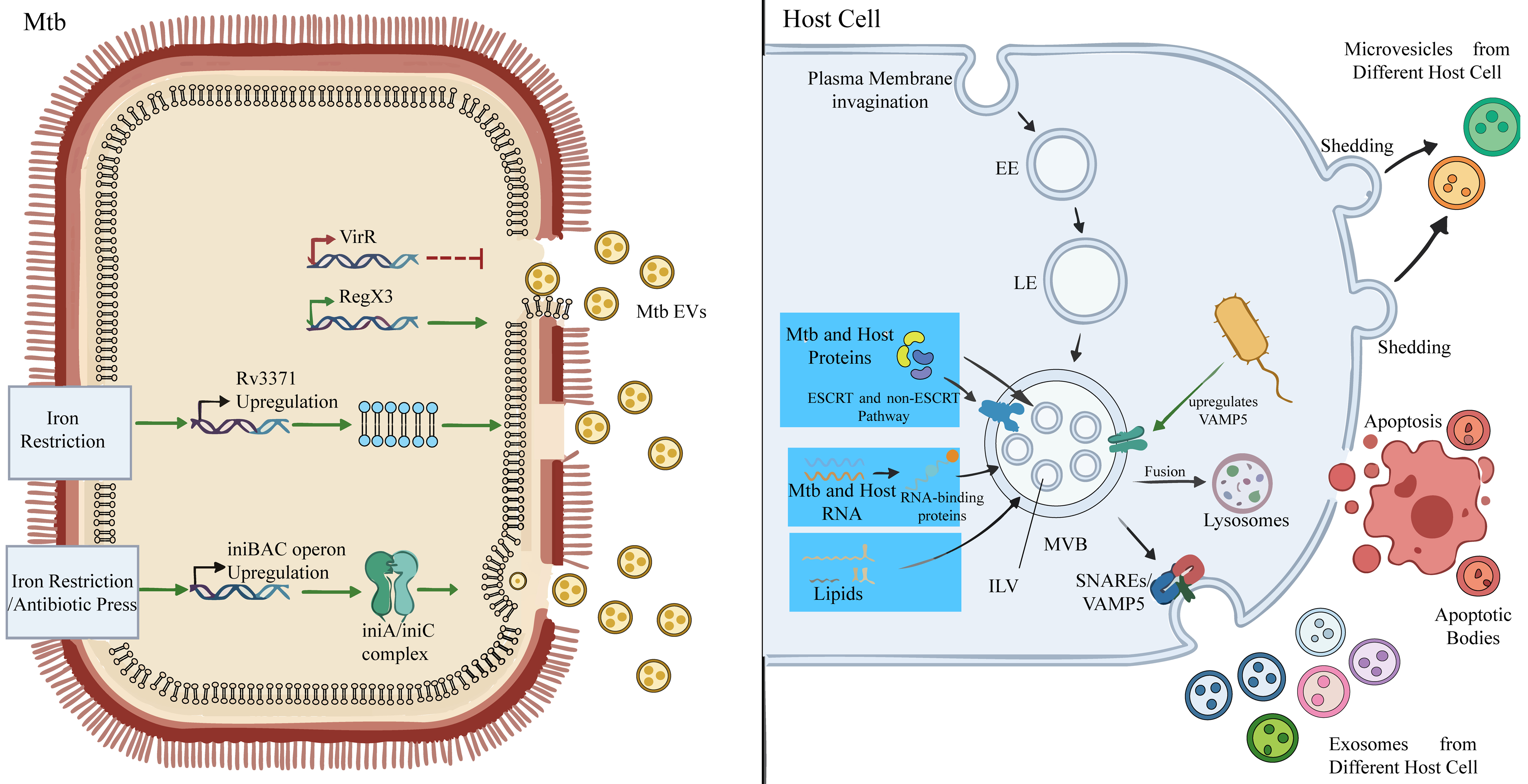
Figure 1. Biogenesis and regulation of EVs from Mycobacterium tuberculosis and infected host cells. On the left side, the biogenesis and release of Mtb EVs are governed by a sophisticated regulatory network. Key regulatory factors include VirR and RegX3: VirR restricts excessive Mtb EVs release by maintaining cell wall integrity, whereas activation of the RegX3 pathway promotes Mtb EVs production. Furthermore, under nutritional stresses imposed by the host, such as iron limitation, Mtb actively upregulates the secretion of Mtb EVs via pathways including Rv3371 and the iniBAC operon (IniA/IniC complex), as a survival strategy to cope with environmental constraints. Ultimately, Mtb EVs carrying diverse bioactive components, including lipids, proteins, and nucleic acids, are released into the extracellular milieu. On the right side, within infected host cells, EE mature into LE and further develop into MVB. ILVs are formed by inward budding of the MVB membrane, a process that relies on both ESCRT-dependent and ESCRT-independent mechanisms for the sorting and loading of host- and Mtb-derived proteins, RNAs, lipids, and other molecules. The fate of MVB determines exosome release: a subset of MVBs fuses with lysosomes for cargo degradation, while another subset fuses with the plasma membrane under the regulation of Rab GTPases and SNARE proteins (e.g., VAMP5), thereby releasing ILVs as exosomes into the extracellular space. In addition, host cells directly release microvesicles through plasma membrane budding and generate apoptotic bodies upon the induction of apoptosis. These distinct classes of HC EVs act as intercellular communication carriers during Mycobacterium tuberculosis-host interactions, participating in critical processes including immune modulation, bacterial survival, and pathogen dissemination. Mtb: Mycobacterium tuberculosis; EVs: extracellular vesicles; Mtb EVs: Mycobacterium tuberculosis-derived extracellular vesicles; HC EVs: host cell-derived extracellular vesicles; EE: early endosomes; LE: late endosomes; MVB: multivesicular bodies; ILV: intraluminal vesicles; ESCRT: endosomal sorting complexes required for transport; VirR: vesiculogenesis and immune response regulator; RNA: ribonucleic acid; SNARE: soluble N-ethylmaleimide-sensitive factor attachment protein receptor; VAMP5: vesicle-associated membrane protein 5.
Proteomic analyses have uncovered another characteristic of the composition of Mtb EVs. A pioneering study by Prados-Rosales et al. was the first to systematically identify the protein composition of Mtb H37Rv EVs, identifying 48 proteins and highlighting the significant enrichment of lipoproteins as a core feature[11]. With advances in mass spectrometry technology, Lee et al. substantially expanded the number of identified Mtb EVs proteins to 287, encompassing diverse functional categories such as metabolism, signal transduction, and lipid metabolism[22]. Notably, these Mtb EVs proteins show substantial overlap with naturally occurring antigen complexes widely used in TB research (e.g., Ag60)[23], providing a solid molecular foundation for understanding the immunological functions of Mtb EVs. Beyond proteins, Mtb EVs also contain a variety of structural and immunomodulatory lipids. Lipids in Mtb EVs are predominantly composed of polar lipids, including phosphatidylinositol, acylated phosphatidylinositol mannosides, cardiolipin (CL), and phosphatidylethanolamine (PE), and carry the key immunomodulatory molecule LAM[11]. These lipid components not only constitute the structure of Mtb EVs but also participate in host immune recognition and regulation. Additionally, bacterial genomic DNA has been identified within Mtb EVs[11]. Although Mtb DNA in the host cytoplasm can initiate innate immune responses, the specific immunological roles and underlying mechanisms of EVs-delivered DNA remain to be further explored. No studies have systematically identified whether Mtb EVs carry RNA molecules; future research clarifying their RNA composition is expected to reveal new layers of bacterial gene regulation and host interactions mediated by Mtb EVs. Simultaneously, it remains unknown whether the cargo profile of Mtb EVs released by Mtb during different infection stages undergoes dynamic changes. Systematic analysis of the cargo of Mtb EVs throughout the entire infection process in future studies may uncover the refined strategies by which Mtb adapts to host immune pressure from a novel “temporal regulation” perspective.
The biogenesis of Mtb EVs is governed by an intricate, multi-factorial regulatory network, whose complexity has been unraveled from diverse research perspectives. A core component of this network is VirR: its deletion enables Mtb to induce heightened immune responses in vivo, resulting in reduced bacterial burden[21]. Subsequent studies revealed that loss of VirR causes profound abnormalities in cell wall structure, increasing its permeability and ultimately driving the massive release of Mtb EVs[24]. Independent VirR-independent pathways have also been reported - for instance, in a mutant strain deficient in the pstA1 gene, over 60 genes sustain RegX3 activation to significantly promote Mtb EVs release[25]. Future research could further identify key effectors driving Mtb EVs secretion from these RegX3-regulated genes. Collectively, these findings indicate that Mtb EVs biogenesis is controlled by specific signaling pathways and responds to distinct intracellular states. Notably, Mtb may restrict excessive Mtb EVs production via factors such as VirR and pstA1, thereby avoiding overexposure of antigens to the host immune system - a potential strategy for immune evasion within the host. In-depth investigations into Mtb EVs regulatory factors in the future will not only uncover the fundamental principles of Mtb EVs biogenesis but also hold promise for disrupting the pathogen’s precise control over Mtb EVs release through interventions targeting key nodes such as VirR or RegX3. Such interventions could enhance host recognition and clearance of the pathogen, offering novel therapeutic targets for anti-TB treatments. Meanwhile, Mtb EVs release represents an active strategy for the pathogen to cope with stress. During infection, the host initiates a “nutritional immunity” strategy to limit Mtb’s iron acquisition, creating an iron-restricted environment critical for Mtb survival[26]. Under this stress, Mtb has evolved multiple countermechanisms, with the role of Mtb EVs in this context increasingly being elucidated. A study by Prados-Rosales et al. observed that Mtb releases significantly higher levels of EVs under iron-depleted conditions compared to iron-replete environments[27]. Iron limitation robustly promotes Mtb EVs secretion, with regulatory mechanisms involving factors such as Rv3371 and the dynamin-like proteins IniA/IniC. Studies have shown that under iron restriction, Rv3371 expression is upregulated, and it promotes Mtb EVs release by altering cell wall lipid composition[28]. Similarly, under iron deprivation or isoniazid stress, the iniBAC operon is upregulated, and its encoded dynamin-like IniA/IniC complex directly mediates membrane fission and Mtb EVs release in a GTP-dependent manner[29]. It is important to recognize that the regulatory pathways described above - particularly the roles of Rv3371 and the IniA/IniC complex in orchestrating stress-induced vesiculogenesis - have been delineated almost exclusively through in vitro experimentation. These culture-based studies have yielded valuable mechanistic insights into the molecular machinery governing Mtb EVs biogenesis. However, a significant knowledge gap persists: whether these same regulatory cascades are operative within the complex and heterogeneous microenvironment of infected host tissues - where Mtb encounters dynamic gradients of iron, oxygen, and other limiting nutrients - has yet to be demonstrated. Direct in vivo evidence confirming that these spatiotemporal dynamics operate inside granulomas, necrotic lesions, or other tissue niches remains conspicuously absent, underscoring the need for future investigations employing physiologically relevant infection models.
Release of Mtb EVs is not a random cellular leakage or evolutionary relic but an active biological program tightly regulated by sophisticated genetic and biochemical networks, serving the pathogen’s survival needs across diverse physiological and stress conditions. The focus of future research should not only be on identifying specific effector molecules and their functions within candidate gene clusters downstream of RegX3 but also on systematically deciphering the topological structure of the global regulatory network connecting key nodes - including VirR, RegX3, iron restriction responses, and cell wall stress sensing - by integrating multi-omics data and quantitative biological approaches from a systems biology and dynamic regulation perspective. Building on this, a quantitative investigation into the dynamic reprogramming of this regulatory network across different infection models and stages will be of great importance for clarifying how environmental signals are sensed, integrated, and ultimately translated into precise regulation of Mtb EVs biogenesis pathways. This will reveal the molecular logic underlying the disruption and re-establishment of the balance between “restricted release” and “promoted release” states. Targeted interventions to modulate this dynamic balance - whether by enhancing inhibitory nodes to impair the pathogen’s environmental adaptability or by relieving inhibition to overexpose pathogen antigens - may disrupt the immune homeostasis established by Mtb within the host, thereby providing new therapeutic breakthroughs to dismantle its persistent infection mechanisms.
HC EVs
As explicitly stated in a key position paper by the International Society for Extracellular Vesicles (ISEV) in 2014, significant terminology confusion existed during the early stages of the field’s development. Terms such as “microparticle”, “ectosome”, and “exosome” were frequently used interchangeably in the literature without uniform standards[30]. Notably, the term “exosomes” - owing to its rapid popularity - was often employed indiscriminately as an umbrella term for all EVs subtypes, obscuring the potential biogenetic and functional differences between distinct HC EVs subpopulations[30]. Therefore, HC EV subtypes have been uniformly classified and named - including exosomes, microvesicles, and apoptotic bodies - based on criteria such as their biogenetic pathways, size, density, and protein composition[5].
As a typical intracellular pathogen, Mtb profoundly alters the physiological state of host cells such as macrophages following infection[31]. Infected cells release a repertoire of HC EVs, which act as complex messengers in host-pathogen interactions, with cargo exhibiting dual host and pathogen characteristics. These vesicles not only carry host proteins, nucleic acids, and lipids that reflect cellular activation or functional status but also frequently encapsulate Mtb antigens and lipids, thereby playing multifaceted roles in mediating immune regulation, promoting bacterial survival, and facilitating long-distance dissemination[32]. Throughout this review, we use the term “HC EVs” as a collective designation encompassing all host-derived vesicle subtypes - exosomes, microvesicles, and apoptotic bodies. However, it is important to note that the literature we cite varies considerably in whether individual studies distinguish among these subtypes. When the original study provides clear evidence for a particular subtype (e.g., through specific isolation methods, marker analysis, or electron microscopy), we adopt the corresponding term in our functional descriptions. When the original work examines total EVs or mixed populations without subtype resolution, we retain “HC EVs” and indicate the composition or methodological limitations accordingly, so that readers can clearly differentiate between subtype-specific and population-level findings.
Among the diverse subtypes of HC EVs, exosomes have emerged as the most extensively studied in TB research, attributed to their unique biogenetic pathway, specific cargo sorting mechanisms, and stability in bodily fluids. Exosomes are nanoscale EVs with a diameter of 30-150 nm, originating from multivesicular bodies (MVB) within the endocytic pathway[33]. Their membrane structure is enriched in tetraspanins (CD9, CD63, CD81), sphingolipids, cholesterol, and unsaturated lipids, and they carry biogenesis-related proteins such as TSG101 and ALIX, as well as immune-associated components including major histocompatibility complex (MHC) molecules[34,35]. During Mtb infection, exosomes released by host cells have been found to be significantly enriched with pathogen-specific cargo, such as bacterial-derived lipoproteins, virulence factors, and host stress-response proteins [Table 1][11,22,27,32,36-44].
Comparison of Mtb EVs and HC EVs: biogenesis, composition, and function
| Mtb EVs | HC EVs | |
| Major subtype | ① Exosomes ② Microvesicles ③ Apoptotic bodies | |
| Biological origin | Mtb | Host cells in the process of TB infection |
| Biogenesis | Current evidence suggests origin from the plasma membrane, though the precise biogenesis site remains under investigation | ① Exosomes: released upon the fusion of MVB with the plasma membrane ② Microvesicles: generated by the direct outward budding and pinching off of the plasma membrane ③ Apoptotic bodies: produced as a result of programmed cell death (apoptosis) |
| Size of the vesicles | ~60-300 nm in diameter | ① Exosomes: 30-150 nm in diameter ② Microvesicles: 100-1,000 nm in diameter ③ Apoptotic bodies: typically > 1 μm in diameter |
| Cargo composition | Prados-Rosales et al., 2011[11] Protein components (48 types): lipoproteins (e.g., 19-kDa, LprA, LprG, LprX, MPB83, etc.), cell wall and cell process-associated proteins (e.g., DnaK, GroEL), metabolic enzymes (e.g., isocitrate lyase), as well as virulence and adaptation-related proteins Lipid components (predominantly polar lipids): PI, PIM2, CL, PE, and others Glycolipid complexes: LAM | ① Exosomes Universal marker: CD9, CD63, CD81, TSG101, ALIX, and others |
| Lee et al., 2015[22] Protein components (287 types): cell wall/membrane-associated proteins, such as lipoproteins (LpqH, LprA, LprG, LppX, PstS1, etc.), FbpA/B/C, and HbHA; secretion system and virulence-related proteins, including ESAT-6 family proteins (EsxA, EsxB), ESX-1 secretion-associated proteins (EspA, EspC), as well as SodB, KatG, and HspX; metabolism-related proteins, such as isocitrate lyase, Fba, Eno, and ATP synthase subunits (AtpA/D); additionally, information pathway proteins (e.g., Hup) and a large number of conserved proteins of unknown function are included Lipid components (predominantly polar lipids): PI, PIM2, CL, PE, and others Glycolipid complexes: LAM | Cargo in TB infection: Giri et al., 2010[32] Mycobacterial proteins in exosomes derived from Mtb-infected J774 cells (41 types): Ag85A, Ag85B, Ag85C, ESAT-6, MPT64, MPT63, GroES, KatG, HspX, GlnA1, PstS1, DnaK, as well as β-1,3-glucanase precursor, LpdC, Mpt32/Apa, Cfp2, and SahH, among others Mycobacterial proteins in exosomes derived from CFP-treated J774 cells (29 types): Ag85A, Ag85B, Ag85C, ESAT-6, MPT64, MPT63, GroES, KatG, HspX, GlnA1, PstS1, DnaK, among others | |
| Prados-Rosales et al., 2014[27] Under iron-limited conditions Siderophores: Mycobactin, Di-deoxymycobactin Lipids: DAG, TAG, PE, DAT | Kruh-Garcia et al., 2014[38] Serum exosomes from patients with TB and LTBI Protein components (20 types): AcpM, Ag85A, Ag85B, Ag85C, Ald, Apa (Mpt32), BfrB, Cfp17, DnaK, Fba, GlcB, GroES, HspX, Icd2, KatG, Mpt51, Mpt53, Mpt63, Mpt64, and SahH | |
| Sharma et al., 2025[36] Under iron-limited conditions Protein components: KatG, Rv2159c, DlaT, PacL, Rv3671c, AcpM, EsxA; NuoG, MoxR1, GroEL2, HbhA, PstS1; LpqH, LprG, LprA, EsxA, MmsA, GroEL2, DnaK, Cfp29, RplJ, RpsO, RpsG, RpsC; LpqH, LprG, LprA, GroEL2; HsaA, Rv3520c, Rv2263, FabG4, FadA3, SerC, DesA1, SecE2, Rv2623 (TB31.7), ClpP1, Rv3671c, Cds1, VapC10, GroEL2, HbhA, LpqE, SahH, Gap Siderophores: Mycobactin | Diaz et al., 2016[39] Exosomes released from Mtb-infected human macrophages Host-derived proteins (41 types): Coronin-1C, Vimentin, HSP90, GAPDH, and L-amino acid oxidase, among others | |
| Davids et al., 2024[37] In the presence of rifampicin and isoniazid Specific lipids: TMM | Ramachandra et al., 2010[40] Exosomes released from Mtb-infected human macrophages Immune-related Cargo: MHC-II molecule invariant chain, costimulatory molecule CD86, and adhesion molecule ICAM-1 | |
| Smith et al., 2015[41] Exosomes from laboratory-cultured macrophages (RAW264.7) and kidney cells (HEK293) Proteins from Mtb: HspX, GroES, KatG, and various antigens | ||
| Biadglegne et al., 2022[42] Serum exosomes from patients with ATB Upregulated proteins: LBP, fibrinogen alpha chain, complement C9, EMILIN-1, and transferrin receptor 1, among others Downregulated proteins: CD36, MHC-I, integrin family members, cytoskeletal proteins, among others | ||
| ② Microvesicles Ramachandra et al., 2010[40] Microvesicles released from Mtb- or LPS-stimulated mouse macrophages Immune-related Cargo: invariant chain, costimulatory molecule CD86, adhesion molecule ICAM-1 | ||
| Vázquez-Flores et al., 2023[43] Microvesicles derived from Mtb-infected neutrophils Host-derived proteins: myeloperoxidase, HLA class I histocompatibility antigen C alpha chain, CD59, neutrophil elastase, matrix metalloproteinase-9, among others Proteins from Mtb (74 types): LprG, HbHA, GroEL, CydA, IF-2, Rv2986c, Rv2091c, Rv1488, among others | ||
| ③ Apoptotic bodies Schaible et al., 2003[44] Lipids from Mtb: LAM, PIM, TDM, MycB, LCL, PE, PG, PI Proteins from Mtb: predominantly proteins < 38 kDa, including lipoprotein p19 Host-derived proteins: CD1b, MMR, MHC-II, among others |
Exosome biogenesis is a highly programmed process involving early endosome formation, MVB maturation, and final secretion. First, the plasma membrane of the cell undergoes initial invagination to form early endosomes (EE)[33]. As EE mature into late endosomes (LE) and further develop into MVB, their membranes bud inward to generate intraluminal vesicles (ILV), with the majority of exosome cargo captured and encapsulated into ILV during this “invagination” process[45]. For protein cargo - including soluble Mtb-derived proteins - sorting and loading into exosomes are primarily mediated by the coordinated action of ESCRT-dependent and ESCRT-independent pathways[46]. Smith et al. have shown that Mtb-derived proteins loaded into exosomes are mainly soluble proteins actively secreted or released by Mtb into host cells[41]. Regardless of how Mtb soluble proteins enter the cell, they can be directed to a ubiquitination-dependent pathway; this ubiquitination tag is subsequently recognized by the ESCRT system, guiding their entry into ILV[41]. Meanwhile, Mtb infection triggers rapid phosphorylation of AKT, activating both ESCRT-dependent and ESCRT-independent pathways. However, whether this represents active hijacking by Mtb or a host cell response to Mtb infection remains to be further elucidated[47]. RNA cargo enters ILV primarily through active sorting mechanisms mediated by RNA-binding proteins recognizing specific sequence motifs[45]. For Mtb RNA in exosomes released by infected host cells, Cheng and Schorey reported that the Mtb SecA2 protein secretion system is crucial for delivering Mtb RNA into host cell-derived exosomes[48]. Mtb infection can alter the composition of both host and bacterial RNA in host cell exosomes[49]. Nevertheless, the underlying logic for the upregulation and downregulation of different RNAs in exosomes released by Mtb-infected host cells remains unclear. The fate of MVB determines exosome release: some fuse with lysosomes for degradation, while others are transported to the plasma membrane under the regulation of Rab GTPases and release exosomes through SNARE protein-mediated membrane fusion[50]. Exosome release is an actively regulated process dependent on Rab GTPase-directed transport, SNARE protein-mediated membrane fusion, and ESCRT complex assistance, with precise control by multiple intracellular and extracellular signals[45]. During Mtb infection, the level of VAMP5 in exosomes from TB patients is significantly increased and decreases following treatment, indicating an association between VAMP5 and disease activity[51]. VAMP5 belongs to the SNARE protein family and acts as a regulator of exosome release[52]. Arya hypothesized that after Mtb infects macrophages, VAMP5 expression on MVB membranes is regulated; it forms specific SNARE complexes by binding to STX1 or STX4 and SNAP47 on the plasma membrane, driving MVB-plasma membrane fusion and thereby promoting exosome secretion[51,53].
Unlike exosomes, the sorting of their luminal cargo relies more on lipid anchoring of proteins (e.g., myristoylation or palmitoylation) rather than the canonical ubiquitination pathway[54]. Anchored proteins can spontaneously accumulate in specific microdomains of the plasma membrane and further selectively encapsulate certain cytoplasmic proteins, RNAs, and other cargo into the ectosome lumen by forming higher-order polymers or protein networks[54]. They carry bioactive molecules such as proteins and nucleic acids, participating in various physiological and pathological processes including immune regulation, cell differentiation, and infection responses[55]. Due to their cargo sorting mechanism that favors membrane-associated proteins, microvesicles may serve as efficient carriers for specific membrane proteins (e.g., co-stimulatory molecules, adhesion molecules), a conjecture supported by studies by Ramachandra et al.[40]. As terminal products of apoptosis, apoptotic bodies function as core “antigen transporters” in TB immunity. Phosphatidylserine exposed on their surface acts as an “eat-me” signal, which can be recognized and phagocytosed by macrophages, enabling gentle antigen delivery without triggering significant inflammation[44,56]. A study has shown that apoptotic bodies produced by macrophages pre-treated with interferon-gamma (IFN-γ) after apoptosis can effectively enhance the antigen-presenting process[57]. However, how Mtb interferes with their formation and cargo sorting, as well as their role in the reactivation of latent infections, remains to be systematically elucidated.
The key characteristics of each HC EVs subtype - including cellular origins, representative cargo molecules, and major immunological functions - are summarized in Table 2 for quick reference[15,17,32,40,43,44,48,49,57-68]. Compared with the rapid development of exosome research, the exploration of microvesicles and apoptotic bodies in translational applications for TB is significantly lagging. How to accurately decipher their specific cargo composition and immunological fate - i.e., whether they cooperate with the host to clear pathogens or are exploited by pathogens to promote immune evasion? Answering these questions is a prerequisite for achieving precise regulation of their functions and comprehensively understanding HC EVs-mediated host-pathogen interactions.
Summary of HC EVs subtypes in tuberculosis: origins, Cargo, and immunological functions
| HC EVs subtype | Major cell sources in current studies | Key Cargo molecules | Major immunological functions | References |
| Exosomes | Macrophages, DCs, MSCs, epithelial cells, NK cells | miRNAs (miR-146a-5p, miR-20b-5p, miR-18a, miR-766-3p); proteins (Hsp70, MHC-II, CD63, CD81, TSG101, ALIX); Mtb antigens (Ag85, ESAT-6, LAM) | Immune suppression via miRNA delivery (inhibiting MHC-II, autophagy, apoptosis) Antigen cross-presentation via Siglec-1 T-cell activation Pro-inflammatory signaling via TLRs | [15,17,32,48,49,58-63] |
| Microvesicles | Macrophages, neutrophils, DCs | CD86, ICAM-1, MHC-II, type I interferon-induced proteins (ISG15, IFIT1-3), Mtb antigens (LprG, HbHA) | Direct T-cell activation via surface CD86/ICAM-1 Macrophage activation and autophagy induction Endothelial barrier modulation Pro-inflammatory cytokine secretion | [40,43,64,65] |
| Apoptotic bodies | Apoptotic macrophages, DCs, epithelial cells | Phosphatidylserine, Annexin A1, CD1b, MHC-II, LAM, PIM, TDM, Mtb proteins (< 38 kDa) | Antigen delivery to DCs via efferocytosis (Annexin A1-mediated) Cross-presentation Gentle antigen transfer without inflammation | [44,57,66] |
| NK-derived exosomes | NK cells | Perforin, granulysin, granzyme A, miR-1249-3p | Inducing apoptosis of infected macrophages via caspase cascade and mitochondrial pathways (caspase-3/PARP activation) Antimicrobial activity | [67,68] |
Isolation and characterization
Isolation
The reliability of EVs research largely depends on efficient isolation and accurate characterization methods. Current isolation techniques face multiple challenges: EVs in biological samples often coexist with impurities such as protein aggregates and lipoproteins, making co-precipitation difficult to avoid[5]; the high heterogeneity of EVs themselves in terms of particle size and surface markers further increases purification complexity[69]. Additionally, the impact of different isolation strategies on EVs integrity, recovery rate, and functional properties reduces the comparability of research results. However, there is currently no universal isolation method applicable to all samples, and appropriate strategies must be selected based on EVs sources and research objectives[34].
For the isolation of Mtb EVs, Rao et al. proposed that their purification primarily involves three steps: induction (culture in minimal medium), collection (centrifugation, filtration, and concentration), and purification (obtaining high-purity Mtb EVs via ultracentrifugation combined with OptiPrep density gradient centrifugation)[70]. Studies have shown that different methods exert significant effects on the yield, composition, and function of Mtb EVs. For instance, Wang et al. demonstrated that commercial kit-based methods yield higher quantities of Mtb EVs, while the EXODUS system produces Mtb EVs with more diverse components[71]; Ryan et al. evaluated four approaches - ultracentrifugation, density gradient ultracentrifugation, qEV size exclusion chromatography, and Capto™Core size exclusion chromatography - among which the combination of qEV and OptiPrep density gradient centrifugation exhibited superior performance in terms of purity[72]. They also noted that although Mtb EVs isolated by different methods are similar in morphology and size, significant differences exist in their protein composition, yield, and reproducibility[72]. Notably, multiple techniques are often combined in practical research to improve the quality of Mtb EVs isolation. For the isolation of specific subpopulations within HC EVs, such as exosomes, more refined strategies are required. Differential centrifugation combined with ultracentrifugation is currently the most commonly used method. Due to differences in size and sedimentation coefficients among exosomes, microvesicles, and apoptotic bodies, exosomes are typically enriched at a centrifugal force of 100,000 × g, while microvesicles and apoptotic bodies can be collected at lower centrifugal forces[40,44]. Furthermore, regarding the accurate separation of Mtb EVs and exosomes, Athman successfully isolated the two in distinct fractions using sucrose density gradient centrifugation[58].
However, achieving higher standards of EVs isolation through advanced technologies remains a current technical bottleneck that urgently requires further exploration. The precise isolation of various EV subpopulations is not only a sophisticated technical operation but also a critical prerequisite for in-depth functional research and the advancement of clinical applications. In accordance with the Minimal Information for Studies of Extracellular Vesicles 2023 (MISEV2023) guidelines issued by ISEV, we have compiled Table 3, summarizing the principles, advantages, disadvantages, and applicable scenarios of commonly used isolation techniques for Mtb EVs and HC EVs in TB research.
Comparative analysis of common isolation techniques for Mtb EVs and HC EVs and their applicable scenarios
| Technology | Principle | Advantages | Disadvantages | Applicable scenarios |
| Density gradient centrifugation | Based on the density differences among various particles, density gradient media (e.g., sucrose) form a gradient layer after ultracentrifugation. EVs then migrate to the region with matching density, enabling their separation from other particles | 1. Enables acquisition of high-purity samples 2. Separates EVs from proteins/non-vesicular extracellular particles by density 3. Distinguishes EVs subtypes of varying densities | 1. Low recovery rate 2. Prolonged centrifugation tends to cause EVs aggregation or damage 3. Complex operation requiring extra steps to remove density media | 1. High-purity EVs requirement 2. Separation of EVs subtypes with different densities 3. Bacterial EVs studies |
| Differential (Ultra) centrifugation, dUC | By gradually increasing centrifugal force, impurities with larger size and density in the sample are sequentially precipitated, and EVs are finally enriched | 1. Simple operation and strong versatility, applicable to multiple EVs sources 2. Preliminarily separate EVs subtypes of different sizes 3. No need for special media | 1. Moderate purity, prone to co-separation of impurities 2. High centrifugal force may cause EVs aggregation or membrane damage 3. Unable to completely separate particles with overlapping size/density | 1. Preliminary separation and concentration of EVs 2. Pre-experiments with low purity requirements 3. Acquisition of large quantities of EVs |
| Size exclusion chromatography, SEC | Using a chromatography column packed with porous media, separation is achieved based on particle size differences. Large-sized EVs are eluted first, while small-molecule contaminants are eluted later | 1. Gentle and non-damaging, preserving EVs structure and activity 2. Effectively separates EVs from small-molecule impurities 3. Scalable operation | 1. Samples are diluted, requiring additional 2. Resolution is affected by column volume and flow rate 3. Lower processing efficiency for large-volume samples | 1. Functional studies requiring preserved EVs activity 2. EVs characterization needing removal of protein contamination |
| Precipitation | Through polymers (e.g., PEG) or salts to reduce the solubility of EVs in solution, causing EVs aggregation and precipitation via low-speed centrifugation for rapid concentration | 1. Simple and fast operation 2. Large processing volume and high concentration efficiency 3. Low cost | 1. Lowest purity, prone to co-precipitation of large amounts of impurities 2. Polymers may affect EVs activity and subsequent analysis 3. Unable to distinguish EVs from other vesicle-like particles | 1. Rapid concentration of EVs 2. Scenarios requiring only volume reduction rather than high-purity separation |
| Kit-based approaches | 1. Polymer precipitation kit: essentially a precipitation-based method 2. Affinity capture kit: integrates probes such as antibodies and peptides to achieve rapid enrichment of EVs | 1. Simple and fast operation 2. Large processing volume and high concentration efficiency 3. No need for professional instruments | 1. The principle of some kits is not transparent, potentially introducing unknown contaminants 2. Precipitation-based kits have low purity 3. Relatively costly | 1. Rapid concentration of EVs 2. Scenarios requiring only volume reduction rather than high-purity separation |
Characterization
Reliable EVs research relies on comprehensive and accurate characterization of isolated products, with strategies tailored to the biological origin of EVs and specific scientific questions. Systematic and precise characterization of Mtb EVs and HC EVs serves as a critical foundation for subsequent mechanistic exploration and translational research. The MISEV2023 guidelines propose a multi-layered characterization framework applicable to EVs from diverse species and cellular sources, but its practical implementation requires adjustments based on the biological characteristics of specific EVs subtypes[73].
Characterization of Mtb EVs should focus on their unique cell wall components, such as the identification of LAM and mycolic acids via specific antibodies (e.g., Western Blot) or lipidomic analysis, alongside confirmation of their lipid bilayer structure using electron microscopy[73]. Additionally, density gradient centrifugation is recommended to reduce interference from co-isolated protein aggregates, with detailed documentation of bacterial culture conditions emphasized - given their significant impact on EVs composition and function[73]. For the characterization of HC EVs, multi-layered, complementary techniques are required to verify their identity, purity, and cellular origin. First, particle concentration and size distribution should be assessed using techniques such as nanoparticle tracking analysis (NTA), while transmission electron microscopy or cryo-electron microscopy can visualize their typical morphology. In terms of biochemical characteristics, the marker framework proposed by Minimal Information for Studies of Extracellular Vesicles 2018 (MISEV2018) should be adopted: detecting at least one transmembrane or glycosylphosphatidylinositol (GPI)-anchored protein (e.g., CD9, CD63, CD81) and one cytoplasmic protein (e.g., TSG101, Alix) as positive EVs markers. Simultaneously, verification of negative or significantly low expression of one intracellular organelle marker (e.g., Calnexin, Grp94, or cytochrome c) is necessary to evaluate sample purity and exclude contamination by cellular debris[73]. Ideally, the proteomic profile of EVs should be compared with that of parental cell lysates, with “blank medium” processed through the same isolation workflow set as a negative control. Only through the integration of the aforementioned physical and biochemical evidence can the isolated particles be confirmed as high-purity HC EVs of host cell origin, providing a reliable basis for subsequent functional studies and clinical applications.
Research on these Mtb EVs and HC EVs - whether the standardization of isolation and characterization techniques or the in-depth dissection of their biogenesis and functions - serves as a critical cornerstone for fully deciphering the pathogenesis of TB and developing novel diagnostic and therapeutic strategies.
DECIPHERING WHY Mtb IS DIFFICULT TO ERADICATE FROM THE PERSPECTIVE OF Mtb EVs AND HC EVs
Traditionally, the academic community’s understanding of its pathogenic mechanism has mostly focused on its “latency” strategies within host cells, such as intracellular survival pathways like resisting lysosomal killing[74]. However, these classic theories are insufficient to fully clarify the overall picture of the clinical intractability of TB. In recent years, with the in-depth research on EVs, Mtb EVs and HC EVs induced by Mtb infection have been gradually revealed as crucial “behind-the-scenes drivers” that promote the occurrence and development of TB. This chapter will systematically elaborate on how Mtb EVs and HC EVs collectively weave a complex pathological network leading to the difficulty in eradicating TB through multiple mechanisms, including mediating immunosuppression, precisely regulating inflammatory responses, promoting bacterial dissemination and persistence, and even potentially participating in the development of drug resistance [Figure 2].
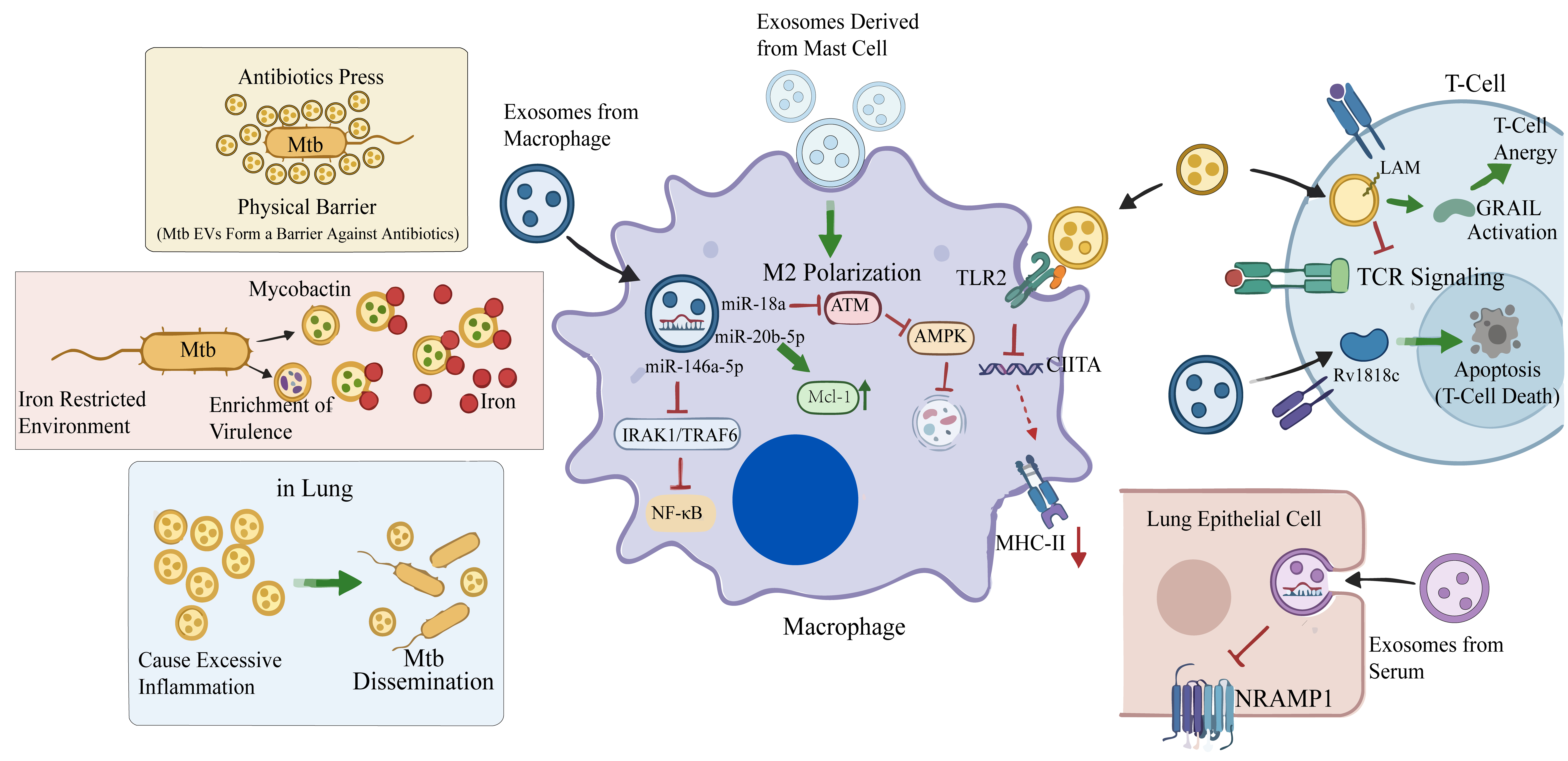
Figure 2. Key mechanisms by which Mtb EVs and HC EVs synergistically mediate Mtb immune evasion, metabolic reprogramming, and pathogen dissemination. Mtb EVs deliver immunomodulatory molecules such as lipoproteins (LprG, LpqH) and LAM to macrophages and T cells, impairing antigen presentation by suppressing MHC-II expression via the TLR2/MYD88/CIITA axis, inhibiting TCR signaling, and inducing T-cell anergy and apoptosis. Meanwhile, Mtb reprograms HC EVs, particularly exosomes, to carry altered profiles of miRNAs (e.g., miR-146a-5p, miR-18a, miR-766-3p) and bacterial proteins (e.g., Rv1818c). These EVs suppress host inflammatory responses, inhibit macrophage autophagy and apoptosis, downregulate the iron-restriction protein NRAMP1 in lung epithelial cells, and induce M2 macrophage polarization, thereby creating an intracellular environment favorable for Mtb survival. Under conditions of iron limitation or antibiotic stress, Mtb releases Mtb EVs enriched in mycobactin and protective lipids, which enhance iron acquisition and form a physical barrier against antibiotics. Collectively, these EVs-mediated processes exacerbate pulmonary inflammation, facilitate systemic bacterial dissemination, and contribute to the establishment of persistent and drug-resistant TB infections. Mtb: Mycobacterium tuberculosis; EVs: extracellular vesicles; Mtb EVs: Mycobacterium tuberculosis-derived extracellular vesicles; HC EVs: host cell-derived extracellular vesicles; TB: tuberculosis; LprG: lipoarabinomannan carrier protein LprG; LpqH: 19-kDa lipoprotein LpqH; LAM: lipoarabinomannan; MHC-II: major histocompatibility complex class II; TLR2: Toll-like receptor 2; MyD88: myeloid differentiation primary response protein 88; CIITA: class II transactivator; TCR: T-cell receptor; miR: microRNA; NF-κB: nuclear factor kappa B; NRAMP1: natural resistance-associated macrophage protein 1; M2: alternatively activated macrophage phenotype; ATM: ataxia-telangiectasia mutated; AMPK: AMP-activated protein kinase; Mcl-1: myeloid cell leukemia 1; IRAK1: interleukin-1 receptor-associated kinase 1; TRAF6: tumor necrosis factor receptor-associated factor 6; GRAIL: gene related to anergy in lymphocytes.
Involvement of Mtb EVs and HC EVs in immunosuppression
Immunomodulatory functions of Mtb EVs in facilitating TB progression
As important immunomodulatory factors, Mtb EVs exert a broad range of effects - they not only influence infected cells but also regulate the functions of distant uninfected immune cells. As will be further discussed in Section “Discussion: allies or foes? Re-evaluating the immunological functions of Mtb EVs and HC EVs”, the net immunological outcome of Mtb EVs exposure is highly context-dependent, varying with ligand dose, cellular target, and infection stage; the immunosuppressive effects described below should therefore be interpreted as one facet of a dynamic and tunable regulatory system, rather than as an intrinsic and invariant property of Mtb EVs. Studies have demonstrated that Mtb EVs can be internalized by macrophages and dendritic cells (DCs), thereby exerting dual regulatory roles: on one hand, they promote DC maturation and antigen presentation through a Toll-like receptor 2 (TLR2)-dependent pathway, characterized by the upregulated expression of MHC-I, MHC-II, and the costimulatory molecule CD86, which in turn induces T-cell responses. This process is associated with pathogen-associated molecular patterns (PAMPs) that act as potent TLR2 agonists[15]; on the other hand, Mtb EVs can significantly inhibit IFN-γ-induced MHC-II expression in macrophages[15]. At first glance, these two observations appear contradictory, as both are mediated via TLR2 signaling. However, as we will systematically dissect in Section “Discussion: allies or foes? Re-evaluating the immunological functions of Mtb EVs and HC EVs”, this apparent paradox can be resolved by considering that the functional outcome of TLR2 engagement depends on ligand dose, duration of exposure, and the cellular context - parameters that vary substantially between the DC maturation assay (short-term, low-dose stimulation) and the macrophage MHC-II suppression model (sustained, high-dose exposure). This effect is likely closely related to lipoproteins such as LprG and LpqH inhibiting CIITA transcription via the TLR2/MYD88 signaling pathway[15,36,58]. As TLR2 agonists, these lipoproteins initiate a negative feedback regulatory loop through sustained TLR2/MYD88 signaling, with the core consequence of suppressing the transcription of CIITA - the “master regulator” of MHC-II molecule expression[75]. Early studies revealed that LAM, a key virulence factor, can directly act on CD4+ T cells and assist Mtb in evading host clearance through multiple potential mechanisms, such as interfering with the T-cell receptor (TCR) signaling pathway, inhibiting CD4+ T-cell activation, and even inducing T-cell anergy[76]. However, how LAM is delivered to T cells remained an unresolved question until Athman et al. proposed, based on their research findings, that Mtb EVs serve as critical carriers responsible for transporting inhibitory components such as LAM[58]. Lipoglycans including LAM carried by Mtb EVs can be directly delivered to CD4+ T cells, inhibiting interleukin-2 (IL-2) secretion and cell proliferation by interfering with the phosphorylation of key molecules (e.g., Lck, ZAP-70, and LAT) in the TCR signaling pathway[58]. Notably, the inhibitory effect of Mtb EVs is significantly stronger than that of purified LAM at the same dose[58]. Given that Mtb EVs also contain other lipoglycans (e.g., LM and PIMs) that have been proven to possess independent inhibitory activities, their potent suppressive effect is likely derived from the synergistic effect of these lipoglycans[58]. Upon delivery of LAM to T cells, it triggers the upregulation of GRAIL protein[77]. GRAIL is an E3 ubiquitin ligase that negatively regulates IL-2 transcription, promoting T cells to enter a state of anergy[77]. Additionally, Mtb EVs may act as carriers to deliver LAM to different compartments to facilitate immune escape, such as phagosomes, to inhibit phagosome maturation[78].
By efficiently delivering their cargo of various lipoproteins and lipoglycans to host immune cells, Mtb EVs play a crucial role in inhibiting macrophage antigen presentation and inducing T-cell exhaustion, providing a new perspective for understanding how Mtb evades host immune clearance. However, it must be acknowledged that although existing studies have initially revealed the potential pathways through which Mtb EVs regulate host immune responses, their underlying molecular mechanisms remain far from fully understood, and many links urgently require in-depth elaboration. As a complex molecular ensemble, the immunosuppressive effects of Mtb EVs are likely not the result of independent actions of a single component but rather stem from the synergy and additive effects among different components. Their total amount, proportion, and spatiotemporal distribution may collectively determine the final outcome of the immune response. As suggested by Athman et al., multiple lipoglycans carried by Mtb EVs may collectively contribute to the inhibition of T-cell activation, potentially producing a stronger suppressive effect than individual components[58]. The specific mode of this “synergistic effect” remains to be clarified. In-depth elucidation of the mechanisms underlying Mtb EVs-mediated immunosuppression is indispensable for comprehensively revealing the pathophysiological processes of TB and explaining the core links of its chronic infection and immune escape.
Mtb-modulated HC EVs: critical players in TB immune evasion
One of the key strategies for Mtb to achieve immune escape is to interfere with multiple links of the host immune system by precisely regulating the composition and function of HC EVs. As key messengers for intercellular communication, HC EVs can accurately deliver a variety of bioactive molecules such as proteins, lipids, and nucleic acids to target cells, thereby reshaping the immune microenvironment at multiple molecular levels. In the complex immune context of TB infection, HC EVs, especially exosomes and microvesicles, collectively form an elaborate immune regulatory network that assists the pathogen in achieving immune escape.
First, in terms of protein composition, Mtb can alter the expression of various antibacterial proteins in host-derived exosomes. For instance, the expression levels of proteins such as CAMP, FCN3, and scavenger receptors are downregulated, which is consistent with previous reports, suggesting that this may be a result of Mtb maintaining intracellular survival, though the specific regulatory mechanisms remain unclear[42]. Second, Mtb can also utilize exosomes to actively eliminate vital immune cells. Infected cells package the bacterial protein Rv1818c into exosomes, which, upon delivery to T cells, induce mitochondria-dependent apoptosis, thereby actively clearing surrounding T cells[79]. Whether Mtb hijacks the exosome system to deliver more immune escape factors can also serve as a direction for future research. Beyond proteins, Mtb’s regulation of nucleic acid components in exosomes is particularly crucial. Transcriptomic data from serum exosomes of active tuberculosis (ATB) patients show significant inhibition of multiple immune-related signaling pathways, indicating that Mtb may achieve immune escape by regulating the composition of exosomal RNA[80]. A recent study confirms this view: miR-146a-5p expression is significantly increased in exosomes derived from BCG-infected macrophages, and uptake of these miR-146a-5p-enriched exosomes by macrophages can suppress inflammatory responses[59]. MiR-146a-5p in exosomes targets the 3’ UTR regions of IRAK1 and TRAF6, inhibits their protein expression, thereby blocking the activation of the nuclear factor kappa B (NF-κB) signaling pathway, and ultimately leading to decreased expression of key pro-inflammatory cytokines such as interleukin-1 beta (IL-1β), interleukin-6 (IL-6), and tumor necrosis factor-alpha (TNF-α), impairing the bactericidal capacity of macrophages[59]. Exosomes are also used to reshape the immune microenvironment. Singh et al. reported that exosomes released by Mtb-infected macrophages can be internalized by uninfected macrophages, activating recipient cells via the TLR2/MyD88 signaling pathway and selectively inhibiting the expression of their critical transcription factor CIITA[81]. Similarly, mannose-capped lipoarabinomannan (ManLAM) of Mtb can induce mast cells to produce chemokine-containing exosomes that recruit macrophages and promote the high expression of M2 macrophage markers[82]. Neutrophil-derived microvesicles can also interfere with the immune function of macrophages. When Duarte et al. treated Mtb-infected macrophages with these microvesicles, the antibacterial activity of macrophages was significantly inhibited[83]. The authors pointed out that this inhibitory effect may be related to the anti-inflammatory properties of neutrophil microvesicles reported previously - namely, they can induce macrophages to produce anti-inflammatory cytokines such as transforming growth factor-β (TGF-β) while suppressing the release of pro-inflammatory mediators such as TNF-α and IL-8[83]. The authors speculate that neutrophil-derived microvesicles may exert different effects in different environments: for example, they can stimulate inflammation in the circulation but exhibit anti-inflammatory properties in the context of Mtb infection[83]. Their specific cargo composition and functional pathways still await systematic elucidation.
By hijacking host exosomes and microvesicles, Mtb has constructed a sophisticated, multi-layered “toolkit” for immune regulation. These HC EVs (including exosomes and microvesicles, with the evidence base for each subtype specified above) not only act as carriers of molecular cargo but also as long-range executors of pathogen instructions, acting synergistically in space and time: exosomes systematically reprogram the host’s innate and adaptive immune responses through the delivery of bioactive molecules such as proteins and nucleic acids, achieving long-term immunosuppression; while neutrophil-derived microvesicles act rapidly at the infection front, directly impairing the antibacterial function of macrophages. Deciphering this complex “communication network” not only holds important theoretical significance for a deep understanding of the pathogenesis of TB but also may provide key targets for the development of novel EVs-based diagnostic biomarkers and immune intervention strategies.
Facilitation of pathogen persistence by Mtb EVs and HC EVs: host metabolic reprogramming
Mtb EVs and pathogen environmental adaptation
As key components for Mtb to regulate the microenvironment and implement immune intervention in the host, the biogenesis and functional diversity of Mtb EVs under iron-limiting conditions have become important entry points for understanding the mechanism of persistent infection by this pathogen. Early studies using lipidomic analysis revealed that Mtb EVs induced by low iron are enriched in mature mycobactin, whereas those under high iron conditions are dominated by precursor forms[27]. Further experiments demonstrated that such Mtb EVs can act as functional siderophores to complement the growth defect of siderophore synthesis-deficient strains in low-iron environments, and this function depends on the mycobactin content in Mtb EVs and the iron-binding capacity of mycobactin[27,84]. This finding not only reveals the direct role of Mtb EVs in iron acquisition but also suggests that they may serve as a group cooperation strategy for Mtb in iron-limited microenvironments, supporting the persistent survival of Mtb in host microenvironments with severe iron restriction[27]. Recent proteomic studies have further expanded the understanding of the functional diversity of Mtb EVs. Quantitative mass spectrometry-based analyses showed that in addition to siderophores, Mtb EVs selectively enrich a wide range of effector molecules, including immune regulatory proteins, anti-apoptotic factors, metabolic enzymes, and various known antigens[36]. These molecules collectively form a multifunctional effector platform, reflecting Mtb’s multi-layered adaptation strategies such as immune evasion, immunosuppression, and immune stimulation under iron-limited host environments[36]. However, current research still has several important limitations. First, existing conclusions are mainly based on in vitro culture models, and the functional integration and spatiotemporal dynamics of Mtb EVs in real infection scenarios have not been directly verified in host cells or animal infection systems. Second, although omics data have revealed the complex composition of Mtb EVs, the interpretation of their functional mechanisms is mainly based on bioinformatics predictions and literature associations, lacking functional experimental verification for key putative factors; nor has it been confirmed in cell or animal infection models whether Mtb EVs components can truly mediate immune escape, regulate host metabolism, or affect infection outcomes. Chiplunkar’s study on Mycobacterium avium also provides evidence for the adaptation mechanism of Mtb EVs under harsh environments in the host[85]. They used an in vitro “phagosome model” simulating the metal ion composition of phagosomes for Mycobacterium avium. In this model, Mycobacterium avium not only significantly enhanced the release of Mycobacterium avium EVs but these EVs also exhibited unique virulence characteristics in protein composition, including Eis protein, PE/PPE family proteins, etc.[85]. Notably, double-stranded DNA (dsDNA) was also detected in the study, and it was proposed that dsDNA may be involved in various biological processes such as biofilm formation, genetic exchange, and host immune regulation[85]. We have observed the specific enrichment of these virulence-related proteins in Mtb EVs under iron-limiting conditions. However, a more in-depth scientific question worthy of exploration is: through what biological mechanisms do these Mtb EVs, with strictly selected cargo, systematically enhance Mtb’s adaptability to extreme host environments?
Notably, this stress-induced nature and cargo plasticity of Mtb EVs are not unique to iron-limiting environments. Recent studies have shown that when Mtb is exposed to sub-inhibitory concentrations of traditional anti-TB drugs (such as rifampicin, isoniazid, ethambutol) or antimicrobial peptides, it also significantly triggers the release of Mtb EVs[37]. Similar to iron limitation-induced EVs enriched in mycobactin, these antibiotic-induced EVs also exhibit stressor-specific lipid and protein compositions. This finding forms an insightful parallel with the response pattern of Mtb EVs under iron limitation. Together, they reveal a core principle: Mtb can assemble and release Mtb EVs according to the specific type of environmental stress encountered. Whether coping with iron starvation (enriching siderophores), antibiotic attack (possibly enriching metabolism-related proteins or lipid barriers), or simulating phagosome environments (enriching virulence proteins such as Eis), Mtb EVs act as a medium for bacteria to export specific stress products extracellularly, perform community cooperation, or interfere with the host. Therefore, combining the study by Davids with research under iron limitation, we can propose a hypothesis: the biogenesis and cargo sorting mechanisms of Mtb EVs may be regulated by a set of core environmental sensing and signaling pathways, which can activate different cargo loading programs in response to different stress signals. When encountering a specific factor that threatens survival, this program is biased towards loading components that counteract the adverse effects of this factor to maintain the basic survival of Mtb. The key to future research lies in clarifying the specialized molecular switches behind this universal stress response platform, deciphering how Mtb precisely perceives the presence of different specific factors and translates them into targeted cargo sorting signals, and verifying how these “customized” EVs synergistically act in the complex in vivo infection environment to ultimately promote the persistent survival and immune escape of Mtb.
Roles of HC EVs in creating a favorable intracellular survival environment
During TB infection, the secretion of HC EVs is regulated by Mtb, and accumulating evidence indicates that exosomes contribute to establishing a favorable intracellular survival environment for Mtb through the delivery of functional molecules. In the early stage of infection, Mtb inhibits host cell apoptosis through multiple mechanisms to maintain a stable intracellular environment required for its replication[86]. As a critical molecular delivery system, exosomes play a central role in this process. Among the various molecules carried by exosomes, miRNAs are pivotal for regulating the host microenvironment[6,87,88]. Mtb infection not only downregulates miR-20b-5p in the cytoplasm of macrophages but, more importantly, triggers the specific sorting and packaging of miR-20b-5p into exosomes by the cells. Zhang et al. hypothesized that Mtb “clears” the pro-apoptotic factor miR-20b-5p from cells by hijacking exosomes, which indirectly leads to the upregulation of its target protein Mcl-1, thereby inhibiting host cell apoptosis[63]. Although this study was the first to report the downregulation of miR-20b-5p in macrophages after Mtb infection and detected the presence of this miRNA in secreted exosomes, it did not clarify the quantitative relationship and causal logic between the two. Therefore, the biological significance of miR-20b-5p in exosomes remains unclear: does it passively reflect the overall decrease in intracellular levels, or does it represent an active hijacking of the exosome system by Mtb to eliminate unfavorable intracellular miRNAs for its own survival? Studies have shown that the total amount of miRNAs in exosomes released by infected cells is significantly reduced, while these miRNAs are not downregulated in the cells themselves[49]. This evidence supports the notion that miRNAs are primarily targeted by Mtb hijacking, but this key mechanism awaits further in-depth investigation. Current research mostly remains at the level of correlative description, and exploring the underlying causal relationships will be an important direction for future studies. Additionally, whether these exosomes carrying specific miRNA cargo, released by infected cells as potential intercellular messengers, can be internalized by surrounding host cells (such as other macrophages, lymphocytes, or lung epithelial cells) to alter the function of recipient cells, regulate the immune microenvironment, and thereby influence the course of infection - all of these require further exploration and verification. This debate over “active hijacking” vs. “passive reflection” is not unique to miR-20b-5p but is further highlighted in Guio et al.’s study on miR-125a-5p. By comparing with healthy controls, Guio’s research identified a specifically dysregulated set of miRNAs in exosomes from patients with ATB and latent tuberculosis (LTB)[89]. Among them, miR-125a-5p was the only miRNA downregulated in blood exosomes from patients with LTB, ATB, and lung adenocarcinoma (PA)[89]. Most differentially expressed miRNAs are highly expressed in normal lung tissue, suggesting their potential origin from lung tissue[89]. Combining existing literature, they hypothesized that this downregulation may inhibit autophagy by relieving its suppression on STAT3 and anti-apoptotic proteins[89,90]. However, miR-125a-5p expression is significantly induced and upregulated in macrophages infected with Mycobacterium avium[90]. If the presence of miR-20b-5p in exosomes is the result of active sorting, it strongly supports the hijacking hypothesis - i.e., Mtb clears this intracellular threat by upregulating its export; if it is merely passive loading, it is just a reflection of the overall decrease in intracellular levels. To clarify this issue, future studies need to accurately compare the relative proportions of miRNAs in infected cells and their secreted exosomes. Moreover, both “hijacking” and “reflection” imply that exosome-mediated intracellular and extracellular distribution of miRNAs is a highly dynamically regulated process during infection, with similar complexity observed in other molecules such as miR-125a-5p. In addition to apoptosis, autophagy - an important mechanism for the host to clear intracellular Mtb - has also become an important target of exosome-mediated immune escape. Yuan et al. found that Mtb infection induces upregulated expression of miR-18a in macrophages and their exosomes[62]. This miRNA targets the ataxia-telangiectasia mutated (ATM) gene, inhibits AMPK phosphorylation, thereby impairing the expression of the key autophagic protein LC3-II and autophagosome formation, ultimately suppressing host protective autophagy[62]. However, there is no evidence yet whether exosomes containing miR-18a can be internalized by surrounding infected cells to create an environment conducive to Mtb survival. In contrast, the latest research has thoroughly investigated and revealed a pathway driven by exosomal miR-766-3p. Mechanistic studies by Cui et al. demonstrated that exosomes isolated from the plasma of ATB patients can be internalized by A549 lung epithelial cells and deliver their cargo miR-766-3p to recipient cells[60]. miR-766-3p can directly bind to and target the 3’-UTR of the mRNA encoding natural resistance-associated macrophage protein 1 (NRAMP1), leading to a significant downregulation of NRAMP1 protein expression[60]. This finding not only reaffirms the cross-cellular transport function of exosome-mediated miRNAs but also provides a mechanistic basis for understanding how Mtb regulates immune responses in distant cells through the host circulatory system. Metabolic reprogramming is another important strategy for Mtb to achieve persistent infection. BCG-infected human monocyte-derived macrophages release a set of specific exosomal miRNAs (e.g., miR-1224, -1293, -484), which mainly target host cell pathways such as fatty acid biosynthesis, central carbon metabolism, and amino acid metabolism. By remodeling the host metabolic network, these miRNAs enhance bacterial survival[91]. Whether the differential expression of exosomal miRNAs during Mtb infection is a passive reflection of host cell metabolic disorders or an active strategy adopted by the pathogen to manipulate the immune microenvironment remains to be determined.
Although current studies have initially revealed the important functions of exosomes in TB infection, several challenges remain: first, the “active hijacking” vs. “passive reflection” mechanisms of exosomal miRNAs require rigorous verification; second, more research is needed to confirm whether exosomes carrying regulatory molecules can be internalized by surrounding cells and exert subsequent effects; finally, the causal relationship between the dynamic changes in exosome content and their functional outputs needs systematic clarification.
Involvement of Mtb EVs in pathogen dissemination
Mtb EVs pave the way for pathogen dissemination by coordinately regulating the survival and death of host cells and host immune surveillance. When infection progresses to the late stage, with the increase in bacterial load, Mtb drives host cells towards necrosis as a mechanism for its escape from originally infected cells and infection of new cells[86]. A study by Prados-Rosales et al. clarified the critical role of Mtb EVs in exacerbating infection and promoting dissemination[11]. The study design confirmed that mice pre-exposed to Mtb EVs derived from Mtb or BCG via the trachea, followed by inoculation with a low dose of Mtb aerosol, exhibited a significantly worse infection outcome. Specifically, compared with the control group, mice in the experimental group not only showed enhanced pulmonary inflammation and more extensive granulomatous lesions but also had significantly increased bacterial loads in the lungs and spleen[11]. The increased bacterial load in the spleen strongly demonstrates that Mtb EVs pre-treatment promoted the dissemination of bacteria from the primary pulmonary lesion to systemic lymphoid organs. Their further mechanistic investigation revealed that this process is dependent on the TLR2 signaling pathway[11]. Subsequent studies found that Mtb EVs simultaneously carry necrotic-promoting or autophagy-inhibiting factors such as EsxA. As a key effector of the Mtb ESAT-6 secretion system 1 (ESX-1), EsxA is essential for inducing phagosomal membrane rupture[36,92]. During the course of TB infection, Mtb EVs may not only facilitate bacterial dissemination but also act as a coordinated delivery system for virulence factors. However, whether Mtb EVs serve as key carriers for these virulence proteins and whether there are synergistic effects between different toxic factors in Mtb EVs require further research for confirmation.
Potential involvement of Mtb EVs and HC EVs in Mtb drug resistance
Bacterial EVs are widely recognized as important tools mediating antibiotic resistance. In recent years, studies have gradually outlined the diverse mechanisms of EVs-mediated bacterial drug resistance, which extend far beyond simple physical barriers. In Gram-negative bacteria, EVs can directly adsorb and neutralize antimicrobial peptides by acting as “decoys”, or provide immediate protection for the entire bacterial community by carrying and disseminating antibiotic-degrading enzymes such as β-lactamases[93]. Similarly, EVs from Gram-positive bacteria have been found to encapsulate antibiotic resistance enzymes and even serve as vectors for horizontal gene transfer to spread resistance genes[94]. These mechanisms collectively form a relatively “direct” mode of EVs-mediated drug resistance. Current research on Mtb has revealed that Mtb EVs may reduce the contact between Mtb and antibiotics through physical sequestration. In a study by Davids et al., Mtb was treated with sub-inhibitory concentrations of traditional anti-TB drugs (e.g., rifampicin, isoniazid, ethambutol) or the antimicrobial peptide NZX[37]. The results showed that these external stresses could significantly induce the formation of Mtb EVs[37]. The study observed that Mtb EVs aggregated around bacteria to form a protective structure[37]. This aggregation of Mtb EVs around bacteria may reduce direct contact between antibiotics and the bacterial cells themselves through physical sequestration, thereby providing protection for the bacteria[37]. However, whether this physical barrier mechanism has significant physiological significance in real infection environments requires further verification. Additionally, Mtb EVs are enriched in non-polar lipids [e.g., trehalose monomycolate (TMM)] and carry a variety of protein cargoes, with their composition showing significant differences depending on the inducing antibiotic[37]. In particular, ethambutol-induced Mtb EVs contain a large number of proteins from cytoplasmic pathways and fatty acid metabolic pathways, suggesting that they may play important roles in stress response and bacterial survival, providing a new perspective for understanding the adaptive mechanisms of Mtb under therapeutic pressure[37]. A critical caveat to note is that the aforementioned observations regarding antibiotic-induced Mtb EVs stem entirely from in vitro drug exposure experiments. While these findings raise intriguing possibilities - such as the potential role of EVs in physical antibiotic sequestration and the stress-responsive enrichment of specific lipids and proteins - their translational relevance to the in vivo setting remains uncertain. Whether Mtb mobilizes these same EVs-mediated adaptive responses within infected tissues, where the pathogen contends with concurrent drug gradients, host immune pressure, and metabolic constraints, is presently unknown. Moreover, it has yet to be established whether antibiotic-triggered Mtb EVs actively contribute to therapeutic failure or facilitate the establishment of persistent infection in animal models or patients. Addressing these questions will require dedicated in vivo studies that move beyond correlative observations toward functional validation.
Host cell-derived exosomes also play a potential role in the drug resistance mechanisms of TB. In a study by Biadglegne et al., it was found that after completing standardized anti-TB treatment, the proteome of exosomes derived from patient plasma did not return to normal, but instead presented a “mixed” state between active pulmonary TB and tuberculous lymphadenitis[42]. This abnormal phenomenon suggests that drug treatment may not have completely eradicated the bacteria, and residual pathogen subpopulations (which may include persisters or drug-resistant strains) are still continuously affecting the host[42]. The study further identified 8 treatment-responsive proteins and proposed an inference based on this: these specific exosomal proteins shaped by treatment that failed to return to normal may themselves actively participate in the establishment and maintenance of drug resistance or persistence[42]. There is an urgent need to confirm the causal role of these exosomal proteins through functional experiments and decipher the precise molecular pathways by which they reshape host cell fate. Another proteomic study revealed more direct evidence: there are 26 differentially expressed proteins between plasma exosomes from drug-resistant and drug-sensitive TB patients. Among these, the downregulation of apolipoproteins and changes in related cholesterol metabolic pathways indicate a functional association between exosome components and drug-resistant states[95]. The latest research also found that the expression profiles of specific miRNAs such as miR-122-5p in exosomes from drug-resistant patients are altered, and there may be an interaction network with host proteins such as KNG1 and complement C3[96]. Although multiple studies have observed significant differences in the protein, miRNA, and lipid compositions of exosomes between drug-resistant and drug-sensitive TB patients, we must clearly recognize that the current evidence chain remains mainly at the “correlative” level. A fundamental unresolved question is: is this a passive result or manifestation caused by drug resistance, or do they actively participate in and promote the generation and development of drug-resistant phenotypes?
Conclusion: multifaceted roles of Mtb EVs and HC EVs in the intractability of TB
Mtb EVs and HC EVs play pivotal multi-layered roles in the intractability of TB, collectively forming a complex pathogenic network through the following mechanisms: As the pathogen’s own virulence weapons, Mtb EVs directly mediate immune regulation and tissue damage: their cargo molecules such as lipoproteins and LAM continuously activate pattern recognition receptors like TLR2, induce an immunosuppressive environment characterized by IL-10, and inhibit T-cell function; meanwhile, effector molecules such as EsxA in Mtb EVs can trigger host cell necrosis to promote bacterial dissemination. In responding to host environmental stresses, Mtb EVs exhibit precise adaptability - for instance, enriching siderophores under iron-limiting conditions to facilitate nutrient uptake, and packaging various antioxidant enzymes and virulence factors in phagosome-mimicking environments to enhance intracellular survival. In contrast, HC EVs (including exosomes and microvesicles) are “hijacked” by the pathogen as long-range regulatory tools to reshape the immune microenvironment during intercellular communication. HC EVs released by macrophages, DCs, and other cells systematically suppress host protective immune responses through the delivery of specific proteins [e.g., Lipocalin-2 (LCN2), Rv1818c] and a series of miRNAs (e.g., miR-146a-5p, miR-20b-5p). These HC EV cargoes can target key signaling pathways, inhibit autophagy, delay apoptosis, and reprogram host metabolism, thereby creating favorable conditions for the persistent colonization of Mtb. Current research has advanced from phenotypic description to mechanistic exploration, but significant challenges remain ahead. The core task lies in deciphering the precise regulatory mechanisms underlying the biogenesis of Mtb EVs and HC EVs (especially exosomes), and validating the causal roles of specific cargoes in these EVs in immune escape, metabolic reprogramming, and drug resistance using in vitro and in vivo models. Ultimately, an in-depth understanding of this complex regulatory network will not only deepen our knowledge of the pathogenesis of TB but also hold great promise for opening up new avenues for the development of EV-based novel diagnostic biomarkers, therapeutic targets, and immune intervention strategies.
Mtb EVs- AND HC EVs-MEDIATED IMMUNE DEFENSE: A CELL-TARGETED REGULATORY NETWORK
Although the preceding sections have focused on the roles of Mtb EVs and HC EVs in mediating immune escape and intractability, it is important to emphasize that the functions of Mtb EVs and HC EVs are not singular. In fact, during Mtb infection, Mtb EVs are not only pivotal for Mtb-mediated TB intractability but also possess the ability to stimulate the host to generate immune defense. A research team utilized a Rab27a-deficient mouse model and found that after Mtb infection, such mice exhibited impaired antigen-specific T-cell activation (e.g., reduced CD69 expression and decreased IFN-γ production) along with increased bacterial load[97]. Rab27a deficiency has been shown to impair MVB docking to the plasma membrane, leading to reduced release of host cell-derived exosomes carrying mycobacterial antigens[97]. These findings indicate that defects in HC EVs (specifically exosomes) secretion directly led to insufficient adaptive immune responses and diminished host capacity to control infection[97]. In-depth exploration of the mechanisms underlying the roles of Mtb EVs and HC EVs in immune defense during TB can provide insights for the future development of vaccines and innovative therapeutic approaches. Recent studies have demonstrated that during Mtb infection, both Mtb EVs and HC EVs participate in innate and adaptive immune responses through multiple mechanisms, constituting an important component of the anti-TB immune defense network [Figure 3].
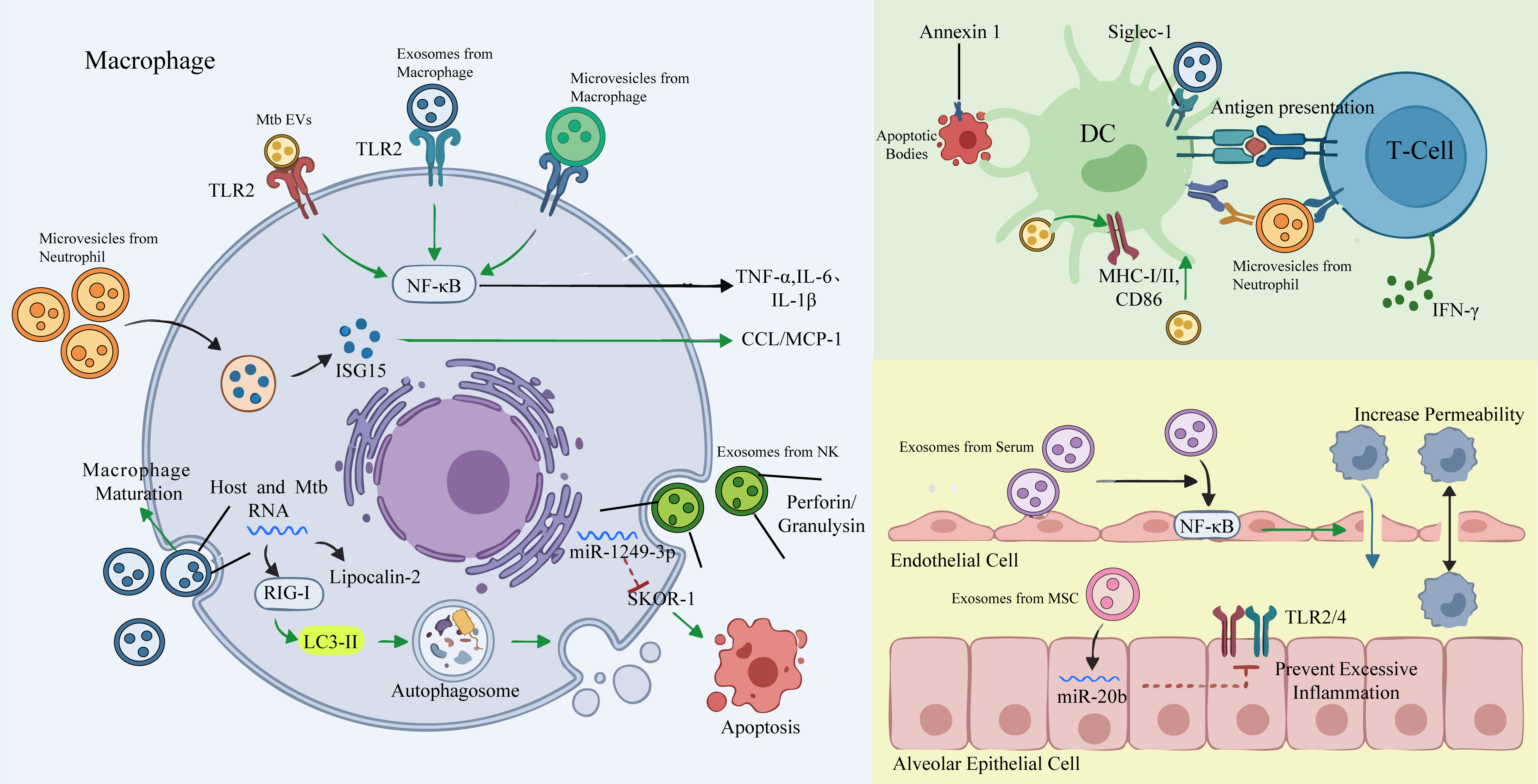
Figure 3. Immune defense regulatory network mediated by Mtb EVs and HC EVs during Mtb infection. Mtb EVs activate macrophages via TLR2 signaling, triggering NF-κB pathway activation, pro-inflammatory cytokine (TNF-α, IL-6, IL-1β) secretion, and chemokine (CCL2, MCP-1) production to recruit immune cells. Host cell EVs, including exosomes and microvesicles, further amplify these defenses: macrophage-derived exosomes deliver host and Mtb RNA, activating the RIG-I pathway to induce autophagy via LC3-II and promote antibacterial protein (LCN2) expression, while NK cell exosomes carry perforin, granulysin, and miR-1249-3p to induce apoptosis of infected macrophages. Neutrophil microvesicles enhance macrophage activation, autophagy, and antigen presentation. DCs capture EVs-associated antigens via Siglec-1, undergo maturation, and present antigens to T cells, driving IFN-γ production and adaptive immunity; apoptotic bodies also deliver antigens to DCs via Annexin A1-mediated uptake. Additionally, endothelial cell permeability is increased by serum exosomes to facilitate immune cell recruitment, while mesenchymal stem cell exosomes deliver miR-20b to alveolar epithelial cells, inhibiting excessive inflammation through TLR2/4-NF-κB signaling suppression. Collectively, these EVs-mediated processes form a dynamic regulatory network that balances effective pathogen clearance with tissue damage limitation. Mtb: Mycobacterium tuberculosis; EVs: extracellular vesicles; Mtb EVs: Mycobacterium tuberculosis-derived extracellular vesicles; HC EVs: host cell-derived extracellular vesicles; DC: dendritic cell; TLR2/4: Toll-like receptors 2 and 4; NF-κB: nuclear factor kappa B; TNF-α: tumor necrosis factor-α; IL-6: interleukin-6; IL-1β: interleukin-1β; CCL2: C-C motif chemokine ligand 2; MCP-1: monocyte chemoattractant protein-1; RNA: ribonucleic acid; RIG-I: retinoic acid-inducible gene I; LC3-II: microtubule-associated protein 1 light chain 3-II; LCN2: lipocalin-2; NK: natural killer cell; miR: microRNA; SKOR1: SKI family transcriptional corepressor 1; Siglec-1: sialic acid-binding immunoglobulin-like lectin 1; MHC-I/II: major histocompatibility complex classes I and II; CD86: cluster of differentiation 86; IFN-γ: interferon-γ; ISG15: interferon-stimulated gene 15; MSC: mesenchymal stem cell.
Regulation of macrophages
Inflammatory effects
Mtb EVs play complex and pivotal roles in host immune regulation. Through their enriched PAMPs such as lipoproteins and lipoglycans, they act as potent TLR2 agonists, directly participating in the activation and modulation of innate immunity[11,15,58,98]. They promote inflammatory responses by activating the TLR2 signaling pathway in macrophages[11]. Yamaguchi et al. further clarified that Mtb EVs, via the binding of TLR2 ligands to receptors in mouse models, activate downstream NF-κB signaling to promote the secretion of inflammatory cytokines such as TNF-α, IL-6, and IL-1β[98]. Beyond pro-inflammatory cytokines, Mtb EVs can also induce the expression of monocyte chemoattractant protein 1 (MCP1/CCL2), effectively driving the migration and chemotaxis of monocytes[98,99]. Collectively, these studies demonstrate that Mtb EVs activate multiple signaling pathways through pattern recognition receptors such as TLR2, not only initiating pro-inflammatory responses but also coordinating the migration and recruitment of immune cells, thereby playing important roles in the immunopathogenesis of TB infection.
In earlier periods, HC EVs (especially exosomes) were considered the main mediators of macrophage inflammatory effects, and numerous studies have provided evidence to support this view. As early as 2007, a study by Bhatnagar et al. revealed the role of exosomes in initiating and amplifying inflammatory
In the immunopathological process of TB, Mtb EVs and HC EVs exert synergistic effects in the initiation, amplification, and regulation of pro-inflammatory responses by mediating intercellular communication. Mtb EVs, enriched in PAMPs such as lipoproteins, mainly activate NF-κB via Toll-like receptor 2-dependent signaling pathways, inducing macrophages to produce key pro-inflammatory cytokines such as TNF-α and IL-6, as well as chemokines such as monocyte chemoattractant protein-1, driving the recruitment of myeloid cells to infection sites. HC EVs, including exosomes and microvesicles, synergistically promote the formation of an inflammatory microenvironment by delivering bacterial components or host-encoded immunomodulatory molecules. Among them, exosomes can carry host molecules such as Hsp70, enhancing the production of pro-inflammatory mediators via the TLR/MyD88 pathway; they can also transport specific microRNAs to limit excessive inflammation by negatively regulating the NFAT5/NF-κB signaling axis. Microvesicles, on the other hand, promote the expression of interferon-stimulated genes by upregulating type I interferon-induced proteins, further enhancing chemokine secretion. Together, Mtb EVs and HC EVs form an elaborate regulatory network that maintains a dynamic balance between establishing effective antibacterial immunity and preventing tissue damage, with their specific molecular mechanisms requiring further clarification.
Immune activation
In the immune process of TB, the precise regulation of various immune cells is crucial for activating effective immune responses. As the primary host cells of Mtb, macrophages play a central role in initiating and controlling anti-TB immunity, and their activation status directly determines the outcome of infection[103]. The host also responds to infection through macrophage death pathways, and macrophage apoptosis has been proven to be an important host defense mechanism[103]. Both Mtb EVs and HC EVs occupy a core position in the regulation of macrophages.
First, we elaborate on the relevant mechanisms of exosomes. On one hand, exosomes lay the foundation for establishing early immune defense lines by promoting the functional maturation and directional recruitment of macrophages. Studies have shown that macrophage-derived exosomes can induce the differentiation of monocytes into macrophages by activating the ERK1/2-MK-2-NF-κB signaling pathway, and upregulate adhesion-related genes to promote cell maturation[47]. Exosomes from immunostimulated macrophages are enriched in endoplasmic reticulum aminopeptidase 1 (ERAP1) and various cytokines, which can significantly enhance the phagocytic activity and NO synthesis capacity of recipient macrophages[104]. On the other hand, exosomes finely regulate cellular autophagy and apoptosis programs by delivering nucleic acid substances. Host and bacterial RNAs carried by host cell-derived exosomes released from Mtb-infected macrophages can be delivered to uninfected macrophages, where these exosomal RNAs can be translated into proteins such as LCN2 to exert antibacterial functions[49]. These exosomes can also directly enhance their ability to clear intracellular bacteria by precisely regulating cellular autophagy and apoptosis programs. Mtb RNA can be delivered into exosomes released by infected macrophages through Mtb’s SecA2 secretion system; after being internalized by recipient macrophages, it can activate RIG-I/MAVS pathway-mediated LC3-associated phagocytosis and enhance phagosome-lysosome fusion[48]. Meanwhile, specific miRNAs precisely target and regulate key autophagic nodes. A study by Xu et al. confirmed that miR-107 delivered by exosomes enhances mitochondrial reactive oxygen species (ROS) production and promotes autophagy by inhibiting Wnt16 to activate the Wnt/β-catenin pathway[105]. In terms of clearing infected cells, natural killer (NK) cell-derived exosomes induce apoptosis by delivering cytotoxic proteins (perforin, granulysin, etc.) to activate the caspase cascade[106]. Although this study confirmed that NK-exo carries typical NK cell cytotoxic proteins such as perforin (PFN), granulysin (GNLY), and granzyme A (Gzm-A), the direct roles and relative contributions of these specific molecules in mediating the aforementioned phenotypes such as pro-apoptosis, anti-inflammation, and ROS elevation require further investigation[106]. The miR-1249-3p carried by NK-exo synchronously activates mitochondrial and death receptor-mediated apoptosis pathways by targeting the SKOR-1 gene, ultimately clearing infected cells through the activation of Caspase-3 and PARP[68]. After being delivered to infected macrophages, miR-1249-3p contained in these cell-derived exosomes directly binds to and inhibits the expression of the SKOR-1 gene, thereby relieving its inhibitory effect on cell apoptosis[68].
The role of microvesicles in the activation and regulation of macrophages has also been gradually revealed. Microvesicles released by Mtb-infected neutrophils significantly activate macrophages by increasing the production of TNF-α, IL-6, and IL-10, as well as upregulating the expression of the co-stimulatory molecule CD86[107]. Alvarez-Jiménez found that macrophages treated with microvesicles from infected neutrophils not only enhance macrophage activation but, more importantly, can significantly upregulate autophagy levels[107]. By inducing the expression of the autophagy marker LC3-II and its colocalization with intracellular bacteria, macrophage clearance of Mtb is enhanced[107].
In the immune regulatory network of TB infection, exosomes and microvesicles collectively form a multi-layered defense system by regulating macrophage differentiation, autophagy, and apoptosis. This provides a vital mechanistic basis for the host to resist pathogens and points out directions for the development of novel immune intervention strategies.
DCs maturation and T-cell activation
DCs serve as the critical bridge connecting innate and adaptive immunity. In the early stage of Mtb infection, DCs undergo maturation by phagocytosing and internalizing pathogens or their antigens, upregulating the expression of MHC class I and II molecules, co-stimulatory molecules, and the chemokine receptor CCR7[108]. Subsequently, DCs migrate to draining lymph nodes and present processed antigen peptides to T cells via MHC molecules, thereby initiating antigen-specific CD4+ and CD8+ T-cell responses - particularly promoting T helper type 1 (Th1) immune responses and producing key cytokines such as IFN-γ to control infection[108]. Recent studies have revealed that this pivotal function of DCs is regulated by Mtb EVs. A study by Jurkoshek et al. demonstrated that after PAMPs enriched in Mtb EVs are recognized and internalized by host DC pattern recognition receptors, downstream pathways are activated to significantly promote DC maturation, characterized by the marked upregulation of MHC-I, MHC-II, and the co-stimulatory molecule CD86. Functional experiments further confirmed that DCs treated with Mtb EVs can efficiently present Mtb antigens[15]. Meanwhile, DCs load processed antigen peptides onto upregulated MHC-II molecules for surface presentation, and mature DCs then migrate to draining lymph nodes to activate CD4+ T cells[15]. Moreover, Mtb EVs themselves possess strong immunogenicity: Prados-Rosales et al. treated mice with Mtb EVs derived from the Mtb H37Rv strain without adjuvants, which induced robust mixed Th1-type cellular and humoral immune responses, resulting in a higher frequency of multifunctional CD4+ T cells[109]. In the context of Mtb infection, exosomes carry Mtb-specific antigens and express host-derived MHC class I/II molecules and co-stimulatory molecules; these exosomes have also been observed to activate DC function[61]. When splenocytes from BCG-infected mice were co-cultured with these exosomes, CD4+ and CD8+ T cells proliferated, upregulated the activation marker CD69, and produced IFN-γ[61]. Nevertheless, the authors noted that exosomes primarily act through a “cross-presentation” mechanism and optimally activate T-cell responses only in the presence of DCs[61]. Current research has identified one pathway for this process: it begins with the specific binding of sialylated glycoconjugates on the exosome surface to the pattern recognition receptor Siglec-1 (CD169) on antigen-presenting cells[110]. When DC Siglec-1 function is deficient, exosome capture efficiency is significantly reduced, leading to delayed TB antigen presentation, impaired activation of CD4+ and CD8+ T cells, decreased IFN-γ production, and ultimately weakened early immune control[110]. In addition to the exosome pathway, infected macrophages can also produce a large number of apoptotic bodies encapsulating various antigens through apoptosis, enabling antigen presentation that bypasses the phagosomal restrictions imposed by Mtb[44]. Subsequent studies revealed that Annexin A1 (ANXA1) expressed on the surface of apoptotic bodies acts as an important phagocytic signal, directly mediating the efficient recognition and internalization of apoptotic bodies by DCs[66]. However, Mtb evades this immune surveillance mechanism by downregulating ANXA1 expression in DCs[66]. Furthermore, microvesicles from infected neutrophils carry a variety of Mtb antigens and PAMPs, which also participate in regulating DC function, significantly upregulating the expression of the co-stimulatory molecule CD80 and the inhibitory molecule PD-L1, and activating Th1 cells as antigen carriers[43]. Interestingly, a study by Ramachandra reported that among the HC EVs subsets (including exosomes and microvesicles) released by macrophages, compared with microvesicles, exosomes - though containing MHC-II molecules - exhibit a weaker ability to directly activate naive T cells and rely on antigen presentation processes to enhance T-cell recognition efficiency[40]. Further analysis showed that microvesicles express higher levels of the co-stimulatory molecule CD86 and ICAM-1 on their surface, which may account for their stronger immune activation capacity[40]. This is well supported by a 2005 study by Segura et al., who pointed out that the presence of ICAM-1 is crucial for HC EVs to exert their immune activation functions[111]. By binding to integrins such as lymphocyte function-associated antigen-1 (LFA-1) on the T-cell surface, ICAM-1 not only enhances adhesion between exosomes and antigen-presenting cells but also promotes stable interactions between T cells and antigen-presenting cells[111].
In the future, when developing exosome-based vaccines, donor cells can be genetically engineered to secrete exosomes co-expressing MHC-II complexes and key co-stimulatory molecules such as CD86 on their membranes, thereby endowing them with the ability to directly and efficiently activate naive T cells. The aforementioned antigen presentation processes mediated by Mtb EVs and HC EVs provide a new perspective for explaining the robust T-cell responses observed in TB granulomas, despite the spatial separation between pathogens and T cells.
Other cells
Although the migration of local tissue-resident cells can supply macrophages to granulomas, a more critical source is the systemic recruitment from the blood circulation - a process traditionally linked to bacterial virulence systems such as RD1/ESX-1 and molecules like 6-kDa early secretory antigenic target (ESAT-6)[112,113], and recently identified to involve HC EVs[114]. HC EVs released by both Mtb-infected and uninfected macrophages enhance the permeability of the endothelial cell monolayer, with those from infected macrophages further promoting macrophage migration across the endothelial barrier[114]. This endothelial activation is mediated primarily via the NF-κB signaling pathway and exhibits a time-dependent pattern, as HC EVs isolated from the serum of infected mice on day 14 display the strongest activating capacity[114]. This aligns with the temporal dynamics of granuloma maturation, suggesting that HC EVs are pivotal factors regulating the timely infiltration of immune cells. Intriguingly, while HC EVs drive endothelial remodeling to support immune cell recruitment - an event inherently linked to pro-inflammatory responses - exosomes also play a counterbalancing role in limiting excessive inflammation, thereby maintaining immune homeostasis. Specifically, mesenchymal stem cell-derived exosomes deliver miR-20b to Mtb-infected type II alveolar epithelial cells[115]. This miR-20b directly targets the nuclear factor NFAT5, inhibiting TLR2/TLR4 dimer formation and downregulating the downstream MyD88/NF-κB signaling pathway, which not only reduces the secretion of pro-inflammatory cytokines and chemokine CCL5 but also enhances the survival of infected cells[115]. This feedback mechanism complements the pro-inflammatory effects of HC EVs, ensuring the localization of inflammatory responses and preventing systemic tissue damage, which serves as a key strategy for the host to maintain immune homeostasis during chronic infection.
Discussion: allies or foes? Re-evaluating the immunological functions of Mtb EVs and HC EVs
Across existing studies, a core and fascinating scientific paradox emerges: why do EVs of the same origin both mediate immunosuppression and effectively activate immune defense during Mtb infection? This seemingly contradictory phenomenon is not an experimental artifact but rather a profound reflection of the functional plasticity and context dependency of Mtb EVs and HC EVs as biological information carriers. Their ultimate immune effects represent a sophisticated balance collectively determined by multiple factors, including vesicle origin, cargo composition, host immune microenvironment, and spatiotemporal exposure characteristics.
Notably, functional attribution within the HC EVs population requires refined subtyping. HC EVs constitute an umbrella term encompassing exosomes, microvesicles, and apoptotic bodies, each with distinct biophysical properties, biogenesis pathways, and cargo repertoires. However, most earlier studies analyzed total mixed EVs populations without precise subtype resolution, which may cause ambiguous or generalized functional interpretation. In this review, we specify exact subtypes whenever original studies adopt definitive isolation protocols, marker validation, or electron microscopic identification. For evidence obtained from unseparated total EVs, we retain the collective term “HC EVs” and explicitly clarify the mixed nature of vesicle populations and corresponding methodological limitations. Readers are encouraged to consult Table 2 for a side-by-side comparison of the origins, cargo, and functions of each HC EVs subtype discussed throughout this review.
First, perhaps the most striking example of this functional duality is the TLR2 signaling cascade. Mtb EVs are enriched with potent TLR2 agonists, including mycobacterial lipoproteins and lipoglycans[11,15,58]. Intriguingly, these identical vesicle-contained ligands drive opposing immunological phenotypes: they facilitate DC maturation and pro-inflammatory cytokine production, yet simultaneously suppress IFN-γ-induced MHC-II expression and trigger T-cell anergy. Emerging evidence resolves this apparent contradiction through a multilayered regulatory framework, in which the final immune outcome is governed by three interconnected determinants: dose dependency, spatiotemporal specificity, and infection-stage dependency. Dose-dependency: Functional analyses of Mtb EVs confirm a dose-dependent TLR2 immune switch. Low concentrations of Mtb EVs and transient TLR2 signaling dominantly activate NF-κB, induce protective TNF-α, IL-6 and IL-1β release, and drive DC cell maturation[11,15,98]. Conversely, prolonged, high-level TLR2 stimulation by abundant Mtb EVs triggers a negative feedback loop mediated by IL-10 and SOCS1, which suppresses the MHC-II master transcription factor CIITA, drastically reducing macrophage antigen presentation capacity[11,15,58,75]. This threshold-dependent switch enables Mtb to fine-tune host immunity dynamically: low-level EVs secretion at early infection stages supports moderate protective immunity to confine bacterial dissemination, whereas sustained high EVs release during persistent infection shifts the immune balance toward tolerance[11,15]. Spatiotemporal specificity: EVs-mediated immune regulation is highly compartment-dependent. In vitro stimulation of macrophages with Mtb EVs predominantly induces robust inflammatory responses[11]. However, when circulating Mtb EVs deliver LAM and lipoglycans to CD4+ T cells, they interfere with the phosphorylation of key TCR signaling molecules (Lck, ZAP-70, LAT), thereby suppressing IL-2 secretion and triggering T cell anergy[58,76,77]. Furthermore, local cytokine milieus profoundly reshape Mtb EVs function: within granuloma structures, Mtb EVs accumulated in necrotic cores promote host cell necrosis and bacterial dissemination, whereas peripheral Mtb EVs sustain T cell exhaustion and local immune suppression[11,92]. This spatial heterogeneity allows similar Mtb EVs cargo to exert divergent effects across distinct anatomical and immunological microenvironments. Infection-stage dependence: The immunological impact of Mtb EVs and HC EVs is not static but evolves over the course of infection. During the acute phase, Mtb EVs and HC EVs predominantly activate innate and adaptive immunity, aiding bacterial containment through the induction of pro-inflammatory cytokines, DC maturation, and effector T-cell responses. However, during chronic or latent infection, the cargo composition of released EVs undergoes notable changes - including enrichment of immunosuppressive miRNAs such as miR-146a-5p and miR-20b-5p, and reduced expression of immunogenic antigenic proteins - and the host immune environment becomes progressively more tolerogenic, favoring immune evasion[59,63]. At the same time, the biogenesis of Mtb EVs itself is upregulated under stress conditions encountered during chronic infection, such as iron limitation or antibiotic pressure, leading to enhanced secretion of EVs enriched with immunosuppressive or pro-survival cargo[27,37]. Conversely, reactivation of latent infection may involve a reversal of this balance, possibly driven by shifts in EVs cargo sorting or secretion rates in response to host immune deterioration or local inflammatory triggers. This stage-dependent dynamics suggests that EVs function as a “rheostat” that continuously adjusts the host-pathogen equilibrium in response to changing conditions.
Collectively, these three regulatory layers - dose, space, and time - argue against a binary “pro-inflammatory vs. anti-inflammatory” classification of EVs functions. Instead, Mtb EVs and HC EVs should be viewed as a multifunctional immune regulatory platform, or a “rheostat”, whose net output is the integrated sum of multiple contextual signals. The ultimate outcome - pathogen clearance, establishment of latent infection, or progression to active disease - depends on the precise balance of these signals at any given moment. This conceptual framework also explains why the same EVs preparation can yield divergent results in different experimental systems or patient cohorts, as variations in dosage, delivery route, host genetic background, and co-existing immune conditions can shift the rheostat in opposing directions.
In terms of origin, Mtb EVs and HC EVs exhibit distinct but complementary roles in immune regulation. Mtb restricts the excessive release of Mtb EVs through factors such as VirR and PstA1, which may suggest that uncontrolled Mtb EVs release is detrimental to bacterial survival - possibly because it exposes too many antigenic stimuli to the host immune system[21,25]. Meanwhile, Mtb EVs possess dual functions of initiating immune defense and eliminating surrounding immune cells. Studies have reported that in the early stage of infection, low-dose Mtb EVs may trigger protective immunity via the TLR2/NF-κB pathway, whereas high-dose exposure during persistent infection may lead to immune tolerance[11]. Future research can explain the functional duality of Mtb EVs by precisely dissecting the threshold dose for the functional switch from “immune initiation” to “immune tolerance”. At the same time, the cargo of HC EVs from different sources (e.g., macrophage culture supernatants vs. patient serum) varies considerably[116,117]. HC EVs derived from
Despite the conceptual progress outlined above, important gaps remain. First, most mechanistic studies on dose-dependency have been performed using purified Mtb EVs in vitro, and it is unclear whether the threshold doses identified in these systems are physiologically relevant in vivo. Second, the spatiotemporal distribution of Mtb EVs within different granuloma compartments has not been directly visualized, nor has it been correlated with local immune phenotypes. Third, longitudinal studies tracking EVs cargo composition across the entire infection continuum - from acute infection through latency to reactivation - are largely lacking. Addressing these gaps will require the development of advanced imaging techniques to track EVs dynamics in vivo, quantitative proteomic and transcriptomic profiling of EVs cargo at sequential time points, and functional validation using in vivo models that allow controlled manipulation of EVs dose and timing. Such efforts will not only deepen our understanding of the EVs-mediated immune switch but also open new avenues for therapeutic intervention by targeting specific nodes of this regulatory network.
APPLICATIONS OF Mtb EVs AND HC EVs IN TB PREVENTION AND CONTROL
Mtb EVs/HC EVs as vaccines
Since the development of BCG, this vaccine has been in use for nearly a century. However, BCG exhibits limited and inconsistent protective efficacy against adult pulmonary TB and cannot effectively prevent the recurrence of latent infections, highlighting the urgent need for developing novel and highly effective vaccines[1]. In recent years, Mtb EVs and HC EVs have emerged as a cutting-edge and highly promising direction in the field of new vaccine development, owing to their inherent enrichment of immunogenic antigens and ability to efficiently activate the host immune system. In EVs-based vaccine development, multiple strategies have evolved, roughly categorized into two technical routes: natural EVs optimization and engineering modification. The major translational applications of Mtb EVs and HC EVs in TB vaccines, diagnosis, and therapy are summarized in Figure 4.
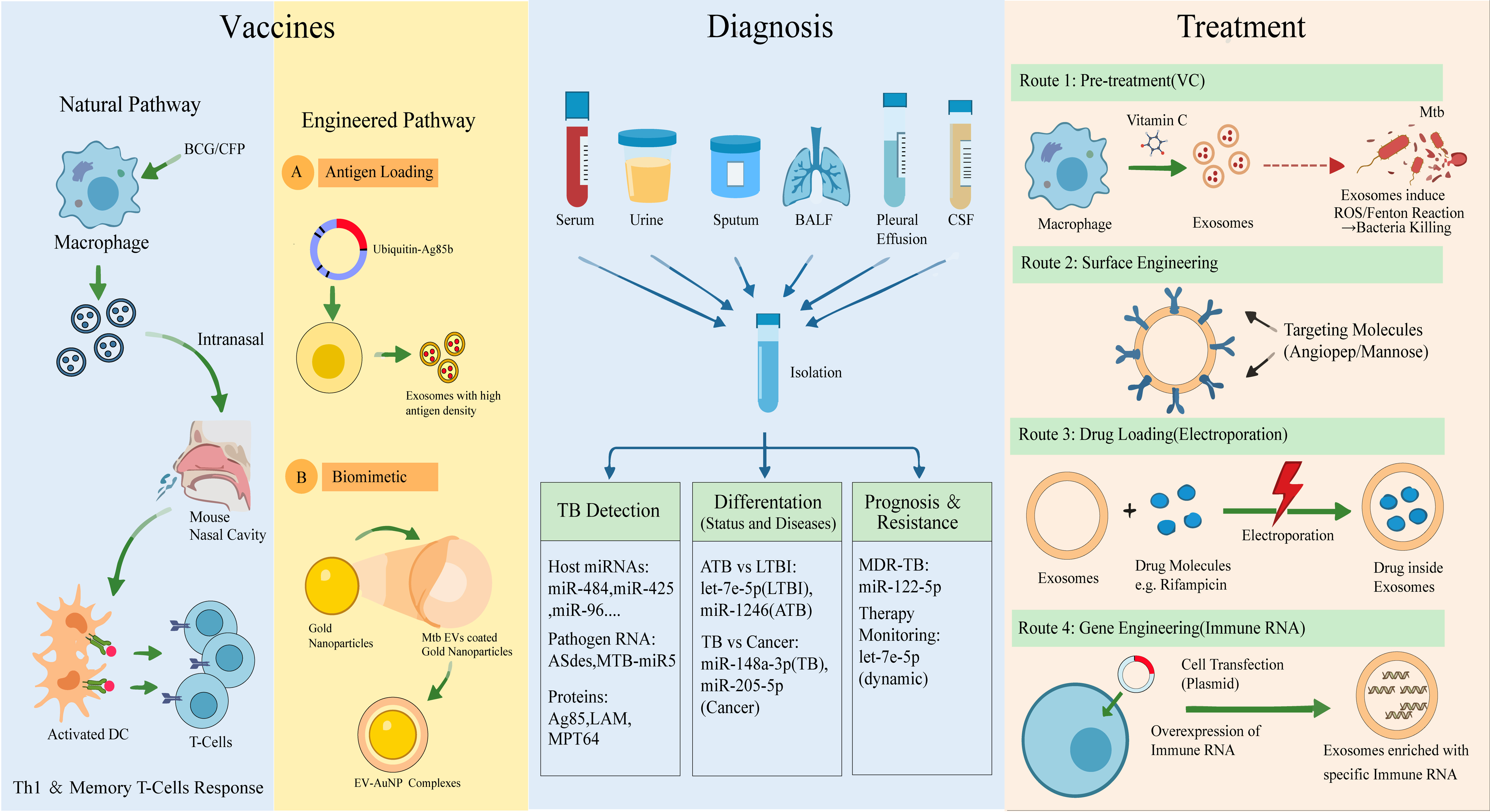
Figure 4. Translational applications of Mtb EVs and HC EVs in TB vaccines, diagnosis, and therapy. For vaccine development, both natural and engineered EVs strategies are employed: natural EVs from BCG-infected macrophages induce protective Th1 and memory T-cell responses, while engineered approaches - including ubiquitin-fusion-mediated antigen enrichment, biomimetic nanocarrier conjugation, and multi-epitope design - enhance antigen stability, immune activation, and scalability. As diagnostic tools, EVs serve as stable carriers of multi-omic biomarkers (host miRNAs, lncRNAs, Mtb-specific nucleic acids, proteins such as Ag85B, LAM, MPT64, and lipids) in accessible bodily fluids, enabling TB detection, differentiation of active from latent infection, discrimination between TB and other pulmonary diseases (e.g., lung cancer), and monitoring of treatment efficacy and drug resistance. For targeted therapy, EVs leverage their biocompatibility, low immunogenicity, and barrier-crossing ability as drug delivery vectors, with strategies including endogenous loading via parental cell engineering, exogenous loading of anti-TB drugs or immunomodulatory molecules, and surface modification to enhance targeting to infected cells, collectively improving therapeutic efficacy while reducing systemic toxicity. These EVs-based strategies collectively offer versatile translational solutions for advancing TB prevention, diagnosis, and treatment. Mtb: Mycobacterium tuberculosis; EVs: extracellular vesicles; Mtb EVs: Mycobacterium tuberculosis-derived extracellular vesicles; HC EVs: host cell-derived extracellular vesicles; TB: tuberculosis; BCG: Bacillus Calmette-Guérin; CFP: culture filtrate proteins; Th1: T helper type 1; DC: dendritic cell; RNA: ribonucleic acid; miRNA: microRNA; lncRNA: long non-coding RNA; Ag85B: antigen 85B; LAM: lipoarabinomannan; MPT64: Mycobacterium tuberculosis protein 64; AuNP: gold nanoparticle; EV-AuNP: extracellular vesicle-coated gold nanoparticle; BALF: bronchoalveolar lavage fluid; CSF: cerebrospinal fluid; ATB: active tuberculosis; LTBI: latent tuberculosis infection; MDR-TB: multidrug-resistant tuberculosis; ROS: reactive oxygen species; VC: vitamin C.
The research and development of exosome-based TB vaccines have progressed from natural extraction to engineered design. An effective TB vaccine needs to induce IFN-γ-producing T cells and long-lasting memory T cells. Early studies confirmed that natural exosomes possess significant vaccine potential[38]. For instance, exosomes derived from BCG-infected macrophages, when administered intranasally, can induce antigen-specific memory T cell responses and directly promote DC maturation, demonstrating their dual properties of antigenicity and adjuvanticity[61]. Subsequent studies further revealed that exosomes obtained by treating macrophages with only Mtb culture filtrate proteins (CFP) are also enriched in various Mtb proteins and can efficiently activate macrophages and DCs[32]. Notably, in mouse models, exosomes treated with CFP not only induce robust Th1-type immune responses with protective efficacy comparable to BCG but also significantly enhance immune memory and protective potency when used as a booster immunization following BCG priming[119]. However, the compositional complexity of Mtb EVs and HC EVs, coupled with challenges in large-scale production, has limited their clinical application. To address this, researchers have developed various engineering and biomimetic strategies to achieve precise regulation of vaccine components. Sharma adopted an immunoinformatics approach: by anchoring immunogenic proteins enriched in exosomes, integrating their B-cell and T-cell epitopes, and fusing a TLR4 agonist as an adjuvant, a multi-epitope vaccine capable of eliciting strong immune responses was constructed[120]. In silico simulations confirmed that this candidate molecule can trigger robust immune responses, theoretically validating the feasibility and great potential of developing novel vaccines targeting exosomal proteins[120]. Cheng and Schorey fused Ag85B and ESAT-6 to the C-terminus of ubiquitin and quantified exosomal loading in stably transfected HEK293 cells[121]. Compared to the unmodified antigens, ubiquitin fusion increased antigen encapsulation by approximately 10-fold, as determined by Western blot densitometry and enzyme-linked immunosorbent assay (ELISA). This marked enrichment directly enhanced immunogenicity, with intranasal immunization eliciting stronger antigen-specific IFN-γ+ T-cell responses in mice[121]. Building on this, Ji et al. further expanded the concept of multi-antigen synergistic immunity. They screened three dominant antigens (Ag85B, ESAT-6, and Rv0580c) from pathogen-derived HC EVs, constructed a chimeric antigen (CA), and enabled its efficient loading into HC EVs through cell engineering. Compared with free antigens, this EVs-delivered chimeric antigen system can more effectively elicit Th1-type immune responses and enhance cytotoxic T lymphocyte activity[122]. The George team coated Mycobacterium smegmatis EVs on the surface of 50-nanometer gold nanoparticles (AuNPs), combining the antigenic diversity of M. smegmatis EVs with the stability and size controllability of AuNPs to construct M. smegmatis EVs-AuNP complexes[123]. These complexes significantly improve the uptake efficiency by macrophages and DCs and promote the expression of pro-inflammatory cytokines[123]. However, the use of non-degradable gold nanoparticles may pose long-term safety concerns, and degradable materials could be employed in future studies. Montalvo-Quiros’ research aimed to mimic the immune activation function of Mtb EVs, developing a biomimetic vaccine platform based on mesoporous silica nanoparticles. By covalently conjugating three Mtb proteins (Ag85B, LprG, and LprA), a biocompatible nanovaccine was constructed that can induce macrophages to produce TNF and IL-10 in vitro[124]. Despite the diverse technical routes, these studies collectively reveal a core principle: a successful Mtb EVs/HC EVs vaccine must possess both proper antigen presentation and effective adjuvant signals. The future development direction lies in integrating the advantages of various strategies - utilizing artificial carriers to ensure the stability of large-scale production, simulating the immune activation characteristics of natural EVs through biomimetic design, and ultimately achieving dual breakthroughs in the safety and efficacy of next-generation TB vaccines.
EVs and biomarkers: diagnosis and clinical implications in TB
Although existing diagnostic methods (such as sputum smear microscopy, GeneXpert MTB/RIF, and Mtb culture) are widely used in clinical practice, they still have limitations in sensitivity, specificity, or timeliness - particularly poor performance in extrapulmonary TB, pediatric tuberculosis, and HIV coinfected patients[125]. Based on this context, numerous studies are currently exploring Advanced materials for TB diagnostics, such as aggregation-induced emission luminogens (AIEgens). Due to their excellent optical properties, AIEgens have been developed for rapid detection and imaging of various infectious pathogens, demonstrating significant potential in point-of-care testing[126,127]. However, achieving highly sensitive and specific detection for TB, particularly its diagnostically challenging forms, still requires the identification of more optimal biomarker carriers and detection targets. As an emerging biomarker carrier, EVs have garnered significant attention in the field of liquid biopsy with their potential as diagnostic biomarkers based primarily on three key aspects: first, accessibility - EVs stably exist in easily obtainable bodily fluids such as blood and urine, providing a sample foundation for non-invasive or minimally invasive diagnosis; second, stability - Their lipid bilayer structure acts as a robust barrier, effectively protecting internal biomolecules such as proteins, nucleic acids, and lipids from degradation, thereby greatly enhancing detection reliability; third, specificity - During biogenesis, EVs actively and selectively load specific “cargo” from their parent cells. This enables them to dynamically and accurately capture the presence of pathogens as well as the host’s specific immune responses during infection[128]. Additionally, the membrane protein profile of EVs allows for tracing their cellular origins[128]. Based on these advantages, researchers are thoroughly exploring the diagnostic potential of EVs in TB.
EVs as diagnostic biomarkers for TB
Nucleic acids
With the implementation of multiple independent studies, a large number of nucleic acids derived from hosts or pathogens have been identified as potential diagnostic biomarkers for TB. Host miRNAs are the most extensively studied biomarkers to date. Multiple independent studies have confirmed that specific miRNAs in the blood exosomes of ATB patients show a significant upregulation trend. For instance, the combined detection of miR-484, miR-425, and miR-96 exhibits superior diagnostic efficacy [area under the curve (AUC) = 0.78] compared to single indicators, demonstrating the advantage of multi-marker combined detection[129]. As research advances, more high-performance single miRNA biomarkers have been successively discovered, such as miR-185-5p (AUC = 0.750) and miR-766-3p (AUC = 0.896), with the latter approaching clinical application standards[60,130]. Diagnostic strategies are evolving from single indicators to multi-dimensional integration. Hu et al. found that six exosomal miRNAs (miR-20a, miR-20b, miR-26a, miR-106a, miR-191, and miR-486) are significantly upregulated in the plasma of TB patients; when combined with electronic health records, this panel exhibits excellent performance in distinguishing pulmonary TB from tuberculous meningitis[131]. Nevertheless, it is important to emphasize that while multi-miRNA panels demonstrate robust statistical associations with disease status, these correlations do not automatically equate to functional causality. The mechanistic interpretations of these miRNA signatures - particularly whether they reflect active bacterial manipulation of host pathways or merely represent general host cellular stress responses secondary to infection - remain largely unresolved. Extensive experimental validation, including loss- and gain-of-function studies in relevant cell types and longitudinal animal models, is still required to determine the functional significance of these EVs-encapsulated molecules. The transition from promising correlative biomarkers to established functional indicators therefore represents a critical gap that future investigations must address. Meanwhile, research focus has expanded from miRNAs to other non-coding RNAs (ncRNAs). Through bioinformatics analysis and experimental validation, Fang et al. identified four significantly downregulated long non-coding RNAs (lncRNAs) (NONHSAT101518.2, NONHSAT067134.2, NONHSAT148822.1, and NONHSAT079957.2) in the serum exosomes of ATB patients, which display high diagnostic value[132]. In addition to host nucleic acids, direct detection of Mtb-derived RNA fragments offers theoretically high specificity. Researchers identified Mtb-specific small RNAs (sRNAs) (ASdes and MTB-miR5) in serum EVs (isolated by size-exclusion chromatography, yielding a mixed population enriched in exosomes and small microvesicles); these biomarkers are exclusively detected in pulmonary tuberculosis patients, including those initially smear- and culture-negative but later converted positive, indicating promising potential for early diagnosis[133]. Similarly, Mtb ribosomal RNA fragments (16S-L1 and 16S-L2) identified from plasma exosomes are significantly upregulated in TB patients, with a combined diagnostic AUC of 0.7197[134].
Proteins and lipids
Mtb-specific proteins and lipids serve as direct evidence of its presence. Targeting these unique molecules enables precise diagnosis of the pathogen. For example, the cell wall lipid LAM has become a key basis for urine testing in HIV-infected individuals[135]; meanwhile, accurate quantification of specific antigen peptides such as the Ag85 complex and ESAT-6/CFP-10 in patients’ blood or urine using high-sensitivity mass spectrometry has also demonstrated excellent diagnostic value[136]. Mtb infection triggers intense immune and inflammatory responses in the human body, which are reflected in changes in protein and lipid metabolism.
As enriched carriers of disease-related information, exosomes provide a highly valuable biomarker library for TB diagnosis, with value manifested at two levels: First, at the host response level, proteomic analysis can screen for host proteins closely associated with infection status. For instance, Zhang performed proteomic profiling of serum exosomes from ATB patients and identified 123 differentially expressed proteins, among which changes in LBP (increased), CD36 (decreased), and MHC-I (decreased) are diagnostically relevant[117]. Wang et al. revealed differential characteristics of infections caused by different mycobacteria: by comparing the plasma exosomal protein profiles of patients infected with M. abscessus, M. intracellulare, and Mtb, they identified protein panels such as C1R/C4B/C9 and SERPINA5/SERPING1 that can effectively distinguish non-tuberculous mycobacterial infections from Mtb infections[137]. Second, more directly, unique components of Mtb itself have been detected in exosomes. Jiang et al. measured three Mtb antigens (MPT64, Ag85A, and CFP-10) in the plasma exosomes of ATB patients, LTB patients, recovered individuals, and healthy controls. They found that these three antigens are specifically enriched in the exosomes of ATB and LTB patients, confirming their diagnostic potential[138]. VAMP5 in HC EVs is significantly upregulated in TB patients and downregulated after treatment, indicating its potential as a TB biomarker[51]. Additionally, the exosome-enriched Hsp16.3 protein is stably present in the plasma of ATB patients but not detected in latent infection or healthy populations, suggesting its value as a highly specific blood biomarker[139]. Notably, this exosome-based detection technology significantly enhances the performance of traditional biomarkers. Dahiya et al.’s study demonstrated that detecting LAM in urine exosomes using high-sensitivity immuno-polymerase chain reaction (I-PCR) exhibits superior diagnostic efficacy compared to traditional ELISA, opening new avenues for non-invasive diagnosis[140].
In the future, organically combining these biomarkers from different levels to construct a multi-dimensional diagnostic model will be the critical direction for achieving ultra-early and high-precision diagnosis of TB.
HC EVs as diagnostic biomarkers for TB: subtype differentiation and cross-disease discrimination
Distinguishing ATB from LTB
Based on WHO guidelines, the accurate distinction between ATB and LTB is a core decision point in TB prevention and control, directly determining drastically different clinical management strategies[1]. This differentiation is crucial for preventing missed diagnoses of active cases that delay treatment, avoiding drug resistance risks caused by single-drug prophylaxis, and optimizing the allocation of public health resources. Emerging EVs research provides a novel perspective to address this issue; by decoding the pathogen- and host-specific molecular information carried by EVs, it is expected to establish a more precise diagnostic pathway in the future.
At the RNA level, Lyu et al. compared the miRNA profiles in serum exosomes from healthy individuals, LTB patients, and ATB patients via small RNA sequencing. They identified 250 differentially expressed miRNAs (including 130 specifically expressed ones), and validated 4 LTB-specific miRNAs (hsa-let-7e-5p, hsa-let-7d-5p, hsa-miR-450a-5p, and hsa-miR-140-5p) as well as 5 TB-specific miRNAs (hsa-miR-1246, hsa-miR-2110, hsa-miR-370-3p, hsa-miR-28-3p, and hsa-miR-193b-5p)[141]. These differentially expressed miRNAs may participate in disease progression by regulating host signaling pathways, providing a molecular basis for distinguishing infection status. Detection of Mtb protein components carried by HC EVs offers more direct evidence. Mehaffy et al. detected multiple Mtb peptides in serum HC EVs (isolated by ultracentrifugation, representing a mixed vesicle population) from individuals with LTB infection using multiple reaction monitoring mass spectrometry (MRM-MS). The results showed that at least one Mtb peptide was detected in 95% of individuals, with the peptide SVF from glutamine synthetase (GlnA1) identified in 82% of samples[142]. A study by the Zheng team in a non-human primate model found that the co-localization pattern of LAM and LprG on the surface of HC EVs has discriminatory value: LTB only shows elevated LprG signals, while ATB exhibits significant enhancement of both LAM and LprG signals[143]. This indicates that their co-localization can serve as a key biomarker panel for distinguishing active disease from latent infection. Finally, at the metabolic response level, host lipid reprogramming also provides important information. Lyu’s team’s lipidomic study identified 7 lipid biomarkers with diagnostic value, among which the free fatty acid FFA (20:1) performed particularly well in distinguishing ATB from LTB, with an AUC of 0.952, 100% specificity, and 88.9% sensitivity[144].
Collectively, these studies demonstrate that HC EVs integrate multi-dimensional information such as pathogen proteins, host miRNAs, and metabolic lipids to construct a molecular diagnostic system capable of accurately distinguishing TB infection status, providing a new technical pathway for optimizing TB clinical management.
Localization, identification, and classification of TB
The accurate diagnosis of different types of TB has long been a clinical challenge, and exosomal miRNAs have shown great potential as promising biomarkers. Multiple studies have revealed their specific expression profiles in distinguishing TB types: Kushwaha et al.’s study on the Indian population found distinct miRNA expression profiles in serum exosomes of patients with HIV-tuberculosis coinfection (HIV-TB), extrapulmonary tuberculosis, and pulmonary tuberculosis alone (PTB). Among them, hsa-let-7i-5p, hsa-miR-24-3p, and hsa-miR-92a-3p were significantly differentially expressed in HIV-TB coinfected patients; hsa-miR-20a-5p, hsa-let-7e-5p, and hsa-miR-26a-5p were specifically downregulated in extrapulmonary tuberculosis (EPTB) patients; while hsa-miR-21-5p, hsa-miR-19a-3p, hsa-miR-19b-3p, and hsa-miR-146a-5p were significantly expressed in PTB patients[145]. Sun et al. identified 62 differentially expressed miRNAs in macrophage exosomes from spinal tuberculosis (STB) patients via high-throughput sequencing, including 28 upregulated and 34 downregulated ones. Further validation by quantitative reverse transcription PCR (qRT-PCR) showed that miR-125b-5p was significantly upregulated in the STB group, with an area under the receiver operating characteristic curve (AUC) of 0.881.[146]. These studies collectively confirm that HC EVs miRNAs, as highly promising biomarkers, hold tremendous exploration value and application potential in achieving precise classification diagnosis of TB.
Distinguishing TB from other pulmonary diseases (e.g., lung cancer)
In clinical practice, the differential diagnosis between TB and lung cancer is of great significance. Due to their potential coexistence or sequential occurrence, coupled with overlapping imaging manifestations, the differential diagnosis and subsequent treatment plans face significant challenges[147]. In this context, identifying novel diagnostic biomarkers capable of effectively distinguishing these two diseases is crucial for improving the accuracy of clinical decision-making.
Molecular analysis based on HC EVs provides a new solution. Studies have shown that the miRNA profiles of HC EVs exhibit significant specificity under different disease states. Guio et al. found via serum exosome miRNA sequencing that miR-125a-5p is perturbed in both TB and lung cancer, while miR-20a-5p and miR-584 are specifically upregulated in LTB and ATB, respectively[89]. Research has also shifted to pleural effusion, a key sample, to achieve precise differentiation between TB and other lesions. The miRNA characteristics of HC EVs in pleural effusion also show discriminatory value: the Zhang team identified 99 upregulated and 91 downregulated miRNAs, among which hsa-miR-200b-3p and hsa-miR-182-5p are highly expressed in malignant pleural effusions, suggesting these exosomal miRNAs may serve as potential biomarkers for distinguishing malignant pleural effusions (MPE) from tuberculous pleural effusions (TPE)[148]; the Wang team found 9 miRNAs (including miR-205-5p, miR-483-5p, miR-375, and members of the miR-200 family) enriched in pleural effusions from lung adenocarcinoma patients, and another 3 miRNAs (miR-148a-3p, miR-451a, miR-150-5p) that can distinguish tuberculous from benign pleural effusions, filling the diagnostic gap of only distinguishing cancerous from non-cancerous pleural
HC EVs show good adaptability in multi-disease differentiation scenarios, and their cargo profiles can effectively reflect disease-specific host responses and pathogen information. However, as noted in the mechanistic discussions in Sections “Roles of HC EVs in creating a favorable intracellular survival environment” and “Potential involvement of Mtb EVs and HC EVs in Mtb drug resistance”, many of the miRNA and protein signatures associated with TB diagnosis currently rest on correlative evidence. While these profiles exhibit promising statistical power in cross-sectional studies, extensive functional and experimental verification is still required to determine whether the observed changes represent active bacterial manipulation of host pathways or are merely secondary manifestations of general host cellular stress. This distinction is not merely academic: it has direct implications for the biological interpretability of diagnostic biomarkers and their potential utility as therapeutic targets. We therefore urge the field to complement continued biomarker discovery with rigorous mechanistic studies that causally link EVs cargo alterations to specific pathogen-host interactions, thereby transforming statistical associations into functionally validated indicators.
Biomarkers for therapeutic efficacy monitoring and prognosis assessment
Prognosis assessment and therapeutic efficacy monitoring of TB are critical links in disease control, which are essential for identifying high-risk individuals with progression potential, accurately evaluating the effectiveness of treatment regimens, and predicting recurrence. However, existing biomarkers still rely on complex technologies and have limitations in application among children, HIV-coinfected patients, and extrapulmonary TB patients, highlighting an urgent need for developing novel non-invasive, high-precision, and easily scalable monitoring tools[152].
In terms of therapeutic monitoring for drug-resistant TB, Carranza et al. identified miR-let-7e-5p with temporal expression characteristics in serum exosomes. The expression level of miR-let-7e-5p undergoes regular changes during treatment progression, especially at and after the time point of sputum culture conversion to negative, demonstrating its potential as a dynamic indicator for therapeutic efficacy monitoring[153]. Meanwhile, studies detected Mtb-specific RNA sequences such as 30 kDa and 5KST in exosomes of 33% of multidrug-resistant tuberculosis (MDR-TB) patients even after the intensive phase of treatment[153]. The authors proposed combining host-derived miR-let-7e-5p with pathogen-derived RNA as a “dual biomarker”: abnormal expression of host miRNA coupled with persistent positivity of pathogen RNA strongly indicates the risk of treatment failure or future recurrence[153].Another study compared the plasma exosomal miRNA expression profiles among MDR-TB patients, drug-susceptible tuberculosis (DS-TB) patients, and healthy controls, identifying 7 miRNAs with significantly differential expression in MDR-TB patients. Further focus was placed on three key molecules: hsa-miR-122-5p_R-1, hsa-miR-23b-3p_R+1, and hsa-miR-15a-5p_R-1[154]. Exosomal miR-122-5p, KNG1, and C3 also exhibit diagnostic value in drug-resistant TB[96]. Beyond nucleic acids and proteins, the exosomal lipidome has emerged as a sensitive indicator of treatment response. Exosomal lipid compositions (e.g., sphingomyelin, triglycerides) undergo significant changes after effective treatment and tend to return to normal, indicating their potential as biomarkers for therapeutic monitoring[42].In LTB, the expression levels of complement component 4 binding protein alpha chain (C4BPA) and S100 calcium-binding protein A9 (S100A9) in plasma exosomes of patients with effective LTB treatment are significantly downregulated. ROC analysis showed that the combined AUC of these two molecules is 0.78, suggesting their potential as biomarkers for evaluating the effectiveness of LTB preventive treatment[155]. Overall, by integrating multi-dimensional signals from both the host and pathogen, exosomes provide an unprecedented systematic monitoring perspective for TB therapeutic efficacy assessment and prognostic management.
Influencing factors, limitations, and clinical translation hurdles
In the development of HC EVs-based diagnostic tools, attention must be paid to the impact of population and regional specificity on diagnostic outcomes. Studies by Kushwaha et al. on the Indian population and Guio et al. on the Peruvian population observed certain regional characteristics[89,145]; miRNA expression profiles, as well as protein and lipid compositions, may be influenced by population genetic backgrounds and regional environmental factors. Whether biomarkers that perform excellently in one cohort will experience diagnostic efficacy drift when generalized to other regions remains unproven. Future research must emphasize external validation in large-scale, multi-center cohorts across different endemic areas to avoid diagnostic bias and ensure the universality and stability of the tools.
The influence of the host’s immune status on diagnosis also deserves attention. Jin’s team evaluated the diagnostic efficacy of six exosomal miRNAs (miR-20a, miR-20b, miR-26a, miR-106a, miR-191, and miR-486) in individuals with HIV-TB coinfection. They found that these miRNAs were significantly upregulated in the plasma of HIV-TB coinfected patients, with more pronounced expression changes in extrapulmonary TB[118]. Subsequent subgroup analysis revealed that the diagnostic performance of these miRNAs was superior in patients with a CD4+ T-cell count ≥ 200 cells/μL, suggesting that the host’s immune status may affect exosomal miRNA expression levels[118]. This finding serves as an important reminder: for populations with comorbidities (e.g., HIV-infected individuals, diabetic patients, or the elderly), HC EVs-based diagnostic models may require specific calibration or the development of independent biomarker panels. This not only relates to diagnostic accuracy but also directly impacts the interpretation of therapeutic efficacy monitoring results. Notably, antigens derived from BCG vaccines or other mycobacteria may cross-react with HC EVs proteins, affecting the specificity of serological tests[23,156]. Whether such cross-reactivity will challenge the specificity of HC EVs-based serological assays or antigen capture experiments, leading to false positives in latent infection or misdiagnosis of ATB, requires further investigation. Additionally, most existing studies focus on cross-sectional comparisons, resulting in limited understanding of the dynamic evolutionary patterns of HC EVs throughout the entire course of TB infection. Comprehensive analysis of the dynamic changes in HC EVs from various sample sources may address many current challenges, such as whether HC EVs exhibit characteristic changes prior to TB recurrence, whether the dynamic changes of HC EVs biomarkers after treatment initiation can predict the final outcome, and whether EVs characteristics after cure can predict recurrence risk.
Beyond these population- and disease-specific considerations, the clinical translation of HC EVs-based TB applications faces profound systemic hurdles rooted in methodological heterogeneity and technical standardization gaps. A fundamental issue lies in the ambiguous subtype attribution that pervades many diagnostic studies: although serum or plasma “exosomal” miRNA panels have shown promising statistical correlations with disease status, the isolation methods commonly employed - typically differential ultracentrifugation or precipitation-based kits - do not achieve complete separation of exosomes from microvesicles and apoptotic bodies. Consequently, markers frequently reported as “exosomal” may actually reflect contributions from mixed vesicle populations, a caveat that does not negate their diagnostic value but critically complicates mechanistic interpretation and cross-study comparability. On a broader level, the lack of standardized isolation and characterization protocols across laboratories represents an equally formidable bottleneck. Different research groups employ vastly divergent methods - including ultracentrifugation, density gradient centrifugation, size-exclusion chromatography, precipitation-based kits, and microfluidic devices - each yielding EVs with substantially different purity, recovery rates, and functional activity. This methodological heterogeneity severely compromises cross-study reproducibility and has been documented to produce divergent conclusions regarding EVs cargo composition and immunomodulatory activity. Although the MISEV2023 guidelines provide a comprehensive minimal information framework encompassing recommended positive and negative markers, characterization requirements, and reporting standards, adherence within the TB field remains inconsistent. Compounding these issues, preanalytical variables - such as blood collection tube types, time from sample collection to processing, freeze-thaw cycles, and storage conditions - can profoundly affect EVs yield, integrity, and molecular cargo profiles, yet most TB studies fail to report these parameters in sufficient detail, potentially explaining some of the discrepant biomarker results between independent cohorts. Furthermore, batch-to-batch variability in EVs preparations poses a major hurdle, particularly for therapeutic applications where consistent dosing and cargo loading are critical, and scaling production from laboratory settings to Good Manufacturing Practice grade introduces additional challenges including cell source consistency, culture condition optimization, and purification reproducibility. Collectively, these challenges are interconnected: the inability to isolate pure EVs subtypes undermines functional attribution, the absence of standardized protocols precludes meaningful inter-laboratory comparisons, and unstandardized preanalytical handling introduces unpredictable variability that obscures true biological signals. To move the field forward, future diagnostic studies should adopt more stringent subtype-specific isolation protocols - such as immunoaffinity capture or optimized density gradient centrifugation - and rigorously adhere to MISEV2023 recommendations for marker verification, ensuring that biomarker candidates can be precisely attributed to their vesicle subpopulations of origin. Concurrently, the establishment of well-characterized EVs reference materials derived from standardized Mtb-infected macrophage cultures or pooled patient plasma, coupled with external quality assessment programs analogous to those in clinical chemistry, would enable benchmarking of isolation efficiency and functional assays across centers, while the adoption of FAIR (Findable, Accessible, Interoperable, Reusable) data principles would facilitate meta-analyses and accelerate the identification of robust biomarkers.
In summary, the value of HC EVs in TB diagnosis and treatment is systematic - not only do they carry pathogen information for direct detection, but they also reflect the host’s immune and metabolic status, and further integrate host and pathogen information in therapeutic efficacy monitoring. However, their future clinical translation must be built on overcoming population heterogeneity, clarifying immune-related influences, avoiding cross-reactivity, and addressing the systemic methodological and standardization challenges outlined above. Only through global collaboration and the establishment of standardized large-scale biobanks and databases can this highly promising technology be transformed into a reliable tool that truly serves the precise prevention and control of TB. Simultaneously, AI-driven HC EVs analysis is emerging as a cutting-edge direction in liquid biopsy for oncology[157]. In the future, leveraging machine learning and deep learning algorithms of artificial intelligence holds the potential to enhance both the analytical efficiency and diagnostic accuracy of exosome-based detection for TB, thereby promising advancements in disease diagnostics overall.
How to utilize exosomes for more effective treatment of TB
Research on biomaterials (such as nanoparticles and liposomes) and bio-inspired nanomaterials in TB treatment is advancing with increasing depth, attracting widespread attention among investigators. Numerous studies have demonstrated that such materials can significantly improve patient compliance, overcome drug resistance, and enhance therapeutic efficacy by optimizing drug delivery, enhancing targeting precision, and prolonging release duration[158,159]. Concurrently, researchers have recognized that the endosomal entrapment of lipid nanoparticles poses a significant challenge, whereas exosomes can effectively overcome this inherent limitation, thereby emerging as ideal carriers for RNA delivery[160]. Exosomes exhibit favorable biocompatibility, low immunogenicity, the ability to cross biological barriers, and intrinsic cell-targeting tropism, offering a highly promising novel strategy to overcome the aforementioned challenges and achieve more intelligent and efficient TB therapy[19]. Nevertheless, how to utilize exosomes for TB treatment remains an underexplored subject. The successful applications of exosomes in oncology have provided novel insights for tuberculosis treatment[161]. For instance, transferrin receptor-targeted multifunctional exosomes, incorporating pH-sensitive linkers, enable selective drug release within the tumor microenvironment, significantly enhancing antitumor activity while reducing toxicity to normal cells[162]. Furthermore, we can also draw upon the successful applications of other biomaterials against Mtb to inform and guide the design of relevant experiments. For instance, studies employed pathogen-pre-stimulated macrophages to coat AIE photothermal agents with macrophage membranes. By leveraging the high expression of corresponding receptors on the membrane surface, this approach enabled targeted accumulation at lesion sites, achieving high-resolution second near-infrared window (NIR-II) imaging and precise photothermal therapy[159,163]. As noted in the preceding context, exosomes secreted by Mtb-infected macrophages also exhibit upregulated expression of receptors such as TLR2 on their surface. This characteristic offers valuable insights for the functional modification of exosomes. Similar functional enhancement or membrane protein engineering can be applied to these exosomes, thereby constructing exosome-based detection or delivery systems with active targeting capabilities.
As an emerging class of bio-derived nanomaterials, exosomes demonstrate particularly prominent potential and advantages as drug delivery carriers. In current exosome loading technologies, two primary strategies are employed: endogenous loading and exogenous loading[164]. Endogenous loading involves genetically engineering parental cells to secrete exosomes that are pre-loaded with target molecules; exogenous loading refers to loading therapeutic agents into isolated exosomes through physical or chemical methods after their purification[164]. Some studies have demonstrated that exosomes derived from host cells not infected with Mtb can be engineered to display targeting molecules on their surface and to encapsulate antibiotic molecules or immunomodulatory RNAs, achieving significant bactericidal effects against Mtb[20,165]. And with the continuous advancement of technology, various novel synthetic drug molecules have been demonstrated to exert synergistic antibacterial effects when combined with antibiotics, such as Nile red derivative-based compounds like NTPA or TPAOMCN[67,166,167]. These molecules may potentially be co-loaded with antibiotics into exosomes, thereby generating more potent therapeutic outcomes. At the same time, exosomes released from Mtb-infected host cells exhibit substantial differences in both their internal cargo and surface composition. We maintain an optimistic perspective that future modifications of such infection-derived exosomes - through the incorporation of targeting moieties, endogenous loading strategies, or the encapsulation of novel therapeutic agents - may, while ensuring safety, exert even more potent antibacterial activity. In the future, exosomes may also be combined with other materials to achieve sustained drug release, thereby enabling multi-faceted targeting of Mtb. Beyond serving as direct drug delivery vehicles, exosomes can be further combined with biomaterial scaffolds such as hydrogels to enhance their therapeutic efficacy and achieve sustained release at target sites, a strategy that has shown considerable promise in tissue regeneration and may offer new avenues for TB-associated tissue repair[168-170].
SUMMARY AND FUTURE PERSPECTIVES
This review systematically summarizes the multiple roles of EVs derived from Mtb and Mtb-infected host cells in the pathogenesis and progression of TB. Mtb EVs are enriched in key virulence factors such as LAM, lipoproteins, and PE/PPE family proteins. Their biogenesis is tightly regulated by multi-layered signaling pathways including VirR, RegX3-SodA, and IniA/C, representing a strategic behavior of the bacterium to actively adapt to host environmental stress. Meanwhile, HC EVs - particularly exosomes, microvesicles, and apoptotic bodies - are subtly “hijacked” by Mtb post-infection. Their cargo sorting is reprogrammed, enabling them to act as tools for spreading bacterial antigens, delivering immunosuppressive miRNAs (e.g., miR-146a-5p, miR-20b-5p), and reprogramming host metabolism. These two sources of EVs collectively form a complex communication network that contributes to the recalcitrance of TB by mediating immunosuppression, regulating autophagy and apoptosis, promoting bacterial dissemination and persistence, and participating in the development of drug resistance. On the other hand, the pivotal role of EVs in TB immune defense provides a solid theoretical foundation and exciting translational prospects for their application in vaccine design and immunotherapy. HC EVs serve as naturally efficient antigen delivery systems and potent adjuvants, playing key roles in antigen presentation, T-cell activation, and pro-inflammatory responses - thus laying the groundwork for novel vaccine design and immunotherapeutic strategies.
HC EVs exhibit broad translational potential: as a stable source of diagnostic biomarkers, their pathogen-specific antigens, host miRNAs, and metabolic lipids offer highly sensitive, non-invasive detection tools for TB screening, latent infection differentiation, drug resistance monitoring, and therapeutic efficacy assessment. In terms of treatment, HC EVs are regarded as ideal drug delivery carriers due to their inherent nanoscale size, excellent biocompatibility, and low immunogenicity. Through genetic engineering, targeted delivery of anti-TB drugs or immunomodulatory molecules can be achieved, promising to overcome bottlenecks of traditional therapies such as poor drug permeability and severe systemic toxicity. However, several critical issues in current research urgently need to be addressed. First, we are confronted with a complex mixture of EVs derived from diverse sources (e.g., Mtb itself, host macrophages, DCs), distinct biogenetic pathways (e.g., exosomes, microvesicles, apoptotic bodies), and varying cargo loading states. Subtype-specific functions of HC EVs, particularly for microvesicles and apoptotic bodies, remain poorly understood and warrant dedicated investigation in future studies. While exosomes have been extensively characterized in TB pathogenesis, the distinct roles of microvesicles and apoptotic bodies - including their cargo sorting mechanisms, target cell specificity, and immunological outcomes - remain largely unexplored. Future investigations should prioritize the isolation and functional characterization of individual HC EVs subtypes using improved separation techniques and side-by-side comparative analyses in physiologically relevant models. Without efficient and controllable isolation of EVs subpopulations with specific origins or functions from complex biological samples (e.g., patient serum, sputum), subsequent omics analyses, functional identification, and biomarker screening may yield conclusions based on averaged signals from different EVs subtypes - leading to ambiguous or even erroneous mechanistic attribution. Simultaneously, establishing standardized isolation and characterization protocols tailored to TB research, supplemented by rigorous quality control standards, is fundamental to enabling data comparison and integration across laboratories and fostering consensus in the field. Despite the availability of various isolation and characterization techniques, efficiently and controllably obtaining functionally specific EVs subpopulations from complex samples remains a core technical challenge. Second, the understanding of the biogenesis, cargo sorting, and in vivo dynamic behavior of Mtb EVs and HC EVs remains incomplete. Most mechanistic studies are based on in vitro models, while the mechanisms governing the generation, transport, and functional regulation of Mtb EVs and HC EVs in complex in vivo microenvironments such as granulomas and necrotic foci remain unexplored. Third, most research findings have not yet entered clinical validation. EV-based diagnostic models and therapeutic strategies need to be validated for stability and universality in multi-center, large-scale populations, with full consideration of the impacts of population genetic backgrounds, regional differences, and comorbidities (e.g., HIV infection, diabetes) on EVs composition and function. Additionally, issues such as the long-term biosafety of engineered EVs, feasibility of large-scale production, and their in vivo metabolic fate must be systematically evaluated prior to clinical translation.
Research on Mtb EVs and HC EVs is gradually advancing from phenotypic description to mechanistic dissection and functional modulation. Despite the substantial progress summarized above, translating these fundamental insights into clinical applications faces formidable hurdles that demand concrete solutions. With the deep integration of multi-omics technologies, nanoengineering, and immunology, breakthroughs are expected in the following directions: first, elucidating the “bidirectional signal transduction” mechanism of EVs in host-pathogen interactions and identifying the regulatory hubs underlying their functional plasticity at different infection stages; second, constructing engineered HC EVs systems with well-defined functions to achieve precise delivery and controlled release of therapeutic payloads such as antigens, drugs, and genes; third, promoting the preclinical-to-clinical translation of EVs-based diagnostic and therapeutic strategies, and establishing standardized, scalable HC EVs detection and preparation protocols; fourth, exploring the roles of Mtb EVs and HC EVs in the maintenance and reactivation of latent infection, providing new targets for intervening LTB. Fifth, the dynamic balance between immune defense and immune evasion mediated by EVs warrants dedicated investigation. As discussed in Section “Discussion: allies or foes? Re-evaluating the immunological functions of Mtb EVs and HC EVs”, the net outcome depends on the integrated effects of dose, spatiotemporal context, and infection stage. However, current knowledge is largely derived from cross-sectional snapshots. Future studies should prioritize longitudinal analyses that quantitatively map EVs cargo ratios (e.g., pro- vs. anti-inflammatory molecules) across the infection timeline, enabling the construction of mathematical models to predict the “immune switch” threshold. Such frameworks would inform the optimal timing and dosing of EVs-based immunotherapies and, when integrated with host genetic and clinical parameters, facilitate personalized risk stratification for early intervention. Sixth, the EVs field urgently needs to address critical standardization gaps that hinder clinical translation. We advocate for the TB community to adopt the MISEV2023 framework as a minimum reporting standard, with a tiered approach: basic characterization (particle concentration, size distribution, positive/negative markers) mandatory for all studies; advanced characterization [subtype-specific markers, cryo-electron microscopy (cryo-EM), proteomics] for studies claiming subtype-specific functions; and rigorous functional controls (e.g., RNase/protease treatment) to confirm EVs-dependent effects. Journals should consider requiring a completed MISEV2023 checklist. Additionally, well-characterized EVs reference materials and external quality assessment programs are needed to benchmark isolation efficiency across laboratories. Raw omics data should be deposited in public repositories with standardized metadata following FAIR principles. Regulatory pathways require early dialogue with agencies, as TB-specific frameworks are currently lacking. Finally, cost-effectiveness and point-of-care adaptability must be prioritized - many EVs isolation methods require expensive equipment and skilled personnel, limiting deployment in high-burden, low-resource settings. Future technological development should focus on simple, robust, and inexpensive platforms (e.g., membrane-based filtration or immunocapture) for peripheral laboratories.
As a multifunctional platform integrating both pathogen characteristics and host response information, Mtb EVs and HC EVs not only deepen our understanding of TB pathogenesis but also open new avenues for the precise prevention, control, and therapeutic innovation of this disease.
DECLARATIONS
Authors’ contributions
Conceptualization: Wang W, Liang Y, Gao Q, Li X
Investigation: Wang W, Liang Y, Gao Q, Li X
Visualization: Wang W, Liang Y, Gao Q, Li X
Funding acquisition: Wang W, Fan Z, Liao Y, Wu Y
Supervision: Li X, Fan Z, Liao Y, Wu Y
Original draft writing: Wang W, Liang Y, Gao Q
Review and editing: Li X, Fan Z, Liao Y, Wu Y
All authors read and approved the final manuscript.
Availability of data and materials
Not applicable.
AI and AI-assisted tools statement
During the preparation of this manuscript, the AI tool DeepSeek (version V3, released 2024-12-26) was used solely for English language polishing and grammatical refinement. In addition, the AI tool Gemini (version 3.0 Pro, released 2025-11-18) was used solely to generate initial conceptual sketches of the graphical abstract and Figures 1-4, which were subsequently thoroughly redrawn, vectorized, modified, and refined by the authors using Adobe Illustrator, with all scientific content, structural details, labeling, and final design decisions being made and fully controlled by the authors. The tool did not influence the study design, data collection, analysis, interpretation, or the scientific content of the work. All authors take full responsibility for the accuracy, integrity, and final content of the manuscript.
Financial support and sponsorship
This research was funded by the Prevention and Control of Emerging and Major Infectious Diseases-National Science and Technology Major Project (2026ZD01913000), the National Natural Science Foundation of China (82472210, 82500159, 22565018), Yunnan Provincial Department of Science and Technology - Kunming Medical University Special Projects (202601AY070001-029), Young Science and Technology Talent Support Program of Guangdong Precision Medicine Application Association (SKXRC2026143), Youth S&T Talent Support Programme of Guangdong Provincial Association for Science and Technology (GDSTA, 2026QNRC143), the National Natural Science Foundation of Ningxia (2025AAC020073), Interdisciplinary Construction Program of College of Laboratory Medicine, Ningxia Medical University (JCXK2025006-FY04), and the Special Talents Start-up Project of Ningxia Medical University (XT2025031).
Conflicts of interest
Fan Z is a Junior Editorial Board member of the journal Extracellular Vesicles and Circulating Nucleic Acids. He was not involved in any steps of the editorial process for this manuscript, including reviewer selection, manuscript handling, or decision-making. The other authors declare that there are no conflicts of interest.
Ethical approval and consent to participate
Not applicable.
Consent for publication
Not applicable.
Copyright
© The Author(s) 2026.
REFERENCES
1. World Health Organization Home Page. Available from https://www.who.int/. [accessed 15 September 2026].
2. Zhai W, Wu F, Zhang Y, Fu Y, Liu Z. The immune escape mechanisms of Mycobacterium tuberculosis. Int J Mol Sci. 2019;20:340.
3. Barry CE 3rd, Boshoff HI, Dartois V, et al. The spectrum of latent tuberculosis: rethinking the biology and intervention strategies. Nat Rev Microbiol. 2009;7:845-55.
4. Brown L, Wolf JM, Prados-Rosales R, Casadevall A. Through the wall: extracellular vesicles in Gram-positive bacteria, mycobacteria and fungi. Nat Rev Microbiol. 2015;13:620-30.
5. Théry C, Witwer KW, Aikawa E, et al. Minimal information for studies of extracellular vesicles 2018 (MISEV2018): a position statement of the International Society for Extracellular Vesicles and update of the MISEV2014 guidelines. J Extracell Vesicles. 2018;7:1535750.
6. Khasabova IA, Khasabov SG, Simone DA. The role of cancer cell-released extracellular vesicles: have we become closer to cancer pain treatment? Extracell Vesicles Circ Nucleic Acids 2024;5:685-7.
7. Li Q, Ding Y, Shi Y, et al. 80 years of extracellular vesicles: from discovery to clinical translation. Extracell Vesicles Circ Nucleic Acids. 2026;7:165-233.
8. Zhou X, Xie F, Wang L, et al. The function and clinical application of extracellular vesicles in innate immune regulation. Cell Mol Immunol. 2020;17:323-34.
9. EL Andaloussi S, Mäger I, Breakefield XO, Wood MJ. Extracellular vesicles: biology and emerging therapeutic opportunities. Nat Rev Drug Discov. 2013;12:347-57.
10. Layre E. Trafficking of Mycobacterium tuberculosis envelope components and release within extracellular vesicles: host-pathogen interactions beyond the wall. Front Immunol. 2020;11:1230.
11. Prados-Rosales R, Baena A, Martinez LR, et al. Mycobacteria release active membrane vesicles that modulate immune responses in a TLR2-dependent manner in mice. J Clin Invest. 2011;121:1471-83.
12. Biadglegne F, König B, Rodloff AC, Dorhoi A, Sack U. Composition and clinical significance of exosomes in tuberculosis: a systematic literature review. J Clin Med. 2021;10:145.
13. Feltri V, Sanfilippo A, Tasso R, Ciferri MC. Extracellular vesicles as modulators of cancer metabolism and microenvironment. Extracell Vesicles Circ Nucleic Acids. 2026;7:51-72.
14. Hashemian SM, Pourhanifeh MH, Fadaei S, Velayati AA, Mirzaei H, Hamblin MR. Non-coding RNAs and exosomes: their role in the pathogenesis of sepsis. Mol Ther Nucleic Acids. 2020;21:51-74.
15. Jurkoshek KS, Wang Y, Athman JJ, Barton MR, Wearsch PA. Interspecies communication between pathogens and immune cells via bacterial membrane vesicles. Front Cell Dev Biol. 2016;4:125.
16. Mukhtar F, Guarnieri A, Brancazio N, et al. The role of Mycobacterium tuberculosis exosomal miRNAs in host pathogen cross-talk as diagnostic and therapeutic biomarkers. Front Microbiol. 2024;15:1441781.
17. Bhatnagar S, Shinagawa K, Castellino FJ, Schorey JS. Exosomes released from macrophages infected with intracellular pathogens stimulate a proinflammatory response in vitro and in vivo. Blood. 2007;110:3234-44.
18. Anand PK, Anand E, Bleck CK, Anes E, Griffiths G. Exosomal Hsp70 induces a pro-inflammatory response to foreign particles including mycobacteria. PLoS One. 2010;5:e10136.
19. Chen Q, Chen J, Liu YN, Qi SH, Huang LY. Exosome-based drug delivery systems for the treatment of diabetes and its complications: current opinion. Extracell Vesicles Circ Nucleic Acids. 2023;4:502-17.
20. Li H, Ding Y, Huang J, et al. Angiopep-2 modified exosomes load rifampicin with potential for treating central nervous system tuberculosis. Int J Nanomedicine. 2023;18:489-503.
21. Rath P, Huang C, Wang T, et al. Genetic regulation of vesiculogenesis and immunomodulation in Mycobacterium tuberculosis. Proc Natl Acad Sci U S A. 2013;110:E4790-7.
22. Lee J, Kim SH, Choi DS, et al. Proteomic analysis of extracellular vesicles derived from Mycobacterium tuberculosis. Proteomics. 2015;15:3331-7.
23. Hanafiah KM, Arifin N, Sanders PR, Othman N, Garcia ML, Anderson DA. Proteomic analysis of antigen 60 complex of M. bovis bacillus Calmette-Guérin reveals presence of extracellular vesicle proteins and predicted functional interactions. Vaccines. 2019;7:80.
24. Salgueiro-Toledo VC, Bertol J, Gutierrez C, et al. Maintenance of cell wall remodeling and vesicle production are connected in Mycobacterium tuberculosis. Elife. 2025;13:RP94982.
25. White DW, Elliott SR, Odean E, Bemis LT, Tischler AD. Mycobacterium tuberculosis Pst/SenX3-RegX3 regulates membrane vesicle production independently of ESX-5 activity. mBio. 2018;9:e00778-18.
26. Hood MI, Skaar EP. Nutritional immunity: transition metals at the pathogen-host interface. Nat Rev Microbiol. 2012;10:525-37.
27. Prados-Rosales R, Weinrick BC, Piqué DG, Jacobs WR Jr, Casadevall A, Rodriguez GM. Role for Mycobacterium tuberculosis membrane vesicles in iron acquisition. J Bacteriol. 2014;196:1250-6.
28. Rastogi S, Singh AK, Chandra G, et al. The diacylglycerol acyltransferase Rv3371 of Mycobacterium tuberculosis is required for growth arrest and involved in stress-induced cell wall alterations. Tuberculosis 2017;104:8-19.
29. Gupta S, Bhagavathula M, Sharma V, et al. Dynamin-like proteins mediate extracellular vesicle secretion in Mycobacterium tuberculosis. EMBO Rep. 2023;24:e55593.
30. Lötvall J, Hill AF, Hochberg F, et al. Minimal experimental requirements for definition of extracellular vesicles and their functions: a position statement from the International Society for Extracellular Vesicles. J Extracell Vesicles. 2014;3:26913.
31. Liu S, Guan L, Peng C, et al. Mycobacterium tuberculosis suppresses host DNA repair to boost its intracellular survival. Cell Host Microbe. 2023;31:1820-36.e10.
32. Giri PK, Kruh NA, Dobos KM, Schorey JS. Proteomic analysis identifies highly antigenic proteins in exosomes from M. tuberculosis-infected and culture filtrate protein-treated macrophages. Proteomics. 2010;10:3190-202.
33. Kalluri R, LeBleu VS. The biology, function, and biomedical applications of exosomes. Science. 2020;367:eaau6977.
34. Zhang Y, Lan M, Chen Y. Minimal Information for Studies of Extracellular Vesicles (MISEV): Ten-Year evolution (2014-2023). Pharmaceutics. 2024;16:1394.
35. Lai JJ, Chau ZL, Chen SY, et al. Exosome processing and characterization approaches for research and technology development. Adv Sci. 2022;9:e2103222.
36. Sharma N, Sharma N, Biswas A, Gupta S, Behura A, Rodriguez GM. Iron-restricted Mycobacterium tuberculosis exports pathogenicity factors packed in extracellular vesicles. PLoS One. 2025;20:e0324919.
37. Davids CJ, Umashankar-Rao K, Kassaliete J, et al. The role of antibiotic-derived mycobacterial vesicles in tuberculosis pathogenesis. Sci Rep. 2024;14:28198.
38. Kruh-Garcia NA, Wolfe LM, Chaisson LH, et al. Detection of Mycobacterium tuberculosis peptides in the exosomes of patients with active and latent M. tuberculosis infection using MRM-MS. PLoS One. 2014;9:e103811.
39. Diaz G, Wolfe LM, Kruh-Garcia NA, Dobos KM. Changes in the membrane-associated proteins of exosomes released from human macrophages after Mycobacterium tuberculosis infection. Sci Rep. 2016;6:37975.
40. Ramachandra L, Qu Y, Wang Y, et al. Mycobacterium tuberculosis synergizes with ATP to induce release of microvesicles and exosomes containing major histocompatibility complex class II molecules capable of antigen presentation. Infect Immun. 2010;78:5116-25.
41. Smith VL, Jackson L, Schorey JS. Ubiquitination as a mechanism to transport soluble mycobacterial and eukaryotic proteins to exosomes. J Immunol. 2015;195:2722-30.
42. Biadglegne F, Schmidt JR, Engel KM, et al. Mycobacterium tuberculosis affects protein and lipid content of circulating exosomes in infected patients depending on tuberculosis disease state. Biomedicines. 2022;10:783.
43. Vázquez-Flores L, Castañeda-Casimiro J, Vallejo-Castillo L, et al. Extracellular vesicles from Mycobacterium tuberculosis-infected neutrophils induce maturation of monocyte-derived dendritic cells and activation of antigen-specific Th1 cells. J Leukoc Biol. 2023;113:588-603.
44. Schaible UE, Winau F, Sieling PA, et al. Apoptosis facilitates antigen presentation to T lymphocytes through MHC-I and CD1 in tuberculosis. Nat Med. 2003;9:1039-46.
45. Yáñez-Mó M, Siljander PR, Andreu Z, et al. Biological properties of extracellular vesicles and their physiological functions. J Extracell Vesicles. 2015;4:27066.
46. Maas SLN, Breakefield XO, Weaver AM. Extracellular vesicles: unique intercellular delivery vehicles. Trends Cell Biol. 2017;27:172-88.
47. Singh A, Das K, Banerjee S, Sen P. Elucidation of the signalling pathways for enhanced exosome release from Mycobacterium-infected macrophages and subsequent induction of differentiation. Immunology. 2023;168:63-82.
48. Cheng Y, Schorey JS. Extracellular vesicles deliver Mycobacterium RNA to promote host immunity and bacterial killing. EMBO Rep. 2019;20:e46613.
49. Singh PP, Li L, Schorey JS. Exosomal RNA from Mycobacterium tuberculosis-infected cells is functional in recipient macrophages. Traffic. 2015;16:555-71.
50. Russell AE, Sneider A, Witwer KW, et al. Biological membranes in EV biogenesis, stability, uptake, and cargo transfer: an ISEV position paper arising from the ISEV membranes and EVs workshop. J Extracell Vesicles. 2019;8:1684862.
51. Arya R, Shakya H, Chaurasia R, Haque MA, Kim JJ. Exploring the role of extracellular vesicles in the pathogenesis of tuberculosis. Genes 2024;15:434.
52. Matsui T, Sakamaki Y, Hiragi S, Fukuda M. VAMP5 and distinct sets of cognate Q-SNAREs mediate exosome release. Cell Struct Funct. 2023;48:187-98.
53. Cocucci E, Racchetti G, Meldolesi J. Shedding microvesicles: artefacts no more. Trends Cell Biol. 2009;19:43-51.
54. Cocucci E, Meldolesi J. Ectosomes and exosomes: shedding the confusion between extracellular vesicles. Trends Cell Biol. 2015;25:364-72.
55. Ismail N, Wang Y, Dakhlallah D, et al. Macrophage microvesicles induce macrophage differentiation and miR-223 transfer. Blood. 2013;121:984-95.
57. Lee J, Kornfeld H. Interferon-γ regulates the death of M. tuberculosis-infected macrophages. J Cell Death. 2010;3:1-11.
58. Athman JJ, Sande OJ, Groft SG, et al. Mycobacterium tuberculosis membrane vesicles inhibit T cell activation. J Immunol. 2017;198:2028-37.
59. Chen HR, Huang HS, Zeng SY, et al. miR-146a-5p targets IRAK1/TRAF6 to promote bacillus Calmette-Guérin survival by exosome-mediated autocrine actions. Mol Immunol. 2025;185:105-15.
60. Cui X, Zhang F, Meng H, et al. Transport of miR-766-3p to A549 cells by plasma-derived exosomes and its effect on intracellular survival of Mycobacterium tuberculosis by regulating NRAMP1 expression in A549 cells. Microbiol Res. 2025;290:127943.
61. Giri PK, Schorey JS. Exosomes derived from M. Bovis BCG infected macrophages activate antigen-specific CD4+ and CD8+ T cells in vitro and in vivo. PLoS One. 2008;3:e2461.
62. Yuan Q, Chen H, Yang Y, Fu Y, Yi Z. miR-18a promotes Mycobacterial survival in macrophages via inhibiting autophagy by down-regulation of ATM. J Cell Mol Med. 2020;24:2004-12.
63. Zhang D, Yi Z, Fu Y. Downregulation of miR-20b-5p facilitates Mycobacterium tuberculosis survival in RAW 264.7 macrophages via attenuating the cell apoptosis by Mcl-1 upregulation. J Cell Biochem. 2019;120:5889-96.
64. Hare NJ, Chan B, Chan E, Kaufman KL, Britton WJ, Saunders BM. Microparticles released from Mycobacterium tuberculosis-infected human macrophages contain increased levels of the type I interferon inducible proteins including ISG15. Proteomics. 2015;15:3020-9.
65. Walters SB, Kieckbusch J, Nagalingam G, et al. Microparticles from mycobacteria-infected macrophages promote inflammation and cellular migration. J Immunol. 2013;190:669-77.
66. Tzelepis F, Verway M, Daoud J, et al. Annexin1 regulates DC efferocytosis and cross-presentation during Mycobacterium tuberculosis infection. J Clin Invest. 2015;125:752-68.
67. Dai J, Fang L, Fan Z, et al. Triphenylamine-substituted nile red derivatives with efficient reactive oxygen species generation for robust and broad-spectrum antimicrobial photodynamic therapy and abscess wound healing. Adv Funct Mater. 2025;35:2421072.
68. Ma F, Wang X, Qiu Z, et al. NK-derived exosome miR-1249-3p inhibits Mycobacterium tuberculosis survival in macrophages by targeting SKOR1. Cytokine. 2024;175:156481.
69. Kowal J, Arras G, Colombo M, et al. Proteomic comparison defines novel markers to characterize heterogeneous populations of extracellular vesicle subtypes. Proc Natl Acad Sci U S A. 2016;113:E968-77.
70. Rao KU, Godaly G. Isolation and purification of mycobacterial extracellular vesicles (EVs). Methods Mol Biol. 2023;2674:55-60.
71. Wang W, Hou Y, Zhang J, Sun Z, Sun H. Improved isolation optimizes downstream application of extracellular vesicles derived from Mycobacterium tuberculosis. Microorganisms. 2024;12:2129.
72. Ryan JM, Shelton K, Dzieciatkowska M, Kruh-Garcia N, Dobos KM. Establishment of minimum protein standards for Mycobacterium tuberculosis-derived extracellular vesicles through comparison of EV enrichment methods. Mycobacteria. 2025;1:3.
73. Welsh JA, Goberdhan DCI, O'Driscoll L, et al. ; MISEV Consortium. Minimal information for studies of extracellular vesicles (MISEV2023): from basic to advanced approaches. J Extracell Vesicles. 2024;13:e12404.
74. Verma A, Ghoshal A, Dwivedi VP, Bhaskar A. Tuberculosis: the success tale of less explored dormant Mycobacterium tuberculosis. Front Cell Infect Microbiol. 2022;12:1079569.
75. Harding CV, Boom WH. Regulation of antigen presentation by Mycobacterium tuberculosis: a role for Toll-like receptors. Nat Rev Microbiol. 2010;8:296-307.
76. Mahon RN, Sande OJ, Rojas RE, Levine AD, Harding CV, Boom WH. Mycobacterium tuberculosis ManLAM inhibits T-cell-receptor signaling by interference with ZAP-70, Lck and LAT phosphorylation. Cell Immunol. 2012;275:98-105.
77. Sande OJ, Karim AF, Li Q, et al. Mannose-Capped lipoarabinomannan from Mycobacterium tuberculosis induces CD4+ T cell anergy via GRAIL. J Immunol. 2016;196:691-702.
78. Fratti RA, Chua J, Vergne I, Deretic V. Mycobacterium tuberculosis glycosylated phosphatidylinositol causes phagosome maturation arrest. Proc Natl Acad Sci U S A. 2003;100:5437-42.
79. Balaji KN, Goyal G, Narayana Y, Srinivas M, Chaturvedi R, Mohammad S. Apoptosis triggered by Rv1818c, a PE family gene from Mycobacterium tuberculosis is regulated by mitochondrial intermediates in T cells. Microbes Infect. 2007;9:271-81.
80. Lv L, Li C, Zhang X, et al. RNA profiling analysis of the serum exosomes derived from patients with active and latent Mycobacterium tuberculosis infection. Front Microbiol. 2017;8:1051.
81. Singh PP, LeMaire C, Tan JC, Zeng E, Schorey JS. Exosomes released from M. tuberculosis infected cells can suppress IFN-γ mediated activation of naïve macrophages. PLoS One. 2011;6:e18564.
82. Tang X, Hu F, Xia X, Zhang H, Zhou F, et al. [Mannose-capped lipoarabinomannan (ManLAM) binding TLR2 activates mast cells to release exosomes and induces M2 polarization of macrophages]. Xi Bao Yu Fen Zi Mian Yi Xue Za Zhi 2021;37:481-6. (in Chinese).
83. Duarte TA, Noronha-Dutra AA, Nery JS, et al. Mycobacterium tuberculosis-induced neutrophil ectosomes decrease macrophage activation. Tuberculosis. 2012;92:218-25.
84. De Voss JJ, Rutter K, Schroeder BG, Su H, Zhu Y, Barry CE 3rd. The salicylate-derived mycobactin siderophores of Mycobacterium tuberculosis are essential for growth in macrophages. Proc Natl Acad Sci U S A. 2000;97:1252-7.
85. Chiplunkar SS, Silva CA, Bermudez LE, Danelishvili L. Characterization of membrane vesicles released by Mycobacterium avium in response to environment mimicking the macrophage phagosome. Future Microbiol. 2019;14:293-313.
86. Rankine-Wilson LI, Shapira T, Sao Emani C, Av-Gay Y. From infection niche to therapeutic target: the intracellular lifestyle of Mycobacterium tuberculosis. Microbiology. 2021;167.
87. Phetkong C, Ariyachet C. MicroRNAs in liver fibrosis: unraveling intercellular crosstalk and the regulatory functions of microRNA-223. BIOI. 2025;6.
88. Trivedi J, Yasir M, Maurya RK, Tripathi AS. Aptamer-based theranostics in oncology: design strategies and limitations. BIOI. 2024;5.
89. Guio H, Aliaga-Tobar V, Galarza M, et al. Comparative profiling of circulating exosomal small RNAs derived from peruvian patients with tuberculosis and pulmonary adenocarcinoma. Front Cell Infect Microbiol. 2022;12:909837.
90. Wang Y, Chen C, Xu XD, et al. Levels of miR-125a-5p are altered in Mycobacterium avium-infected macrophages and associate with the triggering of an autophagic response. Microbes Infect. 2020;22:31-9.
91. Alipoor SD, Mortaz E, Tabarsi P, et al. Bovis Bacillus Calmette-Guerin (BCG) infection induces exosomal miRNA release by human macrophages. J Transl Med. 2017;15:105.
92. Augenstreich J, Arbues A, Simeone R, et al. ESX-1 and phthiocerol dimycocerosates of Mycobacterium tuberculosis act in concert to cause phagosomal rupture and host cell apoptosis. Cell Microbiol. 2017;19:e12726.
93. Schwechheimer C, Kuehn MJ. Outer-membrane vesicles from Gram-negative bacteria: biogenesis and functions. Nat Rev Microbiol. 2015;13:605-19.
94. Fulsundar S, Harms K, Flaten GE, Johnsen PJ, Chopade BA, Nielsen KM. Gene transfer potential of outer membrane vesicles of Acinetobacter baylyi and effects of stress on vesiculation. Appl Environ Microbiol. 2014;80:3469-83.
95. Wu M, Yang Q, Yang C, et al. Characteristics of plasma exosomes in drug-resistant tuberculosis patients. Tuberculosis. 2023;141:102359.
96. Yao X, Zhang L. Exploration of plasma exosomal miR-122-5p and its related targets KNG1 and C3 in the diagnosis of drug-resistant tuberculosis. Sci Rep. 2025;15:26620.
97. Smith VL, Cheng Y, Bryant BR, Schorey JS. Exosomes function in antigen presentation during an in vivo Mycobacterium tuberculosis infection. Sci Rep. 2017;7:43578.
98. Yamaguchi T, Samukawa N, Matsumoto S, et al. BCG-derived acellular membrane vesicles elicit antimycobacterial immunity and innate immune memory. Front Immunol. 2025;16:1534615.
99. Singh S, Anshita D, Ravichandiran V. MCP-1: function, regulation, and involvement in disease. Int Immunopharmacol. 2021;101:107598.
100. Singh PP, Smith VL, Karakousis PC, Schorey JS. Exosomes isolated from mycobacteria-infected mice or cultured macrophages can recruit and activate immune cells in vitro and in vivo. J Immunol. 2012;189:777-85.
101. Wang JJ, Chen C, Xie PF, Pan Y, Tan YH, Tang LJ. Proteomic analysis and immune properties of exosomes released by macrophages infected with Mycobacterium avium. Microbes Infect. 2014;16:283-91.
102. Liu M, Wang Z, Ren S, Zhao H. Exosomes derived from Mycobacterium tuberculosis-infected MSCs induce a pro-inflammatory response of macrophages. Aging. 2021;13:11595-609.
103. Guirado E, Schlesinger LS, Kaplan G. Macrophages in tuberculosis: friend or foe. Semin Immunopathol. 2013;35:563-83.
104. Goto Y, Ogawa Y, Tsumoto H, et al. Contribution of the exosome-associated form of secreted endoplasmic reticulum aminopeptidase 1 to exosome-mediated macrophage activation. Biochim Biophys Acta Mol Cell Res. 2018;1865:874-88.
105. Xu W, Wu Y, Yang M, et al. miR-107-enriched exosomes promote ROS/wnt/autophagy, inhibit intracellular mycobacterial growth and attenuate lung infection. Front Immunol. 2025;16:1567167.
106. Dai Y, Wang X, Du W, et al. NK cell-derived exosomes inhibit survival of Mycobacterium tuberculosis by promoting apoptosis in mice. Cytokine. 2025;185:156820.
107. Alvarez-Jiménez VD, Leyva-Paredes K, García-Martínez M, et al. Extracellular vesicles released from Mycobacterium tuberculosis-infected neutrophils promote macrophage autophagy and decrease intracellular mycobacterial survival. Front Immunol. 2018;9:272.
108. Kim H, Shin SJ. Pathological and protective roles of dendritic cells in Mycobacterium tuberculosis infection: Interaction between host immune responses and pathogen evasion. Front Cell Infect Microbiol. 2022;12:891878.
109. Prados-Rosales R, Carreño LJ, Batista-Gonzalez A, et al. Mycobacterial membrane vesicles administered systemically in mice induce a protective immune response to surface compartments of Mycobacterium tuberculosis. mBio. 2014;5:e01921-14.
110. Benet S, Gálvez C, Drobniewski F, et al. Dissemination of Mycobacterium tuberculosis is associated to a SIGLEC1 null variant that limits antigen exchange via trafficking extracellular vesicles. J Extracell Vesicles. 2021;10:e12046.
111. Segura E, Nicco C, Lombard B, et al. ICAM-1 on exosomes from mature dendritic cells is critical for efficient naive T-cell priming. Blood. 2005;106:216-23.
112. Davis JM, Ramakrishnan L. The role of the granuloma in expansion and dissemination of early tuberculous infection. Cell. 2009;136:37-49.
113. Volkman HE, Pozos TC, Zheng J, Davis JM, Rawls JF, Ramakrishnan L. Tuberculous granuloma induction via interaction of a bacterial secreted protein with host epithelium. Science. 2010;327:466-9.
114. Li L, Cheng Y, Emrich S, Schorey J. Activation of endothelial cells by extracellular vesicles derived from Mycobacterium tuberculosis infected macrophages or mice. PLoS One. 2018;13:e0198337.
115. Yan K, Xu G, Li Z. MicroRNA-20b carried by mesenchymal stem cell-derived extracellular vesicles protects alveolar epithelial type II cells from Mycobacterium tuberculosis infection in vitro. Infect Genet Evol. 2022;101:105292.
116. Tyagi P, Pal VK, Agrawal R, Singh S, Srinivasan S, Singh A. Mycobacterium tuberculosis reactivates HIV-1 via exosome-mediated resetting of cellular redox potential and bioenergetics. mBio. 2020;11:e03293-19.
117. Zhang M, Xie Y, Li S, et al. Proteomics analysis of exosomes from patients with active tuberculosis reveals infection profiles and potential biomarkers. Front Microbiol. 2021;12:800807.
118. Jin Y, Liu Y, Yu W, et al. Exosomal microRNAs associated with tuberculosis among people living with human immunodeficiency virus. J Clin Tuberc Other Mycobact Dis. 2024;36:100453.
119. Cheng Y, Schorey JS. Exosomes carrying mycobacterial antigens can protect mice against Mycobacterium tuberculosis infection. Eur J Immunol. 2013;43:3279-90.
120. Sharma R, Rajput VS, Jamal S, Grover A, Grover S. An immunoinformatics approach to design a multi-epitope vaccine against Mycobacterium tuberculosis exploiting secreted exosome proteins. Sci Rep. 2021;11:13836.
121. Cheng Y, Schorey JS. Targeting soluble proteins to exosomes using a ubiquitin tag. Biotechnol Bioeng. 2016;113:1315-24.
122. Ji L, Fu Y, Xiong S. Chimeric antigen carried by extracellular vesicles induces stronger protective immunity against Mycobacterium tuberculosis infection. Immunobiology. 2024;229:152834.
123. George E, Goswami A, Lodhiya T, et al. Immunomodulatory effect of mycobacterial outer membrane vesicles coated nanoparticles. Biomater Adv. 2022;139:213003.
124. Montalvo-Quirós S, Vallet-Regí M, Palacios A, et al. Mesoporous silica nanoparticles as a potential platform for vaccine development against tuberculosis. Pharmaceutics. 2020;12:1218.
125. Kasule GW, Hermans S, Semugenze D, et al. ; Stool4TB Global Partnership. Non-sputum-based samples and biomarkers for detection of Mycobacterium tuberculosis: the hope to improve childhood and HIV-associated tuberculosis diagnosis. Eur J Med Res. 2024;29:502.
126. Wang W, Wang J, Hu Z, et al. Advancing aggregation-induced emission-derived biomaterials in viral, tuberculosis, and fungal infectious diseases. Aggregate. 2025;6:e715.
127. Wang W, Liu Z, Li M, et al. AIEgen-based proactive early warning system and precise treatment strategy for monkeypox prevention and control. Adv Sci. 2026;13:e15865.
128. Hu Z, Wang W, Lin Y, et al. Extracellular vesicle-inspired therapeutic strategies for the COVID-19. Adv Healthc Mater. 2024;13:e2402103.
129. Alipoor SD, Tabarsi P, Varahram M, et al. Serum exosomal miRNAs are associated with active pulmonary tuberculosis. Dis Markers. 2019;2019:1907426.
130. Kaushik AC, Wu Q, Lin L, et al. Exosomal ncRNAs profiling of mycobacterial infection identified miRNA-185-5p as a novel biomarker for tuberculosis. Brief Bioinform. 2021;22:bbab210.
131. Hu X, Liao S, Bai H, et al. Integrating exosomal microRNAs and electronic health data improved tuberculosis diagnosis. EBioMedicine. 2019;40:564-73.
132. Fang Y, Zhao J, Wang X, et al. Identification of differentially expressed lncRNAs as potential plasma biomarkers for active tuberculosis. Tuberculosis. 2021;128:102065.
133. Lu G, Jiang X, Wu A, et al. Two small extracellular vesicle sRNAs derived from Mycobacterium tuberculosis serve as diagnostic biomarkers for active pulmonary tuberculosis. Front Microbiol. 2021;12:642559.
134. Gu W, Huang Z, Fan Y, et al. Peripheral blood microbiome signature and Mycobacterium tuberculosis-derived rsRNA as diagnostic biomarkers for tuberculosis in human. J Transl Med. 2025;23:204.
135. Lawn SD, Kerkhoff AD, Burton R, et al. Diagnostic accuracy, incremental yield and prognostic value of Determine TB-LAM for routine diagnostic testing for tuberculosis in HIV-infected patients requiring acute hospital admission in South Africa: a prospective cohort. BMC Med. 2017;15:67.
136. Liu C, Zhao Z, Fan J, et al. Quantification of circulating Mycobacterium tuberculosis antigen peptides allows rapid diagnosis of active disease and treatment monitoring. Proc Natl Acad Sci U S A. 2017;114:3969-74.
137. Wang L, Zheng X, Ma J, Gu J, Sha W. Comparative proteomic analysis of exosomes derived from patients infected with non-tuberculous Mycobacterium and Mycobacterium tuberculosis. Microorganisms. 2023;11:2334.
138. Jiang Y, Yan L, Zhou B, et al. Identifying plasma exosome antigens as a potential diagnostic biomarker for tuberculosis disease. BMC Infect Dis. 2025;25:65.
139. Huang C, Pan L, Shen X, et al. Hsp16.3 of mycobacterium tuberculosis in exosomes as a biomarker of tuberculosis. Eur J Clin Microbiol Infect Dis. 2021;40:2427-30.
140. Dahiya B, Khan A, Mor P, et al. Detection of Mycobacterium tuberculosis lipoarabinomannan and CFP-10 (Rv3874) from urinary extracellular vesicles of tuberculosis patients by immuno-PCR. Pathog Dis. 2019;77:ftz049.
141. Lyu L, Zhang X, Li C, et al. Small RNA profiles of serum exosomes derived from individuals with latent and active tuberculosis. Front Microbiol. 2019;10:1174.
142. Mehaffy C, Kruh-Garcia NA, Graham B, et al. Identification of Mycobacterium tuberculosis peptides in serum extracellular vesicles from persons with latent tuberculosis infection. J Clin Microbiol. 2020;58:e00393-20.
143. Zheng W, LaCourse SM, Song B, et al. Diagnosis of paediatric tuberculosis by optically detecting two virulence factors on extracellular vesicles in blood samples. Nat Biomed Eng. 2022;6:979-91.
144. Lyu L, Jia H, Liu Q, et al. Individualized lipid profile in urine-derived extracellular vesicles from clinical patients with Mycobacterium tuberculosis infections. Front Microbiol. 2024;15:1409552.
145. Kushwaha S, Yadav R, Kumar R, Kumar S, Chauhan DS, Vir Singh A. Expression profiling & functional characterization of candidate miRNAs in serum exosomes among Indians with & without HIV-tuberculosis coinfection. Indian J Med Res. 2024;159:653-62.
146. Sun Z, Pang X, Wang X, Zeng H. Differential expression analysis of miRNAs in macrophage-derived exosomes in the tuberculosis-infected bone microenvironment. Front Microbiol. 2023;14:1236012.
148. Zhang X, Bao L, Yu G, Wang H. Exosomal miRNA-profiling of pleural effusion in lung adenocarcinoma and tuberculosis. Front Surg. 2022;9:1050242.
149. Wang Y, Xu YM, Zou YQ, et al. Identification of differential expressed PE exosomal miRNA in lung adenocarcinoma, tuberculosis, and other benign lesions. Medicine. 2017;96:e8361.
150. Jindal N, Sharma P, Punia S, et al. Utility of pleural fluid-derived extracellular vesicles as a source of Mycobacterium tuberculosis antigens MPT51 and MPT64 for pleural TB diagnosis: a proof-of-concept study. Tuberculosis. 2025;150:102578.
151. Luo P, Mao K, Xu J, et al. Metabolic characteristics of large and small extracellular vesicles from pleural effusion reveal biomarker candidates for the diagnosis of tuberculosis and malignancy. J Extracell Vesicles. 2020;9:1790158.
152. Goletti D, Lee MR, Wang JY, Walter N, Ottenhoff THM. Update on tuberculosis biomarkers: from correlates of risk, to correlates of active disease and of cure from disease. Respirology. 2018;23:455-66.
153. Carranza C, Herrera MT, Guzmán-Beltrán S, et al. A dual marker for monitoring MDR-TB treatment: host-derived miRNAs and M. tuberculosis-derived RNA sequences in serum. Front Immunol. 2021;12:760468.
154. Zhang Y, Zhang L, Shi L, et al. Plasma exosomal miR-122-5p_R-1, miR-23b-3p_R + 1, and miR-15a-5p_R-1 are associated with multidrug-resistant tuberculosis. BMC Infect Dis. 2025;25:1063.
155. Du Y, Xin H, Cao X, et al. Association between plasma exosomes S100A9/C4BPA and latent tuberculosis infection treatment: proteomic analysis based on a randomized controlled study. Front Microbiol. 2022;13:934716.
156. Palacios A, Sampedro L, Sevilla IA, et al. Mycobacterium tuberculosis extracellular vesicle-associated lipoprotein LpqH as a potential biomarker to distinguish paratuberculosis infection or vaccination from tuberculosis infection. BMC Vet Res. 2019;15:188.
157. García-Barberán V, Gómez Del Pulgar ME, Guamán HM, Benito-Martin A. The times they are AI-changing: AI-powered advances in the application of extracellular vesicles to liquid biopsy in breast cancer. Extracell Vesicles Circ Nucleic Acids. 2025;6:128-40.
158. Wang W, Guo H, Lin S, et al. Biosafety materials for tuberculosis treatment. Biosaf Health. 2022;4:258-68.
159. Li B, Wang W, Zhao L, et al. Photothermal therapy of tuberculosis using targeting pre-activated macrophage membrane-coated nanoparticles. Nat Nanotechnol. 2024;19:834-45.
160. Hagedorn L, Jürgens DC, Merkel OM, Winkeljann B. Endosomal escape mechanisms of extracellular vesicle-based drug carriers: lessons for lipid nanoparticle design. Extracell Vesicles Circ Nucleic Acids. 2024;5:344-57.
161. de Rezende CP, de Lima Alves D, de Almeida Chuffa LG, Pires de Campos Zuccari DA. Extracellular vesicles and miRNA-based therapies in triple-negative breast cancer: advances and clinical perspectives. Extracell Vesicles Circ Nucleic Acids. 2025;6:54-71.
162. Amreddy N, Srivastava A, Riedinger N, et al. Tumor-targeted multifunctional extracellular vesicles as drug carriers for lung cancer therapy. Extracell Vesicles Circ Nucleic Acids. 2025;6:1054-78.
163. Wang W, Li B, Wu Y, et al. Macrophage-derived biomimetic nanoparticles for light-driven theranostics toward Mpox. Matter. 2024;7:1187-206.
164. Zhang H, Wang S, Sun M, et al. Exosomes as smart drug delivery vehicles for cancer immunotherapy. Front Immunol. 2022;13:1093607.
165. Pi J, Ma Y, Wang W, et al. Exosomal co-delivery of CircRNA-TRAPPC6B and rifampicin for enhanced immunotherapy against tuberculosis. Int Immunopharmacol. 2025;167:115696.
166. Fang L, Wang W, Dai J, et al. Engineered nile red derivatives-mediated efficient phototherapy against drug-resistant bacteria and monkeypox virus. Aggregate. 2025;6:e70130.
167. Wang W, Mo W, Xiao X, et al. Antibiotic-loaded lactoferrin nanoparticles as a platform for enhanced infection therapy through targeted elimination of intracellular bacteria. Asian J Pharm Sci. 2024;19:100926.
168. Li D, Li S, He S, He H, Yuan G, et al. Restoring tendon microenvironment in tendinopathy: macrophage modulation and tendon regeneration with injectable tendon hydrogel and tendon-derived stem cells exosomes. Bioact Mater. 2025;47:152-69.
169. Fan MH, Pi JK, Zou CY, et al. Hydrogel-exosome system in tissue engineering: a promising therapeutic strategy. Bioact Mater. 2024;38:1-30.
Cite This Article
 Open Access
How to Cite
Wang W, Liang Y, Gao Q, Li X, Fan Z, Liao Y, Wu Y. Revisiting tuberculosis through the Mtb and host cell extracellular vesicles. Extracell Vesicles Circ Nucleic Acids. 2026;7:1705-50. https://dx.doi.org/10.20517/evcna.2026.100
Download Citation
Export Citation File:
Type of Import
Tips on Downloading Citation
Citation Manager File Format
Type of Import
Direct Import: When the Direct Import option is selected (the default state), a dialogue box will give you the option to Save or Open the downloaded citation data. Choosing Open will either launch your citation manager or give you a choice of applications with which to use the metadata. The Save option saves the file locally for later use.
Indirect Import: When the Indirect Import option is selected, the metadata is displayed and may be copied and pasted as needed.
About This Article
Special Topic
Copyright

Data & Comments
Data
0







Comments
Comments must be written in English. Spam, offensive content, impersonation, and private information will not be permitted. If any comment is reported and identified as inappropriate content by OAE staff, the comment will be removed without notice. If you have any queries or need any help, please contact us at support@oaepublish.com.