

fig6
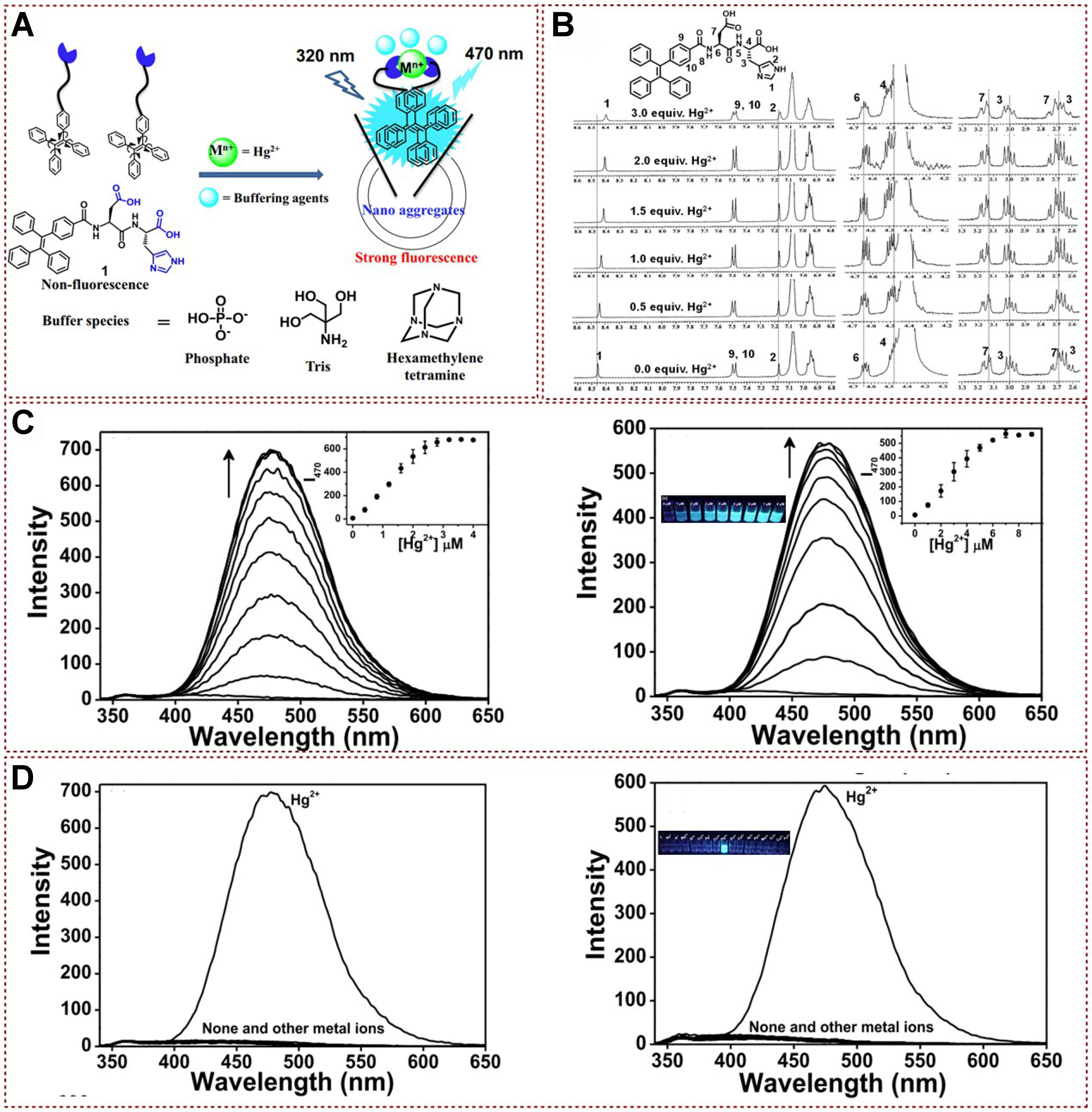
Figure 6. (A) Proposed binding mechanism of fluorescent peptidyl bioprobe with Hg2+; (B) 1H NMR spectra analysis of peptidyl bioprobe with Hg2+; (C) Fluorescence spectra of peptidyl bioprobe with different concentration of Hg2+ in distilled water (left), and phosphate buffered aqueous system (right); (D) Fluorescence spectra of peptidyl bioprobe with different kinds of metal ions in distilled water (left), and phosphate buffered aqueous system (right)[52]. Copyright 2017, Elsevier B.V. NMR: Nuclear magnetic resonance.


