



Capsule biopsy and targeted sampling techniques: current status and future perspectives
 0
Abstract
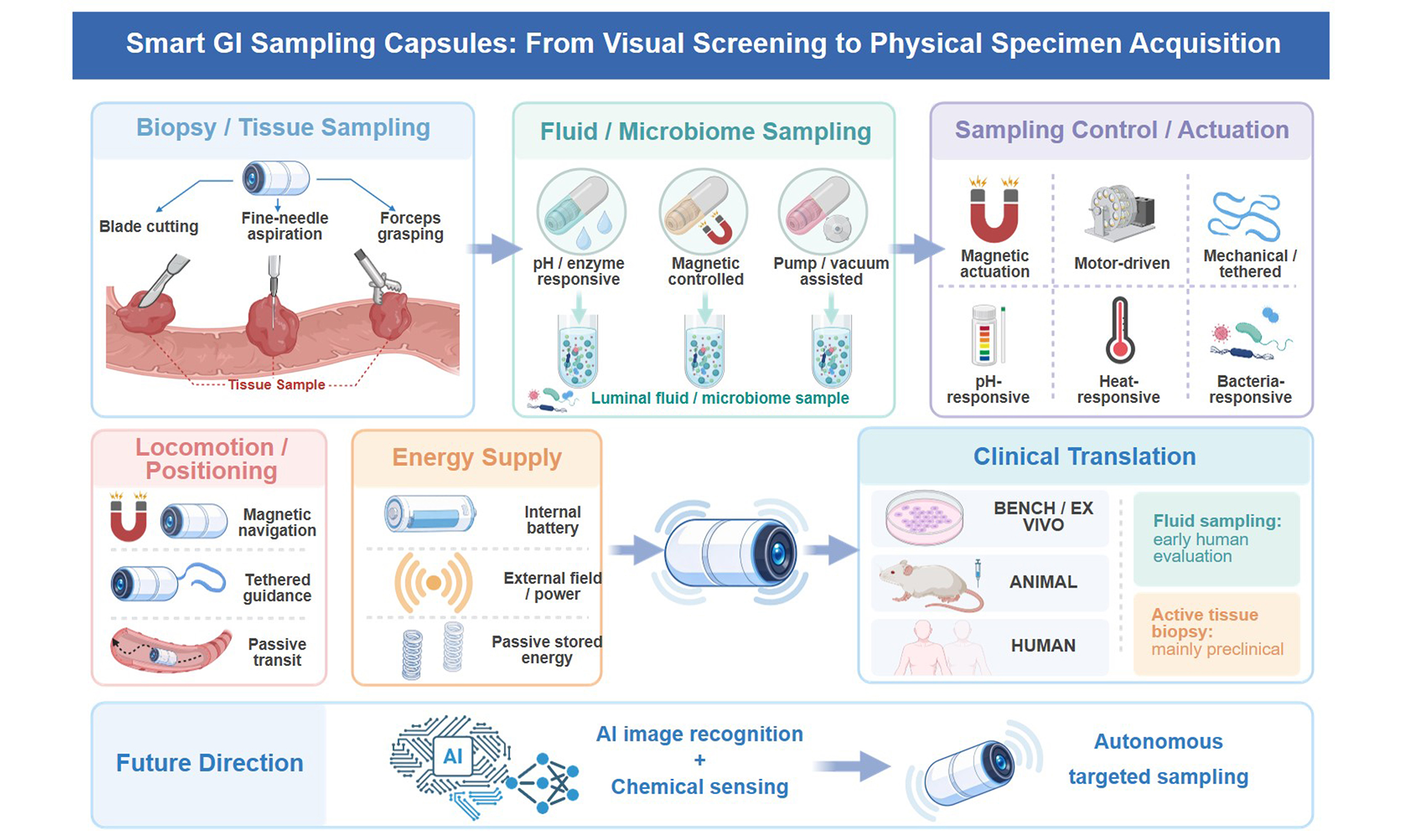
Early screening and definitive diagnosis of gastrointestinal (GI) diseases remain major clinical challenges. Although wireless capsule endoscopy enables patients to undergo minimally invasive imaging of the GI tract, it cannot obtain tissue or fluid samples; therefore, a separate invasive endoscopic procedure is still required when a suspicious lesion is identified. The next logical step is to develop capsule systems capable of both visualizing and sampling lesions during a single swallowable examination. This review summarizes recent advances in smart GI sampling capsules, categorized according to the type of specimen collected (solid tissue vs. luminal fluid or microbiome) and the mode of device control. Miniaturized electromechanical systems (MEMS), stimuli-responsive materials, and magnetic actuation have substantially expanded the technical capabilities of sampling capsules; however, most active tissue-biopsy platforms remain at the preclinical stage. In contrast, several passive or environment-responsive luminal-sampling systems have progressed to early human evaluation. Major barriers to clinical translation include reliable lesion targeting, adequate and reproducible specimen acquisition, prevention of cross-contamination, device miniaturization, and demonstration of procedural safety. Artificial intelligence (AI)-assisted and autonomous targeting remain areas for future investigation rather than established clinical practices.
Keywords
INTRODUCTION
Gastrointestinal (GI) diseases are among the leading causes of morbidity and mortality worldwide. Early detection of GI cancers and accurate characterization of alterations in the gut microbiome remain important unmet clinical needs[1]. Since its introduction in 2000[2], wireless capsule endoscopy has transformed GI diagnostics by allowing patients to swallow a camera-equipped capsule for minimally invasive visualization of the GI tract, particularly the small intestine, which is difficult to access using conventional endoscopic techniques.
However, visual inspection of tissue cannot substitute for pathological examination. Although capsule endoscopy provides direct visualization of the mucosa, imaging alone cannot replace histopathological assessment when evaluation of tissue architecture, microscopic inflammation, dysplasia, or neoplasia is required[3,4]. Histological examination of endoscopic biopsy specimens remains an integral component of the diagnostic work-up for inflammatory bowel disease and other GI disorders requiring microscopic characterization[5]. At present, when capsule endoscopy identifies a suspicious lesion, the patient must undergo a separate conventional endoscopic procedure to obtain a biopsy specimen[6]. This two-step process diminishes the convenience and minimally invasive advantages of capsule examination and may reduce patient adherence to cancer screening.
The limitations of conventional capsule endoscopy extend beyond the assessment of solid lesions. Stool-based methods for gut microbiome analysis predominantly reflect microbial communities of the distal colon and cannot capture the distinct, site-specific microbial communities of the small intestine[7]. By the time stool samples are collected, ongoing fermentation and DNA degradation may also compromise specimen quality, thereby limiting the accuracy of multi-omics studies. There is therefore a compelling clinical rationale for developing capsules capable of collecting uncontaminated biological samples from specific locations within the GI tract.
Recent engineering advances in miniaturized electromechanical systems (MEMS), stimuli-responsive coating materials, and magnetic control technologies have brought this objective closer to realization. Emerging biopsy capsules incorporating magnetic torsion springs (MTSs)[8], microcutters[9], and high-speed rotating blades[10] are capable of obtaining mucosal and submucosal tissue while minimizing tissue injury. For luminal fluid and microbiome sampling, capsules equipped with magnetically controlled microfluidic pumps[11] and pH-sensitive coatings can collect sealed specimens from targeted intestinal locations while limiting cross-contamination. Collectively, these technologies represent a shift from purely visual screening toward diagnostic platforms capable of physically acquiring biological specimens at the point of detection.
Despite this progress, substantial challenges remain before sampling capsules can be incorporated into routine clinical practice. Generating sufficient mechanical force within the highly constrained capsule volume, reliably targeting specific lesions without the direct control and visualization available during conventional endoscopy, and preventing cross-contamination among specimens all require close collaboration between clinicians and engineers. For the purposes of this review, a GI sampling capsule is defined as an ingestible, tethered, or capsule-scale device designed to physically acquire biological material - including solid tissue, luminal fluid, mucus, or microbiome-containing material - from the GI tract. Devices that perform only in situ sensing without physically acquiring specimens are not considered primary sampling devices, although representative systems are discussed separately when they provide insights relevant to the future development of integrated sampling-and-analysis platforms. Similarly, studies focusing primarily on localization, pose sensing, posture adjustment, or optimization of sampling parameters are classified as enabling technologies rather than complete sampling systems.
METHODS
Search strategy
This article is a narrative review supported by a structured literature search [Figure 1]. PubMed/MEDLINE, Embase, Web of Science Core Collection and IEEE Xplore were searched from database inception to March 1, 2026, with records restricted to those published in English. The reference lists of all included articles and of relevant reviews were hand-searched, and forward citation tracking was performed to capture recent work citing key device papers. Search strings combined two concept blocks using the Boolean operator AND, with synonyms within each block combined using OR: (i) device terms - “capsule endoscopy”, “capsule robot”, “ingestible device”, “smart capsule”, “swallowable device”, “wireless capsule”; and (ii) sampling terms - “biopsy”, “fine-needle aspiration”, “forceps”, “microgripper”, “gastric juice”, together with combinations of sampling or collection terms and tissue, luminal fluid, gut or microbiome terms.
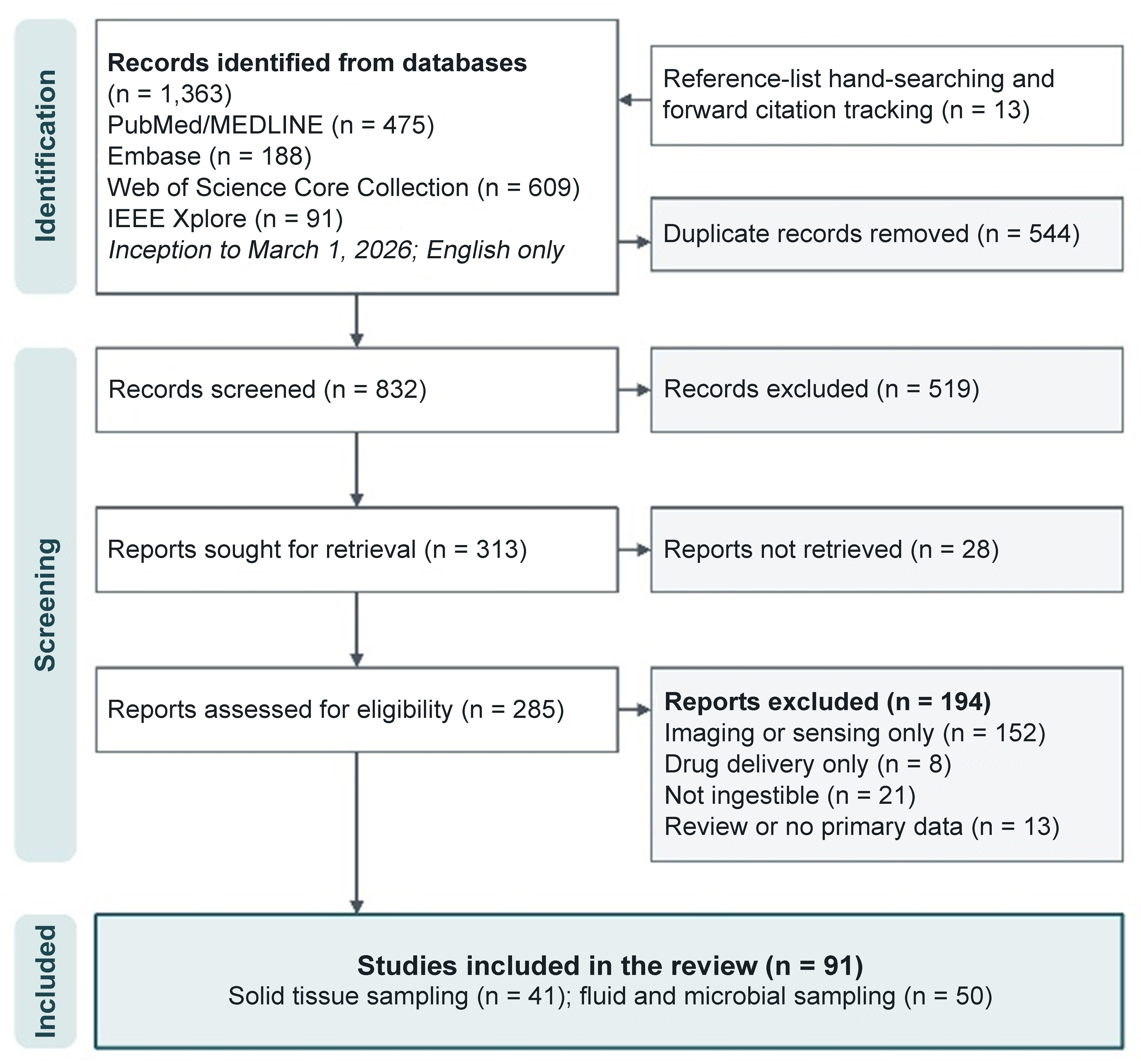
Figure 1. Flow diagram of the literature search and study selection.
Eligibility criteria
Records were eligible for inclusion in the device tables if they (i) were peer-reviewed journal articles, full-length conference papers, or clinical meeting abstracts reporting primary device or clinical sampling data, published in English; (ii) described an ingestible device, or a device-level mechanism explicitly intended for integration into an ingestible device, whose function includes the physical acquisition and retention of a specimen - solid tissue, luminal fluid, or mucosa-associated material - from the GI tract; and (iii) reported experimental data on sampling performance at bench-top level or above. Devices combining sampling with drug delivery were included provided that a sampling function was described and characterized. We excluded (i) imaging- or sensing-only capsules with no specimen-acquisition function; (ii) drug-delivery-only devices; (iii) instruments introduced through the working channel of a conventional endoscope, and other non-ingestible endoscopic accessories; and (iv) reviews, editorials and abstracts that did not report primary device data. Studies that did not themselves describe a complete sampling device were nonetheless retained in the narrative where they addressed enabling technologies for such devices - capsule localization, pose and needle-extension sensing, attitude control, actuation-parameter optimization, and fabrication methods - including studies reporting only computational modeling or simulation without a physical prototype. Because such work supports sampling capsules without itself constituting one, it is discussed in the text but is not entered in the device tables; the same applies to in situ detection devices, which analyze luminal contents inside the body without retaining a specimen for subsequent ex vivo analysis.
Study selection and data extraction
Titles and abstracts were screened independently by two authors (Wan Y, Han Z); full texts of potentially eligible records were then assessed against the criteria above, and disagreements were resolved by discussion with a third author (Tian Y). For each included device, we extracted capsule dimensions, sampling capacity, locomotion or control method, sampling trigger, energy supply, the highest stage of validation reported (bench-top, ex vivo, animal, or human), and distinctive design features. Where a study did not report a given parameter, this is recorded as “not reported” rather than inferred.
SAMPLING MECHANISMS
Solid tissue sampling
Obtaining tissue for histopathological examination is essential for diagnosing GI cancers and submucosal lesions. The challenge is that the intestinal wall is soft and slippery, making it difficult to grasp or cut without causing tearing or perforation. Three main approaches have been developed: high-speed blade cutting, fine-needle aspiration (FNA), and forceps-based grasping [Table 1].
Mechanism for solid tissue sampling
| Study | Capsule size (mm) | Sampling capacity | Control method | Triggered sampling | Energy supply | Features | Validation stage |
| Straker et al. 2025[20] | NR | 19 ± 8 mg | Passive GI transit; no active capsule locomotion demonstrated | Electrothermal trigger (microheater-mediated EVA melting) | Internal battery | Low power consumption | Bench-top |
| Wang et al. 2025[34] | 17 × 14 | 3.8 × 2.8 mm | External magnetic navigation + tether-assisted retrieval | Operator-triggered manual mechanical actuation of tethered commercial biopsy forceps | External magnetic and manual mechanical energy; no onboard battery reported for locomotion or biopsy actuation | Large sample size; can be directly recovered | Ex vivo - fresh porcine stomach |
| Hu et al. 2025[28] | 34 × 18.8 | NR | External gradient magnetic-field actuation | Motor-driven mechanical trigger | External electrical power in prototype; onboard battery not yet integrated | Biopsy/drug delivery switchable | Bench-top + gastric phantom/liquid-environment validation |
| Li et al. 2025[29] | 26.8 × 14.8 | 0.6 mm3 | External permanent-magnet actuation; robotic magnetic manipulation | Motorized lead-screw trigger for needle deployment; magnetic thrust for tissue penetration | External magnetic energy for locomotion and biopsy thrust; electrical power for internal motors, with onboard battery integration not specified | Biopsy/drug delivery switchable | Ex vivo - fresh porcine stomach with PDX tumor model |
| Xu et al. 2025[4] | 27 × 13 | NR | External magnetic actuation | Pulsed electrical current applied to the onboard micro-coil generates transient magnetic fields that drive stepwise biopsy-needle insertion | External magnetic energy for whole robot locomotion + externally supplied; tethered electrical power for onboard micro-coil biopsy actuation | Post-puncture thermal hemostasis | Ex vivo - porcine stomach |
| Zheng et al. 2025[100] | 29 × 14 | NR | External rotating magnetic-field actuation; gradient field locking during biopsy | Rotating magnetic field triggered Magnet B–lead screw actuation for biopsy needle deployment | External electro-magnetic energy; battery-free onboard actuation | Reducing coupling interference between motion and biopsy | Ex vivo - porcine small intestine |
| Rashid et al. 2024[24] | 32.2 × 12 | NR | External electro-magnetic actuation of internal PM1 | External electromagnetic force triggered needle deployment via PM2, with spring mediated needle retraction | External electro-magnetic energy; battery-free magnetic actuation for locomotion/biopsy | Dual permanent magnet independent decoupled control | Bench-top only |
| Zheng et al. 2024[23] | 28 × 18 | NR | External rotating magnetic-field actuation | External rotating magnetic field trigger | External electro-magnetic energy; battery-free magnetic actuation for locomotion/biopsy | Threshold control based on magnetic field strength enables the decoupling of motion and biopsy | Bench-top only |
| Zarifi Khameneh et al. 2024[38] | 30 × 12 | NR | External magnetic actuation; uniform-field orientation control | Threshold external magnetic field actuation; field application deploys the forceps and field removal causes automatic retraction for tissue cutting | External magnetic energy for locomotion and biopsy actuation + internal batteries for imaging electronics | A safety mechanism for the automatic retraction of biopsy forceps upon power loss | Bench-top only |
| Xu et al. 2023[79] | 34.3 × 18.8 | NR | External permanent-magnet actuation with vision-guided orientation control | Motor-driven needle deployment + magnetic puncture thrust | External magnetic + internal electrical motor power; onboard power source not specified | Precise visual dynamic feedback positioning; multiple biopsies or delivery of multiple drugs | Ex vivo - porcine stomach |
| Ye et al. 2022[25] | 32 × 15 | 0.35 mm3 | External electro-magnetic actuation: rotating uniform-field rolling + gradient-field 3D positioning | Gradient magnetic field triggered PM3 needle deployment; spring-mediated retraction | External electromagnetic energy; battery-free for locomotion and biopsy actuation | Combining planar and three dimensional motion, the patient requires no repositioning | Ex vivo - fresh porcine stomach + simulated tumor model |
| Liu et al. 2022[41] | NR | NR | External rotating magnetic-field actuation: PM3-driven four-blade propeller locomotion with PM2-mediated magnetic anchoring | External magnetic field triggered PM1–CAM actuation for biopsy-forceps deployment | External electro-magnetic energy; battery-free magnetic actuation for locomotion, anchoring, and biopsy | The four-bladed propeller offers higher propulsion efficiency than two- or three-bladed designs; it integrates propulsion, anchoring, and biopsy capabilities via a three-stage magnetic triggering mechanism | Bench-top only |
| Song et al. 2022[16] | 40 × 18 | 2.5-5.5 mm3 | External permanent-magnet actuation for magnetic locomotion and posture control | Operator-triggered Bowden cable mechanical release | External magnetic energy for locomotion + preloaded spiral-spring mechanical energy for biopsy cutting | High-speed cutting reduces the risk of soft tissue deformation and tearing; an external force gauge monitors cable tension in real time | Ex vivo - porcine large intestine |
| Guo et al. 2021[36] | NR | NR | External rotating magnetic field actuation | External magnetic field switching triggers PM1–CAM actuation for biopsy-forceps deployment | External electro-magnetic energy; battery-free magnetic actuation for locomotion and biopsy | Propeller fluid propulsion; anti detachment microbarb design | Bench-top only |
| Guo et al. 2020[40] | 52 × 20 | ≥ 1 mm3 | External rotating magnetic-field actuation with spiral-body propulsion | Directional magnetic field triggered magnetic switch; biopsy completed during field-switch mediated forceps closure | External electro-magnetic energy; battery-free magnetic actuation for locomotion and biopsy | Enables 360° sampling | Bench-top only |
| Leon-Rodriguez et al. 2020[37] | NR | ≥ 5 mm3 | External electro-magnetic actuation | Operator-controlled external magnetic field strengthening + rotational actuation | External electro-magnetic energy for locomotion/biopsy actuation + internal battery for imaging electronics | Safety of folding tools | Animal - in vivo porcine stomach |
| Hoang et al. 2020[21] | 30 × 12 | 1.95 mm3 | External electro-magnetic actuation | Low-amplitude rotational magnetic field actuates PM2 | External electro-magnetic energy for locomotion/biopsy actuation + internal battery for imaging electronics | Independent decoupled control of motion and biopsy | Ex vivo - porcine small intestine |
| Son et al. 2020[74] | NR | NR | External electro-magnetic actuation | Strong magnetic gradient triggered collapsing motion | External electro-magnetic energy for locomotion/biopsy | Enables access to deep subsurface tissues; possesses potential for magnetic localization | Ex vivo - porcine stomach |
| Zhang et al. 2019[26] | 24 × 16 | 1-5 mm3 | Passive intestinal peristalsis; magnetically actuated anchoring for target-site stabilization | External rotating magnetic field trigger | External electro-magnetic energy for anchoring/biopsy | Decoupling of anchoring and biopsy functions using a single actuator | Bench-top only |
| Ye et al. 2019[39] | 32 × 14 | NR | External electro-magnetic actuation | Uniform magnetic field triggered MTS actuation | External electro-magnetic energy for locomotion/biopsy actuation + internal battery for imaging electronics | Repeatable triggering; forward extension and direct-view monitoring | Bench-top only |
| Song et al. 2019[15] | 36 × 20 | 1-5 mm3 | External magnetic control | Manual external-wire/lever mechanical trigger releasing a stop-baffle latch | Preloaded spiral-spring elastic energy for biopsy cutting; external magnetic control for capsule locomotion | Performing multiple biopsy samplings during a single examination | Ex vivo - manually triggered component-level biopsy validation on porcine skin |
| Tovmachenko et al. 2019[32] | NR | 3 mm3 | External electro-magnetic control | DC motor–triggered threadless-ballscrew gripper actuation | Internal battery for biopsy motor + external electro-magnetic for locomotion | A novel actuation method utilizing a threadless ballscrew has been introduced | Bench-top only |
| Hoang et al. 2019[10] | 32 × 12 | 1-5 mm3 | External EMA: uniform-field orientation + gradient field 5-DOF active locomotion | Strong rotating magnetic field trigger overcoming the small–large magnet holding force, enabling rotary biopsy-blade actuation | External electro-magnetic energy for locomotion and biopsy actuation; biopsy mechanism is battery-free | The four blades provide ample cutting power | Ex vivo - porcine small intestine |
| Le et al. 2018[22] | 32 × 12 | 5 mm3 | External electro-magnetic actuation | Rotating magnetic field triggered screw translation of the biopsy punch | External electro-magnetic energy for locomotion/biopsy actuation + internal battery for imaging electronics | Replaced traditional clamps with a punch module | Ex vivo - extracted intestinal segment |
| Pan et al. 2018[31] | ≤ 15 × 30 | NR | Not reported | Controller-triggered micro-stepper motor actuation via gear–rack transmission | Electrical power for micro-stepper motor; specific power source not reported | Dual-layer housing and sampling groove design; miniature barbed sampling structure | Ex vivo - porcine intestine |
| Le et al. 2017[17] | 30 × 11 | 5.4 mm3 | External electro-magnetic actuation | Rotating uniform magnetic field triggered rotational biopsy-blade actuation | Electro-magnetic energy for locomotion/biopsy actuation; onboard battery reserved for capsule electronics | In-situ storage | Ex vivo - fresh porcine intestine |
| Le et al. 2017[35] | 32 × 13 | 5 mm3 | External electro-magnetic actuation | Strong rotating magnetic field triggered permanent-magnet/gearbox actuation of biopsy gripper opening and closure | External electro-magnetic energy for locomotion/biopsy actuation + internal battery for imaging electronics | Miniature gearbox amplifies cutting force | Ex vivo - fresh porcine intestine |
| Son et al. 2017[27] | NR | NR | External electro-magnetic actuation | Strong magnetic-gradient force triggered Sarrus-linkage collapse for fine-needle deployment | External electro-magnetic energy for locomotion/needle insertion | Targeted deep-tissue fine-needle biopsy with integrated locomotion and magnetic tracking | Ex vivo - fresh porcine adipose tissue in an anatomical stomach phantom |
| Le et al. 2016[14] | 32 × 12 | 5 mm3 | External electro-magnetic actuation | Threshold external magnetic-field trigger | External electro-magnetic energy for locomotion + onboard battery for SMA trigger | Spring mechanism ensures powerful cutting force | Ex vivo - fresh porcine intestine |
| Le et al. 2015[9] | 12 × 33 | 6 mm3 | External EMA provides active 5-DOF locomotion | Electrical activation of SMA wire opens the biopsy razor | Onboard battery for SMA actuation + external electro-magnetic energy for locomotion and rotational tissue cutting | Ultra-sharp, low-resistance blades; resolves issues with insufficient grip | Ex vivo - porcine small intestine |
| Yim et al. 2014[42] | 31.5 × 18 | 200-300 μm | External electro-magnetic actuation | Body temperature triggered autonomous self-folding at 37 °C | External magnetic + stored residual-stress elastic energy | Parallel multi-point sampling | Ex vivo - fresh porcine stomach |
| Simi et al. 2013[8] | 24 × 9 | > 1 mm3 | Passive peristaltic locomotion with external magnetic anchoring during biopsy | Decrease of the external magnetic field triggers MTS release and blade rotation | Externally magnetically loaded MTS elastic energy; no onboard power supply or electronics | Charge the MTS and provide sufficient anchoring force to secure the capsule to the intestinal wall | Ex vivo - porcine small intestine |
| Chen et al. 2013[33] | 73 × 19 | NR | Motor-driven inchworm locomotion with alternating pneumatic balloon anchoring and axial extension | Operator-commanded RF/electrical activation of the biopsy micro-motor | Inductive WPT | Secure flexible balloon anchoring and comprehensive safety feedback monitoring | Ex vivo - porcine colon/intestine |
| Kong et al. 2012[13] | 40 × 15 | NR | No integrated locomotion; SMA anchoring + SMA rotational alignment | Electrical activation of a chip resistor melts its solder joint (~200 °C), releasing the mechanical latch and the spring-loaded inner biopsy razor | External electrical power (prototype) + preloaded spiral-spring cutting energy | Directly observe and monitor the entire alignment and cutting process in real time through side openings | Ex vivo - porcine large intestine |
| Bourbakis et al. 2010[43] | 26 × 11 20 × 10 | NR | Motorized bidirectional wheeled locomotion + expandable-wing stabilization + gripper orientation control | Motorized gripper control; exact sampling trigger not specified | Onboard battery electrical power for the micro-motors and capsule electronics | Bionic active perching | Conceptual design |
| Park et al. 2008[19] | 26 × 10 | NR | Passive peristalsis; active locomotion not integrated | Electrical activation of an SMA heating wire melts a polymer retaining string | Internal battery (trigger) + preloaded torsion-spring mechanical energy | LiGA process breaks through the bottleneck of micro-nano manufacturing | Ex vivo - porcine small intestine |
| Kong et al. 2005[12] | Diameter 10 | 1.0 mm | Not reported | Electrical thermal-release trigger | Electrical trigger power + preloaded torsion-spring cutting energy | One-stop automated operation: sampling, sealing, and fixing | Ex vivo - cow small intestine + animal - small intestine in etherized rabbit |
High-speed blade cutting
Conventional biting or pulling mechanisms may cause tearing of fragile mucosal tissue. In contrast, high-speed rotary cutting mechanisms can mitigate this risk by storing energy in a pre-compressed spring and releasing it in a rapid, controlled burst, thereby driving a sharp blade to excise a relatively clean tissue sample.
Early prototypes primarily relied on thermal triggering to activate the cutting mechanism. Kong et al. used electrically heated wax to trigger a torsion spring, driving a blade 1.5 mm into tissue for superficial sampling, with the device then automatically sealing and fixing the specimen in preservative fluid[12]. A later version added modules for tissue monitoring and capsule anchoring, using electrical heating to melt solder and release a double-layered cutting blade. However, this device still required an external power cable, limiting its applicability as a fully wireless capsule system[13]. Le et al. subsequently integrated a shape memory alloy (SMA) wire, which contracts upon heating, into a locomoting capsule (ALICE)[9]. A later version incorporated a thermal-fuse trigger to activate the biopsy mechanism[14]. Although these thermally triggered systems demonstrated reliable actuation, their irreversible triggering mechanisms generally permit only a single sampling event, while the heating process can impose a substantial energy burden on the capsule battery.
To avoid electromagnetic interference, some groups used a physical wire (Bowden cable) to mechanically trigger blade release, allowing multiple independent samples to be stored in a rotating multi-slot compartment[15,16]. This works well but sacrifices the fully wireless nature of capsule endoscopy.
Wireless magnetic triggering has become one of the most extensively investigated approaches in preclinical capsule biopsy research. In one design, interaction between two permanent magnets within the capsule was used to control blade opening and closure; however, the relatively large magnet dimensions required by this mechanism pose challenges for integration into commercially sized capsules[17]. A widely adopted strategy employs an MTS. In this configuration, an external magnetic field displaces an internal rotating magnet relative to a fixed magnet, thereby storing magnetic potential energy; removal of the external field subsequently releases this stored energy and drives rapid blade rotation[8]. A related design locks the blade magnetically during transit and releases it by applying a stronger external field[10]; this required an additional robotic arm system to adjust the capsule’s orientation so that the side-mounted biopsy port aligns with the lesion[18]. Despite their advantages in wireless actuation, current MTS-based capsule systems remain largely limited to single-shot sampling and generally produce relatively small tissue specimens.
FNA
Superficial blade biopsy may fail to reach submucosal lesions and can cause tearing of the overlying mucosa. FNA offers a potential solution to these limitations by using a small needle to penetrate deeper tissue layers, analogous to the FNA technique routinely employed during clinical endoscopic ultrasound-guided sampling.
Early needle-based capsule designs primarily relied on thermally triggered actuation. Park et al. used electrically heated SMA wire to deploy a biopsy needle[19], whereas the SCUUP device employed a microheater to melt an adhesive bond, releasing a spring-driven gear system that enabled the acquisition of relatively large mucosal specimens suitable for downstream genetic analyses, including polymerase chain reaction (PCR)[20]. Both approaches work reliably but are limited to a single use because thermal melting cannot be reset.
External electromagnetic actuation (EMA) has emerged as a widely investigated preclinical approach for remotely controlled needle deployment without requiring local electrical power for the actuation step. In these systems, external magnetic coils generate a magnetic field that drives an internal magnet to rotate, with the rotational motion subsequently converted into reciprocating linear motion of the biopsy needle[21,22]. Hoang et al. further refined this concept by incorporating an automatic needle-retraction mechanism that retracts the needle during capsule locomotion, thereby reducing the risk of inadvertent mucosal injury. A front-facing camera was also integrated to provide real-time visualization and monitoring during the procedure[21].
A major challenge is to decouple magnetic capsule locomotion from magnetic needle deployment. Several strategies have been proposed to address this issue. One approach uses a gradient magnetic field to first anchor the capsule against the intestinal wall, followed by a rotating field to independently drive needle insertion[23]. Another employs two internal magnets, in which the anterior magnet controls the capsule steering while a second magnet, actuated by an independently controlled external magnetic field, drives the needle forward and permits automatic retraction when the field is removed[24,25]. A third strategy uses an eccentric wheel-ratchet mechanism, in which reversing the direction of rotation of an external magnet independently controls both the anchoring legs and needle extension, substantially reducing the required internal space[26]. The B-MASCE, a soft-bodied capsule using a concertina-like linkage structure, achieves both movement and needle guidance through axial magnetic compression, though it is currently limited to collecting cells rather than tissue cores[27].
More advanced MEMS-integrated systems have combined needle biopsy with immediate post-procedure hemostasis. Xu et al. developed a capsule incorporating a graphene-carbon nanotube heating film that can cauterize the biopsy tract immediately after needle withdrawal, providing integrated tissue sampling and hemostatic control[4]. Multi-chamber “roulette-style” designs with dual built-in motors can cycle through several biopsy needles and drug delivery compartments in a single procedure[28,29]. However, further miniaturization of the internal motors while maintaining sufficient torque and reliability remains a major engineering challenge for integration into a swallowable capsule platform.
Beyond the sampling mechanism itself, accurate real-time localization of the biopsy needle is critical for procedural safety, particularly when deep tissue penetration is required. Liu et al. addressed this by embedding three aligned permanent magnets within the capsule body and combining external magnetic sensors with a computational algorithm, achieving 1.83 mm positional accuracy while simultaneously tracking how far the biopsy needle has extended[30]. This approach has not yet been tested in the complex electromagnetic environment of a clinical setting.
Forceps-based grasping
Forceps-based biopsy most closely replicates the tissue-acquisition technique used by clinicians during conventional endoscopy. Miniaturized forceps jaws can be opened, positioned against the target tissue, and closed to pinch off a specimen.
Several designs employ miniature electric motors to actuate the forceps mechanism. These include micro-barbed forceps driven by a stepper motor via a gear rack[31], a three-jaw gripper incorporating a ball-screw drive to minimize mechanical backlash[32], and a caterpillar-like colon biopsy capsule that receives power wirelessly, thereby eliminating the need for an onboard battery, to operate both a balloon anchoring system and a forceps-driving lead screw[33]. The integration of commercially available biopsy forceps into a magnetically steered capsule has also been demonstrated, with tissue acquisition achieved through mechanical traction after the capsule reaches the target site[34]. The main drawback of all motor-based forceps designs is that motors and drive mechanisms take up substantial space inside the capsule.
Battery-free forceps mechanisms actuated by external magnetic fields offer a potential solution to this space constraint but require precise magnetic-field control. The ALICE capsule incorporates a miniature gearbox to amplify the magnetic torque required to operate micro-forceps[35]. However, the fabrication of such high-precision gearboxes, together with the difficulty of preventing the rotating magnetic field from inadvertently rotating the entire capsule, has thus far limited practical translation and commercialization. Simpler magnetic designs include a coaxial “magnetic switch” that separates capsule movement from biopsy activation[36], and a passive folding tool that deploys when the capsule rotates against the intestinal wall[37], though the cutting force of the former decreases significantly if the magnet is made smaller. Safety improvements include a fail-safe magnetic spring that automatically closes the forceps if the external magnetic field is interrupted, protecting the bowel wall from injury[38]. A modified MTS design further deploys the forceps from the front face of the capsule, allowing the integrated camera to monitor the entire procedure without blind spots, with gravity retracting the forceps when the field is removed[39]. However, the combined space requirements of the MTS and forceps mechanism remain considerable, and actuation delays may occur at certain magnetic-field orientations.
Other actuation strategies have also been investigated. Propeller-driven biopsy capsules actuated by an internal motor and triggered through a cam mechanism, which work well in fluid-filled bowel segments but lose propulsion in drier regions[40,41], and a swarm of heat-activated micro-grippers deployed in large numbers to compensate for the difficulty of accurate targeting[42], though retrieval rates are very low at around 3%. A capsule incorporating a rubber odometer wheel has also been proposed to estimate travel distance and thereby improve targeting accuracy for subsequent biopsy procedures[43]. Nevertheless, the safety and reliability of prolonged contact between the rubber wheel and delicate GI tissue require further evaluation.
Fluid and microbial community sampling [Table 2]
Mechanism for sampling liquids and microbial communities
| Author | Size (mm) | Sampling capacity | Control method | Triggered sampling | Energy supply | Features | Validation stage |
| Yun et al. 2026[69] | 32 × 12 | 1,020 μL | External permanent magnet guidance | Distance and polarity controlled external magnetic actuation; magnetic force opens selected chamber when exceeding sealing force | Battery-free | Dual sampling, high capacity, cross-contamination prevention | Ex vivo - porcine small intestine |
| Nema et al. 2026[87] | 7 × 2.7 | 20-25 µL | Passive GI peristalsis; no active steering | pH-responsive activation + fluid-triggered hydrogel closure | Battery-free | Extremely compact size; enables simultaneous acquisition of microbiome and protein biomarkers | Animal |
| Shahadha et al. 2026[61] | 30 × 11.4 | 133 μL | Passive GI peristalsis; electronic communication for site-specific activation | Wireless electrical activation of microheater causing thermal melting of wax valve | Internal button-cell battery | Clog-resistant porous filtration membrane design; in situ sample nucleic acid preservation technology | Ex vivo - porcine intestine |
| Zhang et al. 2025[75] | 16.3 × 24.4 | 230 μL | Passive GI peristalsis; external magnetic orientation control + magnetic localization | Increased external magnetic field strength triggers magnetic spring deployment | Battery-free | High sampling volume | Ex vivo -fresh porcine colon |
| Gupta et al. 2025[67] | 25 × 10 | NR | External permanent magnet active steering | External magnetic field triggered opening via magnetic torque | Battery-free | Multifunctional targeted drug delivery, sampling, and transport | Bench-top only |
| Wu et al. 2025[80] | 9.8 × 20 | 40-50 μL | Active rolling locomotion driven by a uniform rotating magnetic field | Directional gradient magnetic field triggered valve opening; oscillating gradient field enhances fluid sampling | Battery-free | pH-sensitive gel linkages enhance maneuverability in narrow intestinal environments; operation is possible under ultrasound guidance | Ex vivo - porcine stomach |
| Li et al. 2025[96] | 14 × 42 | 200 μL | Passive GI peristalsis; no active steering | Preprogrammed autonomous electronic trigger controlling stepper-motor rotation | Internal coin-cell batteries | Multi-site, multiple liquid sampling; capable of distinguishing between gastric, small intestinal, and colonic samples | Animal - in vivo canine model |
| Lee et al. 2025[68] | 11 × 8 | 290 µL | Active rolling locomotion via low strength rotating external magnetic field | Higher strength external magnetic field produces magnetic torque that snap-opens capsule by overcoming sealing/hinge restoring torque | Battery-free | Large sample storage capacity | Ex vivo - swine stomach |
| Tronel et al. 2025[86] | 8 × 22 | 150 µL | Passive GI peristalsis; no active steering | pH-triggered module release; fluid-induced polymer expansion triggers valve closure | Battery-free | In vivo human validation; multi-point sampling across three modules; 87% recovery rate of all modules | First-in-human - monocentric interventional study in 15 healthy volunteers |
| Sarnaik et al. 2025[51] | 20 × 9.5 | NR | Passive GI peristalsis; no active steering | pH + bacteria-triggered enteric coating; hydrogel-swelling closure | Battery-free | Bacteria + pH dual triggering to overcome colonic pH fluctuations | Animal - in vivo pig model |
| Wang et al. 2025[46] | NR | 1,000 μL | External magnetic guidance with manual string-length adjustment | Operator-triggered pneumatic detachment followed by manual syringe aspiration | Sampling actuation: externally powered; onboard electrical power of the MCE capsule not specified | Visualized precision sampling; rapid diagnosis via PCR detection bypassing bacterial culture | Pilot clinical feasibility study (n = 5 patients) |
| Sadeqi et al. 2025[95] | 27 × 9 | 250 μL | Passive GI peristalsis; no active steering | pH-dependent enteric exposure followed by on-demand magnetic reed-switch activation | Internal battery | Sampling on demand; capable of handling highly viscous contents | Animal - preliminary in vivo pig feasibility (n = 1) |
| Krishnakumar et al. 2025[89] | 20 × 9.5 | 400 μL | Passive GI peristalsis; no active steering | pH-dependent enteric-coating dissolution; hydrogel swelling provides passive sample uptake and valve closure | Battery-free | Simple structure, low cost | Animal - in vivo pig model |
| Park et al. 2024[64] | 21 × 8 | NR | Active external magnetic positioning and orientation control | External magnetic field actuates internal magnetic driving unit to deploy and retract one of six sampling tools | Battery-free sampling actuation; camera module power not specified | 6 sampling channels | Animal - in vivo swine stomach |
| Li et al. 2024[62] | NR | 6.8 μL | Active magnetic rolling; permanent magnet guidance for bulb-cage capsule | High strength external magnetic field deforms magnetic bulb | Battery-free | Digital light processing integrated printing, cage structure with high compressive strength | Bench-top only |
| Lai et al. 2024[7] | A 19 × 8 B 21 × 8 | 31 μL (Type A); 41 μL (Type B) | External rotating magnetic field for rotation; field-plane tilting for steering | pH-responsive hydrogel swelling; magnetic latch release; piston sampling | Battery-free onboard | Compact size; automatic sampling | Bench-top only |
| Dong et al. 2024[63] | 1.8 × 1.6 | 0.7-4 μL | Active rolling locomotion driven by rotating external magnetic field with magnetic gradient pulling | External magnetic field exceeding a preset threshold opens the magnetic inlet valve | Battery-free | Capable of entering highly confined spaces; medical imaging guidance | Ex vivo - porcine liver |
| Del-Rio-Ruiz et al. 2024[90] | 20 × 9 | 400 µL | Passive GI peristalsis; no active steering | pH-triggered inlet exposure followed by fluid-induced polyacrylate-bead swelling and mechanical self-locking | Battery-free | Soft structures; small intestine-specific sampling; self-locking protection against colonic contamination | Animal - in vivo pig and hound |
| Mandsberg et al. 2024[91] | 20.5 × 7.3 | 93 µL | Passive GI peristalsis; no active steering | Sequential gastric fluid-triggered dissolution of pullulan housing and blocking disk; disk dissolution releases rubber-band closing force | Battery-free | Suitable for highly fluid gastric fluids; sample pH is relatively stable within 24-48 h | Animal - in vivo pig model |
| Nejati et al. 2024[92] | 20 × 9.5 | NR | Passive GI peristalsis; no active steering | pH-responsive enteric-coating dissolution (> 5.5) | Battery-free | Integrated metal tracer ring, approximately 10 cm reading distance; can determine emission time | Animal - in vivo pig model |
| Bundalovic-Torma et al. 2024[84] | 25.4 × 8 | 105 µL | Passive GI peristalsis; X-ray tracking used to verify sampling location | Small intestinal pH-dependent coating dissolution | No onboard power source described; passive sampling mechanism | X-ray-based condition monitoring | Clinical validation |
| Wang et al. 2024[85] | 25.4 × 8 | 105 µL | Passive GI peristalsis; X-ray tracking used to verify sampling location | pH-triggered opening + time controlled spring closure | Battery-free | Excellent sealing performance; built-in preservative facilitates DNA stabilization after recovery | Early clinical performance (n = 20) |
| Khachornsakkul et al. 2024[101] | 15 × 7 | 315 μL | Passive GI peristalsis; no active steering | pH-dependent enteric coating dissolution | Battery-free | can target specific bacteria; can connect to a mobile phone to read fluorescently monitored bacterial water samples; in vivo validation results are consistent with ELISA results | Animal - in vivo dog |
| Menard et al. 2023[88] | 23.4 × 8.6 | 80 μL | Passive GI peristalsis; no active steering | pH-triggered opening + time controlled autonomous sealing | No onboard electrical power described; passive sampling mechanism | Direct comparison and differences between small intestine and fecal samples revealed; non-invasive tracking | Animal - in vivo dog |
| Rehan et al. 2022[58] | 45 × 12 | 250 μL | Passive GI peristalsis; no active steering | Externally commanded 433-MHz wireless trigger | Internal battery | Enables simultaneous collection of luminal and mucosa-associated microbiota | Bench-top |
| Nam et al. 2022[66] | 26 × 12.4 | 1,500 μL | No active locomotion demonstrated; magnetic self-orientation only | Resonant alternating magnetic field actuation | Battery-free onboard; externally powered alternating magnetic field | Utilizing the resonance effect to significantly reduce driving magnetic field requirements | Ex vivo - fresh sheep small intestine |
| Park et al. 2022[11] | 11 × 26 | 45 μL | External EMA: + gradient field propulsion | Precessional magnetic field triggered impeller rotation and centrifugal micropump suction | External electromagnetic energy for locomotion, and micropump-driven sampling; battery-free mechanical actuation | Allows for 3 samplings | Ex vivo - porcine colon |
| Nejati et al. 2022[50] | 11 × 7 | NR | Passive GI peristalsis; no active steering | Sequential dual pH-dependent enteric-coating dissolution | Battery-free | Dual pH-responsive coating, targeting the proximal colon | Ex vivo |
| John et al. 2022[45] | Diameter: 8/10/15/20 mm | 350 μL (8 mm); 850 μL (10 mm); 1,350 μL (15 mm); 1,600 μL (20 mm) | Manual tethered control via retrieval string | Gelatin dissolution + manual string retrieval | Battery-free | Capable of simultaneously collecting gastric fluid, pathogens, and esophageal tissue samples | Animal - in vivo porcine targeted proximal-colon sampling (n = 2) |
| Xin et al. 2022[76] | 40 × 16 | NR | Magnetic rotational control | Motorized leg deployment + magnetic rotational sampling + motorized retraction | External magnetic field for rotation + onboard electrical power for micro motor; specific onboard source not specified | Capable of distending the intestinal wall to expose hidden lesions | Ex vivo - porcine esophageal tissue |
| Finocchiaro et al. 2021[71] | 30.5 × 11.5 | ≥ 18 μL | External permanent magnet provides attraction and remote magnetic actuation | External magnet distance controlled gate opening + brush rotation | Battery-free | Two samples can be collected simultaneously in a single operation | Ex vivo - fresh porcine intestine |
| Rehan et al. 2021[57] | 30 × 12 | 500 μL | Passive GI peristalsis; no active steering | Externally commanded 433-MHz wireless trigger | External battery connected by wire | Active sampling of small intestinal mucosa-associated microbes achieved for the first time | Bench-top only |
| Shokrollahi et al. 2021[65] | 11 × 8 | 42 μL | Passive GI peristalsis; external magnetic self-orientation and blind actuation | External magnetic field activation | Battery-free | Smaller than most previous sampling capsules | Ex vivo |
| Ding et al. 2021[60] | 32 × 11.6 | 400 μL | Active external magnetic positioning and orientation | Operator-issued electronic command activating thermal microvalve | Internal battery | Enables targeted sampling under direct visualization; capable of distinguishing between the jejunum and ileum, as well as disease models | Animal - pig model |
| Waimin et al. 2020[49] | 15 × 9 | NR | Passive GI peristalsis; no active steering | pH-dependent CAP dissolution | Battery-free | 3D-printed, simple structure, low cost | Animal |
| Rehan et al. 2020[56] | 30 × 12 | 500 µL | Passive peristaltic transit intended; movement manually simulated in ex vivo experiments | Current triggered SMA actuation | External electrical power supply in current prototype | In-situ sealing mechanism to prevent downstream contamination | Bench-top only |
| Banis et al. 2019[52] | NR | NR | Passive transit intended; no active locomotion | Local pH-dependent polymer dissolution | Rechargeable 14-mAh 3.3-V Li-polymer battery | Pancreatic secretions can be collected by setting a specific pH level; it can be paired with mobile devices via bluetooth low energy for real-time sensing during sampling | Ex vivo |
| Banis et al. 2019[53] | NR | NR | Passive GI transit intended; no active locomotion or steering demonstrated | pH-dependent polymer dissolution | Onboard 14-mAh Li-polymer battery, 3.3 V | The triglyceride-based coating monitors the lipase content in the collected solution, and can be sensed in real time during sampling via bluetooth low energy | Bench-top only |
| Rezaei Nejad et al. 2019[54] | 28 × 7 | 120 µL | Passive GI peristalsis; optional external magnetic holding for regional targeting | pH-dependent polymer dissolution | Battery-free | In vivo validation in higher mammals | Bench-top only |
| Du et al. 2018[59] | 14 × 20 | 300 µL | Passive GI peristalsis; no active steering | External magnetic field + local mucus presence | External 1.5-V electrical power | Magnetic field + mucus dual-condition triggering mechanism | Animal |
| Salem et al. 2018[48] | ≤ 26.1 × 9.9 | 200 µL | Passive GI peristalsis; no active steering | pH-triggered opening followed by fluid-induced sponge-swelling triggered bistable closure | Battery-free | Bistable mechanism | Bench-top only |
| Hajima et al. 2015[70] | 26 × 10 | NR | External magnetic orientation control | Sequential external magnetic actuation | Externally powered electromagnetic field; no onboard actuator battery reported | Coordinated control of rotating magnetic field, direct current magnetic field and alternating current magnetic field | Bench-top only |
| Leon-Rodriguez et al. 2014[97] | NR | NR | Magnetic rolling + orientation control | External magnetic field drives needle jabbing | Wireless external magnetic actuation; no onboard battery | Soft Sarrus linkage + magnetic actuation | Bench-top |
| Cui et al. 2008[73] | 30 × 10.2 | 510 µL | Passive GI transit; real-time localization using an internal magnetic marker and external Hall-sensor array | Externally issued RF remote control command | Three onboard Sony button cells | Simultaneously performs targeted drug delivery and fluid sampling; features real-time 3D positioning | Bench-top |
| Muretto et al. 2003[44] | 14 × 5 | NR | Passive swallowing into stomach; no active steering; tethered retrieval with nylon thread | Gastric dissolution of gelatin outer envelope + fixed exposure time | Battery-free | Well-tolerated by the human body; no adverse reactions observed in 62 cases; low cost | Animal |
Physical tethered retrieval
The simplest approach to gastric fluid collection is to attach an absorbent material to a retrieval string, swallow the assembly, and subsequently withdraw it. Gelatin capsules containing absorbent paper connected to a nylon thread have been used to non-invasively collect gastric fluid for the analysis of cancer biomarkers[44]. Replacing the absorbent paper with a three-dimensional polycaprolactone (PCL) nanofiber sponge, which can absorb up to 60 times its dry weight in fluid, substantially increases the volume of fluid collected and enhances the capture of bacterial and viral particles[45]. Wang et al. further integrated this concept with magnetically controlled capsule endoscopy, enabling the capsule to be visually navigated to the desired sampling site before fluid collection. Importantly, the retrieval string can be detached from the capsule in situ and withdrawn separately, thereby avoiding the discomfort associated with pulling a rigid capsule back through the esophagus and pharynx[46]. The fundamental limitation of all string-based approaches is that the length and entanglement risk of the string restricts sampling to the stomach; reaching the small intestine or colon is not feasible.
Stimuli-responsive coatings and targeted triggering
To enable deeper GI sampling without a retrieval string, researchers have developed capsule coatings that dissolve or change properties in response to the local chemical environment, such as the pH of the intestinal fluid, specific enzymes, or particular bacteria.
The IMBA capsule uses a conventional enteric coating that dissolves under intestinal pH conditions, thereby releasing a spring-loaded latch that opens the capsule and allows intestinal fluid to flow in, before a piston closes and permanently seals the sample chamber[47]. A related approach employs a 3D-printed bistable mechanical structure that snaps between two stable positions to passively seal the sample without any battery or electronics[48]. These designs are simple and mechanically robust in bench testing, but the sampling site is wherever the coating dissolves, which the device cannot control.
Beyond triggering sampling at the desired anatomical location, preventing contamination of the collected specimen by downstream intestinal contents during subsequent transit is a critical design requirement. pH-responsive hydrogels combined with magnetic actuation allow the hydrogel trigger to be physically separated from the sample chamber itself, so that triggering is autonomous while the sample remains isolated[7]. Sealing the sampling port with a polydimethylsiloxane (PDMS) membrane provides a self-locking physical barrier while retaining gas permeability, thereby supporting the viability of captured microorganisms[49]. This principle has been further extended using a dual-layer pH-sensitive coating to improve targeting of the proximal colon[50]. The principal limitation of pH-based triggering is the substantial interindividual and temporal variability in GI pH, which can be influenced by factors such as diet and intestinal transit time. Consequently, the precise anatomical location of capsule activation may remain unpredictable. Using two simultaneous triggers, both a specific pH threshold and the presence of certain bacteria, reduces this uncertainty[51]; adding capacitive sensors that detect the local environment and communicate sampling status provides additional control[52,53], though the battery and electronics needed for sensing take up space that would otherwise hold the sample.
Osmotic pumps and on-site bacterial detection
Rather than collecting a discrete sample at a single point in time, osmotic pump-based capsules continuously draw small volumes of fluid throughout their transit along the GI tract. This approach enables spatially resolved sampling, allowing different portions of the collected sample to be associated with distinct intestinal locations and thereby facilitating whole-gut microbiome mapping[54]. However, these devices cannot collect a large volume at a specific site and their spatial resolution depends on how quickly the capsule moves.
In situ sensing systems represent an adjacent but conceptually distinct technology. The Smart Capsule Bacterial Detection System (SCBDS), for example, is designed to detect metabolically active bacteria within the GI environment rather than to recover a specimen for external laboratory analysis[55]. Although such platforms fall outside the primary definition of sampling capsules used in this review, their integration with physical sample acquisition could ultimately enable combined sampling and on-board analysis.
Internal thermal actuation and vacuum-assisted collection
Capsules equipped with internal power sources and heating elements can actively aspirate large volumes of GI fluid. SMA-spring-powered scrapers can generate forces of up to 800 mN against the intestinal wall, enabling the collection of mucosal material together with the microbes attached to it[56-58]. Du et al.’s soft capsule uses an external magnetic field for positioning and an internal heater to melt a wax seal, releasing vacuum pressure that draws in a large fluid sample without mechanically damaging the mucosa[59]. Similarly, capsules incorporating microheaters can melt wax or metal valves to open a pre-evacuated collection chamber, enabling rapid fluid aspiration through a pressure differential. Integrated filters can exclude food particles, while preservatives help stabilize microbial DNA and RNA during collection[60,61]. Despite their sampling efficiency, thermally actuated designs share several limitations. The heating element occupies valuable internal space, elevated temperature may pose a potential risk of thermal injury to surrounding tissues, and the additional electronic components increase system complexity, thereby constraining further miniaturization.
Magnetically controlled sampling without internal electronics
A particularly space-efficient approach to miniaturization is to use an external magnetic field to do all the work, eliminating the need for any internal battery or electronics.
Millimeter-scale magnetic collection devices fabricated by 3D printing can operate without any onboard electronics[62]. Combining super-absorbent polymer pads with magnetically controlled valves has enabled multi-capsule cluster collection even in very narrow spaces such as the bile and pancreatic ducts[63]. Magnetically driven microfluidic capsules can further enable the collection of multiple independent microbial samples from different GI locations during a single transit, with separate channels minimizing cross-contamination[11]; A six-channel magnetically controlled design has also been proposed for sequential microbiome sampling[64]; however, magnetic control of capsule orientation should be distinguished from the energy source required by the internal sampling mechanism. The blindly controlled magnetically actuated capsule (BCMAC) was designed to simplify clinical operation, allowing a clinician to trigger sampling by manually moving a strong external magnet over the patient’s abdomen, without requiring a robotic magnetic control system[65]. A resonant magnetic spring design reduces the field strength required to as little as 6 mT by exploiting the resonance of an alternating magnetic field, though the resonant response can be dampened by intestinal fluid[66]. Other spring-magnet configurations have also been developed in which deformation of a flexible internal membrane generates negative pressure for fluid aspiration[67].
Maximizing the volume of sample collected per swallow has been a particular research focus for microbiome studies that require large amounts of material. Optimizing internal architecture has produced a chamber capacity of 290 mg (52% of the capsule’s total volume), sealed by a 2.8 N magnetic force[68]. A dual-chamber design incorporating a distance-modulation mechanism can achieve a sampling capacity of 1,020 µL per chamber, representing one of the largest reported passive sampling volumes; however, its operation requires highly accurate real-time localization of the capsule[69].
Beyond luminal fluid, many clinically relevant gut microorganisms reside preferentially on or within the mucosal surface. Magnetically controlled brush capsules that sweep the intestinal wall, modeled on the brush cytology used in conventional endoscopy, address this[70,71]. A sequential dual-brush configuration incorporating a nonlinear internal spring can increase the scraping force while reducing mechanical interference between the brushing motion and capsule locomotion. Nevertheless, achieving smooth, reproducible, and predictable brush motion remains a key technical challenge.
CONTROL STRATEGIES
To avoid conflating different engineering functions, capsule operation in this review is described along three independent dimensions. Locomotion/positioning refers to how the capsule reaches and maintains a target location; sampling actuation or triggering refers to the mechanism that initiates specimen acquisition; and energy supply refers to the source of energy required to operate the sampling mechanism and other onboard components. Magnetic fields may contribute to one or more of these functions, but magnetic navigation does not necessarily imply magnetic sampling actuation or a completely battery-free device.
Externally controlled and magnetically actuated systems
External magnetic fields have been extensively investigated for capsule navigation, orientation, anchoring, and, in selected devices, activation of the sampling mechanism. Importantly, these functions should be distinguished. Some systems use magnetic fields only for locomotion while relying on an internal motor, heater, or battery-powered mechanism for sampling; others use the external field itself to provide mechanical actuation and therefore require no local electrical energy for the sampling step. Accordingly, the energy architecture of each device must be assessed independently from its locomotion and trigger strategy. Controlling a capsule with an external magnetic field is attractive because the capsule itself needs no battery: the magnetic field can both propel the capsule and actuate the sampling mechanism[72]. Proof of concept was demonstrated in a porcine model in 2008, with a 10.2 mm × 30 mm capsule that could be wirelessly commanded, located in three dimensions using onboard magnetic sensors, and used to collect 0.25 mL of fluid[73]. Since then, animal experiments have demonstrated that magnetically actuated cutting tools can obtain mucosal tissue specimens[37]. More recently, combining magnetic navigation with capsule imaging has enabled targeted sampling from specific regions of the jejunum and ileum, yielding microbial and metabolomic profiles closely matching those from surgically obtained specimens; however, in that study, the capsule was placed surgically rather than swallowed[60]. Capsules connected to a thin tether represent an intermediate approach, enabling sample retrieval and external power delivery while limiting access to the upper GI tract. This strategy has been applied to FNA[27,74], puncture biopsy with integrated thermal hemostasis[4], and forceps biopsy[34]. Fully wireless prototypes have also demonstrated controlled movement and sampling in laboratory models[17,18,40,67,75,76]. However, these systems have not yet been evaluated in humans.
One of the most important unsolved challenges in capsule endoscopy, including sampling capsules, is precisely determining the location of the capsule within the body[77]. Camera images can provide information about local orientation[38,78,79] but reliable in vivo positional confirmation has only been achieved in one study[60]. Ultrasound guidance[80], magnetic sensor localization[75], and a multi-magnet tracking algorithm that simultaneously estimates capsule orientation and needle extension with 1.83 mm accuracy[30] have been explored, but the majority of magnetically actuated sampling capsules reported in the literature have no integrated localization at all[7,11,24,37,56,78].
The external magnetic field can also serve as the sampling trigger, eliminating the need for onboard electronics. The most common approach is a threshold mechanism: when the field exceeds a certain strength, it overcomes a retaining spring and deploys the biopsy tool or opens the collection chamber[24,25,37,64,65,68]. Changing the direction of rotation of the field to exploit rotational torque is used in other designs[22,23,26,40,67], and proximity-based magnetic latching enables sequential dual-chamber collection[69]. More unconventional triggering mechanisms include heat-sensitive micro-grippers[42], a hydrogel latch that opens when intestinal pH exceeds 6[7], and a mucus-activated electrical circuit that melts a wax seal[59].
A fundamental physical limitation of magnetic control is that the force from a single external magnet drops steeply with distance, roughly as the fourth power of distance[81]. At the typical separation of 70 mm between the abdominal wall and the intestinal capsule, even small changes in position can produce substantial and difficult-to-predict variations in magnetic force, affecting locomotion, localization, and triggering simultaneously. Despite these limitations, magnetic control has enabled fluid sampling in humans using tethered and magnetically navigated capsule systems. In contrast, untethered magnetic tissue sampling, which requires more precise force control, has not yet been demonstrated in vivo in humans.
Passively triggered capsules
Unlike magnetically actuated designs, passively triggered capsules require no external control system: the capsule responds automatically to the chemistry of the intestinal environment, opening when the local pH reaches a predetermined threshold. This simplicity has enabled human feasibility and validation studies in this field.
Shalon et al. evaluated four capsule variants with coatings designed to dissolve at different pH values (5.5, 6.0, 7.5, plus a time-delay variant) targeting segments from the duodenum to the ascending colon in 15 healthy adults. Among the 240 capsules administered, 218 yielded more than 50 µL of analyzable fluid, enabling multi-omic profiling of the intestinal contents[82], and a companion metabolomic study identified 1,909 metabolites varying markedly along the gut[83]. The SIMBA capsule (Nimble Science) has been evaluated in the largest clinical study to date, including 50 participants (20 with irritable bowel syndrome) compared against endoscopically obtained fluid samples[84], and Wang et al. confirmed successful pre-colonic small intestinal sampling in 35 of 36 capsules across 20 healthy volunteers[85]. Tronel et al. further validated a three-module capsule in 15 healthy adults, achieving 87% complete recovery and demonstrating multi-omic analytical capability[86]. Additional pH-triggered sampling capsules have also been tested in animal models[51,87-92].
Despite this clinical progress, a persistent challenge is determining precisely where along the GI tract the capsule releases its sample. Shalon et al.’s main study inferred sampling location from the pH coating’s dissolution threshold and post-hoc molecular signatures[82]; Wang et al. used serial abdominal X-rays, confirming small intestinal collection in 97.2% of devices but unable to distinguish between duodenum, jejunum, and ileum[85]; Tronel et al. used bile acid profiling as a biochemical indicator of small intestinal origin[86]. These indirect approaches highlight an inherent limitation of pH-based targeting: intestinal pH varies between people and can be influenced by diet, disease, and transit time, making the precise sampling location difficult to predict.
A further practical limitation is the approximately 30-minute dissolution time of enteric coatings, during which ongoing peristalsis can carry the capsule further along the GI tract, shifting the actual sampling location away from the intended site[93]. To reduce this variability, strategies include dual-layer coatings that require both a specific pH and the presence of colonic bacteria to dissolve[50,51], using a magnetic field to steer the capsule past the stomach before allowing the coating to trigger release[94], combining pH triggering with a magnetically activated pump to collect fluid on demand[95], using a pre-programmed timer-based motor to initiate sampling independently of intestinal pH[96], and wirelessly activated spring mechanisms for mucosal brushing[57,58]. Despite these advances, all of these hybrid approaches still rely on natural GI movement to deliver the capsule to the right region. By decoupling sample collection from intestinal pH, however, they can improve the consistency of the sampling location.
POWER SUPPLY
How a sampling capsule is powered has major implications for its size, the force it can generate, how many times it can sample, and whether it needs to be wirelessly controlled. Four broad strategies have been developed.
Internal battery: motor drive and thermal actuation
Capsules with an internal battery and electric motor can generate comparatively high and repeatable mechanical force in bench-top testing, without interference from external magnetic fields. Motorized designs include conical grippers collecting 3-10 mm3 specimens per cycle[32], rack-and-pinion forceps with micro-barbs[31], and multi-chamber capsule robots for sequential sampling and drug delivery[29].
Thermally actuated designs use electrical energy to heat a material that subsequently triggers mechanical sampling, enabling substantial actuation forces within a compact form factor. SMA wire, which contracts when heated, is used in several designs: the TMM/AM/BM integrated capsule uses approximately 300 J to heat its SMA anchoring mechanism[13]; SMA heating to melt a polymer and release a barbed needle requires as little as 66.67 µWh and yields samples over 400 µm in size[19]; Le et al. developed an SMA-triggered blade that retrieves tissue visible to the naked eye[9]. Bidirectional SMA actuators have also been used for microbiome scraping, providing up to 800 mN of force and yielding samples of 107-134 µL[57,58]. An alternative strategy uses the heater primarily as a switch: electrical heating melts a wax or adhesive seal, releasing a pre-compressed spring that provides the force required for cutting. Wax-triggered designs obtain approximately 1.0 mm tissue samples[12], while adding a gear system to amplify spring force has enabled mucosal specimens of up to 19 mg per cut, sufficient for PCR analysis[20]. For fluid collection, a small vacuum chamber can be sealed in advance and subsequently opened by thermally melting the seal, generating a pressure differential that aspirates luminal contents. Early designs used MEMS heating elements for microfluidic-scale collection[73], while more recent versions using nichrome wire to melt a paraffin seal collect volumes proportional to the chamber size[59] or use screen-printed microheaters to melt wax valves[61]. All thermally actuated designs share the limitation that once the wax or adhesive has melted, the mechanism cannot reset, restricting them to a single sampling event.
Wireless power transfer (WPT) can overcome the limited energy capacity of onboard batteries by delivering power transcutaneously through the abdominal wall[33], enabling increasingly sophisticated on-site analysis. An integrated sensing capsule coated with pH-sensitive polymer uses onboard power to drive capacitive sensors and Bluetooth data transmission[52]. The SCBDS uses internal power for in situ bacterial detection rather than physical specimen retrieval[55]. As capsule functions evolve towards real-time chemical analysis, energy demand will increasingly challenge the capacity of miniaturized power sources.
Externally actuated systems
External magnetic fields can reduce or eliminate the need for local electrical power for selected mechanical functions, but the presence of magnetic actuation should not be interpreted as evidence that the entire capsule is battery-free. Accordingly, devices in this section are classified according to the energy source of the sampling mechanism itself, while onboard imaging, sensing, communication, or auxiliary actuation may still require an internal battery. The most space-efficient approach is to use an external magnetic field to directly actuate the internal mechanism, thereby eliminating the need for onboard power.
For solid tissue biopsy, battery-free magnetic actuation supports a broad range of specimen sizes and collection strategies. Smaller, cellular-level samples are obtained by needle-based approaches: a rotating external magnetic field drives a nut-and-screw mechanism to push a 23G needle 10 mm into tissue[23]; micro-threaded structures convert magnet rotation to linear needle motion for 1-5 mm3 specimens per procedure[21]; and spring-loaded designs with automatic needle retraction on field removal prevent accidental injury during transit[24,74]. For specimens large enough for routine histological analysis (1-5 mm3), reported designs include magnetically triggered micro-biopsy punches; the ALICE capsule with EMA-triggered forceps and cutting blades[14,35]; high-speed rotating blade and folding tool designs[10,37]; commercial 2.3 mm biopsy forceps[34]; an attitude-adjustment system that aligns the capsule circumferentially before cutting[18]; and crank-slider four-blade capsules that convert external field rotation directly into cutting force[40,41]. The classic MTS capsule stores mechanical energy by displacing an internal magnet with an external field and delivers approximately 1 mm3 per sample[8]. A modified version deploys forceps from the front face for camera-guided biopsy[39]. Independent control of anchoring and cutting is achieved by a pawl-ratchet mechanism[26], and a fail-safe design ensures the needle retracts automatically if the external field fails[38].
For fluid and microbiome collection, battery-free magnetic systems include magnetically activated negative-pressure aspiration (up to 0.24 mL per operation)[75], 3D-printed magnetic suction balls[62], EMA-driven centrifugal micro-pumps and hydraulic pump systems for quantitative fluid extraction[11,97], flexible valve and super-absorbent polymer combinations for sampling in confined ducts[63], and BCMAC-type hinged magnet designs collecting 18-61 mg per operation[65]. Magnetically controlled brushes have also been developed to collect adherent mucosal bacteria[70,71], including six-channel rotating designs intended to reduce cross-contamination[64]. Recent innovations include a resonant design reducing the required trigger field to
Complementing these approaches, tethered pull-string collection has evolved from simple gelatin capsules containing absorbent paper[44] to MCE-integrated devices capable of enabling PCR-based H. pylori typing and antibiotic resistance profiling after gastric fluid collection[46]. Stimuli-responsive coatings can provide another battery-free strategy: the IMBA capsule achieves an average of 74 mg per collection[47]; pH-hydrogel and enteric polymer capsules yield 31-41 µL of sequencing-quality samples[7,49,50]; and dual bacterial-pH triggered capsules improve targeting accuracy in the deep small intestine[51].
DISCUSSION
The clinical case for sampling capsules is straightforward. Early GI cancers, submucosal tumors, and gut dysbiosis cannot always be definitively diagnosed by imaging alone, yet the current standard of care requires patients who have already undergone capsule endoscopy to return for a separate invasive endoscopic biopsy. A capsule capable of both visualization and sampling during the same procedure could reduce patient burden, lower barriers to participation in cancer screening, and enable uncontaminated, site-specific microbiome sampling under physiological conditions that cannot be achieved using stool-based tests[69].
In practice, however, sampling capsules have not yet achieved the reliability or versatility of conventional endoscopy. A conventional endoscope can receive a continuous power supply, transmit sufficient force through its instrument channel to reliably and grasp tissue, and be positioned under direct visualization to obtain repeated samples throughout the accessible GI tract. Sampling capsules face several fundamental constraints that do not apply to conventional endoscopy.
Because a capsule must be sufficiently small to be swallowed, only limited space is available for high-power motors or large-capacity batteries, and many current designs generate insufficient cutting force to reliably excise tissue from the resilient and slippery intestinal wall. Equipping the existing capsules with micro-mechanical biopsy tools might also be the solution to this problem[98]. Systems that rely on high-intensity external magnetic fields may require expensive robotic equipment that is not yet widely available in clinical practice[21,99]. Without the direct visual control available during conventional endoscopy, many current sampling capsules rely on blind activation or pH-triggered mechanisms, the performance of which may vary substantially among patients, making precise sampling of a specific lesion challenging. The smooth surface of the capsule further complicates stable anchoring, and maintaining sufficient contact with the intestinal wall to permit cutting without slippage remains problematic[12]. Most existing capsules are capable of only a single sampling event, and the amount of material collected is often insufficient for standard histological analysis or metagenomic sequencing[27]. Finally, although various sealing strategies have been developed and subjected to rigorous multidimensional validation [Table 3], ensuring that the sample compartment remains effectively sealed against contamination by downstream intestinal contents throughout the remainder of GI transit remains an unresolved engineering challenge. The translational readiness of the above major capsule-based sampling classes is summarized in Figure 2.
Comparison of sealing mechanisms
| Author | Sealing strategy | Validation methods | Years | Ref. |
| Yun et al. | Silicone rubber O-ring seal + magnetic attraction for sealing | Ex vivo cross-contamination resistance test + vortexing contamination test | 2026 | [69] |
| Shahadha et al. | Meltable wax-based opening valve + cellulose membrane-based closing valve | None | 2026 | [61] |
| Nema et al. | pH-responsive coating + polyacrylate hydrogel seal | In vivo integrity check | 2026 | [87] |
| Li et al. | Flexible membrane + motor-driven piston with seal | Leakage test | 2025 | [96] |
| Tronel et al. | pH-responsive coatings | Multi-omic profiling comparison | 2025 | [86] |
| Sadeqi et al. | pH-responsive coating + one-way valve | Leakage test + in vivo integrity check | 2025 | [95] |
| Sarnaik et al. | PDMS membrane | Leakage test + in vivo integrity check | 2025 | [51] |
| Zhang et al. | One-way valve + magnetic spring locking mechanism | Leakage test | 2025 | [75] |
| Wu et al. | Modular magnetic compliant valves with directional stiffness | Sealing performance test via weighing + multi-site sampling pH consistency | 2025 | [80] |
| Lee et al. | Silicone rubber O-ring seal + magnetic attraction for sealing | Leakage test | 2025 | [68] |
| Park et al. | Mechanical piston-suction sealing | Ex-vivo tests and 16s rRNA sequencing | 2024 | [64] |
| Del-Rio-Ruiz et al. | pH-responsive coating + silicone-based valve | Microbiome specificity analysis + leakage test + in vivo integrity check | 2024 | [90] |
| Dong et al. | Flexible magnetic valves | Leakage test | 2024 | [63] |
| Wang et al. | pH-responsive coating + dissolvable latch/seal mechanism | Leakage test + bioinformatic comparative validation | 2024 | [85] |
| Bundalovic-Torma et al. | pH-responsive coating | Clinical validation against gold standards | 2024 | [84] |
| Mandsberg et al. | pH-responsive coatings + dissolvable disk + PDMS membrane | Leakage test + in vivo integrity check | 2024 | [91] |
| Nejati et al. | pH-responsive coating + hydrogel-driven self-sealing | None | 2024 | [92] |
| Lai et al. | Vegetable oil liquid piston seal + silicone rubber O-ring seal | Static liquid seal test + O-ring seal pressure limit test | 2024 | [7] |
| Menard et al. | pH-responsive coating + time-controlled dissolvable latch | Leakage test | 2023 | [88] |
| Shalon et al. | pH-responsive coating + one-way valve | Statistical contamination screening | 2023 | [82] |
| Nam et al. | Silicone rubber O-ring seal + magnetic spring for sealing | Leakage test | 2022 | [66] |
| Nejati et al. | Dual-layer seals + PDMS membrane | Leakage test + in vivo integrity check | 2022 | [50] |
| Rehan et al. | Flexible material seal | Leakage test | 2022 | [58] |
| Rehan et al. | SMA spring-driven + silicon seal | Leakage test | 2021 | [57] |
| Shokrollahi et al. | Inter-magnet attraction force + compliant silicone rubber gasket | Leakage test + in vivo integrity check | 2021 | [65] |
| Ding et al. | Low-melting-point metal microvalve + absorbent resin ball | Colorimetric leakage test + long-term agitation leak-proof test | 2021 | [60] |
| Rehan et al. | SMA spring-driven + silicon seal | Leakage test | 2020 | [56] |
| Waimin et al. | pH-responsive coating + PDMS membrane | Electrical conductivity leak test + UV-visible spectroscopy dye leak test + harsh environment survival assessment | 2020 | [49] |
| Banis et al. | pH-responsive coating | Real-time capacitive sensing visual confirmation via dye | 2019 | [53] |
| Banis et al. | pH-responsive coating | Capacitive-sensing leakage detection | 2019 | [52] |
| Salem et al. | pH-responsive coating + self-locking bistable mechanism | None | 2018 | [48] |
| Du et al. | Wax seal + valve-like structure | Leakage test | 2018 | [59] |
| Cui et al. | Mechanical piston-suction sealing + silicon seal | Leakage test | 2008 | [73] |
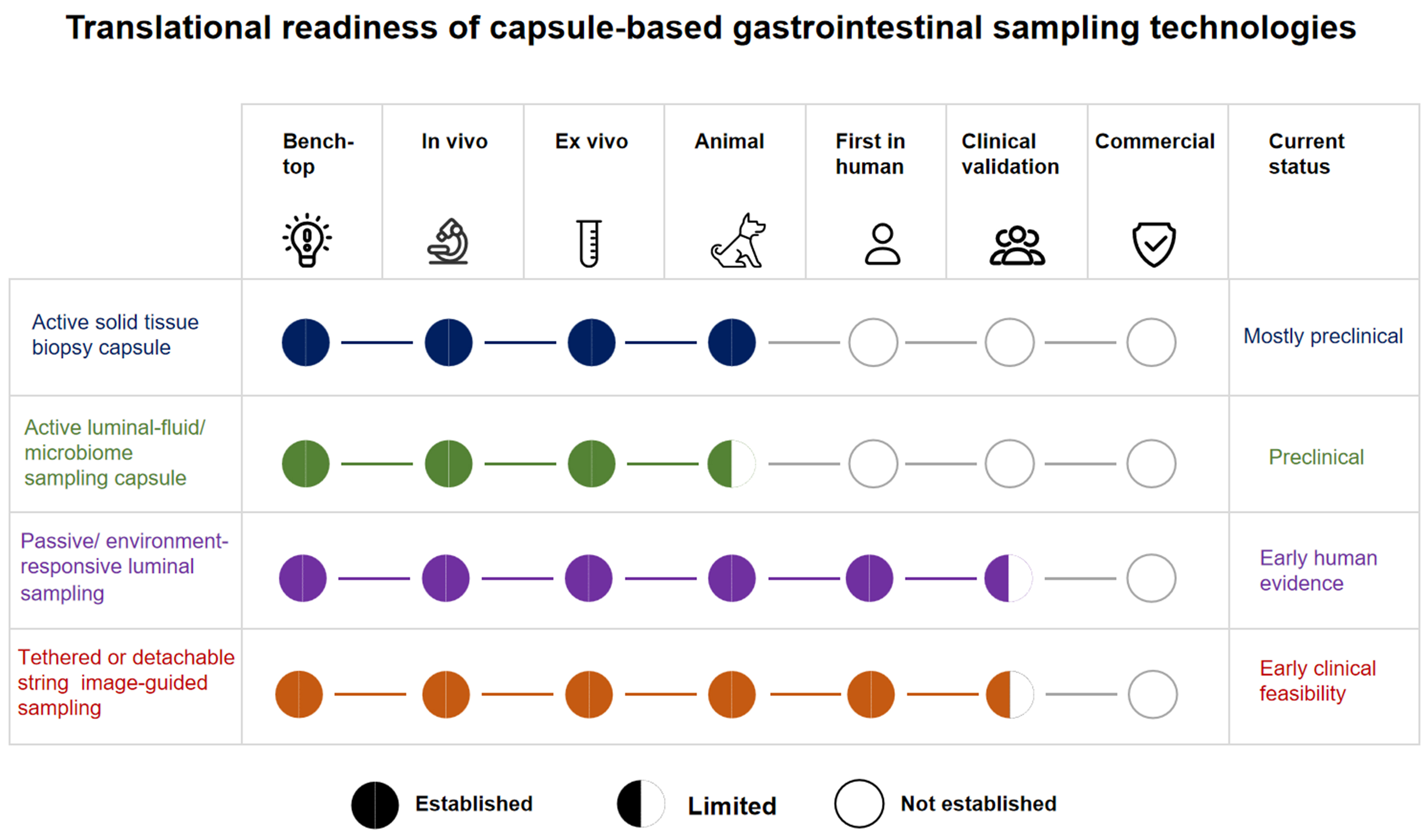
Figure 2. Translational readiness of capsule-based GI sampling technologies across bench-top, ex vivo, animal, first-in-human, clinical-validation, and commercial stages. The classifications are descriptive summaries of the validation stage reported in the reviewed literature. GI: Gastrointestinal.
Three directions are particularly important for further progress. First, sampling capsules need greater capability to determine where and when samples should be acquired. Integration of artificial intelligence (AI)-based image recognition to identify suspicious lesions within the camera field, together with onboard chemical sensors capable of detecting disease-associated metabolites, could potentially enable autonomous targeted sampling without relying on blind activation or variable pH triggers. However, AI-guided autonomous sampling currently remains a conceptual framework and a future research direction; to our knowledge, fully integrated physical prototypes have not yet been realized. The envisioned pathway toward such integration builds on the rapidly developing foundation of diagnostic AI. Deep learning models trained on large, high-quality upper and lower GI endoscopic video datasets have demonstrated high performance in automated lesion detection and anatomical recognition. The next conceptual step from diagnostic to interventional capsules would involve coupling real-time image-analysis algorithms with onboard electromechanical actuation systems. Second, sealing mechanisms require rigorous and standardized validation. Ensuring that the sample chamber remains hermetically sealed despite variations in intestinal pressure and pH throughout GI transit is essential for maintaining specimen integrity and preventing cross-contamination. Third, the field must progress from animal models toward clinical evaluation in humans. Passively triggered capsules have already demonstrated safety and feasibility in small human studies[85,86], whereas magnetically actuated and other active sampling capsules have not yet undergone human testing and remain largely confined to laboratory settings or porcine intestinal models. Regulatory pathways, biocompatibility requirements, and safety frameworks for evaluating active sampling capsules in humans will therefore need to evolve alongside the technologies themselves.
CONCLUSIONS
Smart capsule robots for GI biopsy and sampling have advanced considerably in recent years. Innovations in MEMS, stimuli-responsive materials, and magnetic control have enabled initial progress toward in vivo collection of tissue and microbiome samples without conventional endoscopy. The central challenge is no longer merely to demonstrate technical feasibility in the laboratory, but to achieve the reliability, safety, and precision required for clinical application. Capsule-based GI sampling has progressed from proof-of-concept engineering toward early translational evaluation, although the maturity of different device classes remains highly uneven. Several passive or environment-responsive fluid-sampling platforms have demonstrated feasibility in humans, whereas active solid-tissue biopsy systems remain predominantly at the bench-top, ex vivo, or animal stage. Future progress will therefore depend not only on further miniaturization and improved actuation efficiency but also on reproducible specimen adequacy, precise localization, robust sealing, standardized safety evaluation, and prospective validation in humans. AI-assisted autonomous targeting may ultimately integrate lesion recognition with intervention, but it remains a future objective rather than an established clinical capability.
DECLARATIONS
Acknowledgments
The Graphical Abstract was created using BioRender (https://BioRender.com/0q5ny81).
Authors’ contributions
Made substantial contributions to the conception and design of the study: Tian Y, Rong L
Performed data acquisition: Wan Y, Han Z
Performed data analysis and manuscript drafting: Wan Y, Han Z
Contributed to manuscript revision and critical review: Tian Y, Rong L
Availability of data and materials
Not applicable.
AI and AI-assisted tools statement
During the preparation of this manuscript, the AI tool ChatGPT (version 5.5, released 2026-04-23) was used solely for language editing. The tool did not influence the study design, data collection, analysis, interpretation, or the scientific content of the work. All authors take full responsibility for the accuracy, integrity, and final content of the manuscript.
Financial support and sponsorship
The work was supported by the National High Level Hospital Clinical Research (Research Achievement Transformation Project of Peking University First Hospital) (No. 2025CX03) and the National Natural Science Foundation of China (No. 82570784).
Conflicts of interest
All authors declared that there are no conflicts of interest.
Ethical approval and consent to participate
Not applicable.
Consent for publication
Not applicable.
Copyright
© The Author(s) 2026.
REFERENCES
1. Cao W, Chen HD, Yu YW, Li N, Chen WQ. Changing profiles of cancer burden worldwide and in China: a secondary analysis of the global cancer statistics 2020. Chin Med J. 2021;134:783-91.
3. Shi H, Pang S, Ming F, Yangdai T, Tian S, Lin R. A novel intelligent chromo capsule endoscope for the diagnosis of neoplastic lesions in the gastrointestinal tract. Gastroenterol Rep. 2023;11:goad021.
4. Xu X, Gao J, Tong D, Zhao Y, Fan X, Ge W. A multifunctional capsule-like puncture biopsy robot for the gastrointestinal system. Micromachines. 2025;16:589.
5. Kucharzik T, Taylor S, Allocca M, et al. ECCO-ESGAR-ESP-IBUS guideline on diagnostics and monitoring of patients with inflammatory bowel disease: part 1. J Crohns Colitis. 2025;19:jjaf106.
6. Ciuti G, Caliò R, Camboni D, et al. Frontiers of robotic endoscopic capsules: a review. J Microbio Robot. 2016;11:1-18.
7. Lai YP, Lee T, Sieben D, Gauthier L, Nam J, Diller E. Hybrid hydrogel-magnet actuated capsule for automatic gut microbiome sampling. IEEE Trans Biomed Eng. 2024;71:2911-22.
8. Simi M, Gerboni G, Menciassi A, Valdastri P. Magnetic torsion spring mechanism for a wireless biopsy capsule. J Med Devices. 2013;7:041009.
9. Le VH, Hernando LR, Lee C, et al. Shape memory alloy-based biopsy device for active locomotive intestinal capsule endoscope. Proc Inst Mech Eng H. 2015;229:255-63.
10. Hoang MC, Le VH, Kim J, et al. Untethered robotic motion and rotating blade mechanism for actively locomotive biopsy capsule endoscope. IEEE Access. 2019;7:93364-74.
11. Park S, Lee H, Kim D, Kee H, Park S. Active multiple-sampling capsule for gut microbiome. IEEE/ASME Trans Mechatron. 2022;27:4384-95.
12. Kong K, Cha J, Jeon D, Cho DD. A rotational micro biopsy device for the capsule endoscope. In: 2005 IEEE/RSJ International Conference on Intelligent Robots and Systems, Edmonton, Canada. Aug 02-06, 2005. IEEE; 2005. pp. 1839-43.
13. Kong K, Yim S, Choi S, Jeon D. A robotic biopsy device for capsule endoscopy. J Med Devices. 2012;6:031004.
14. Le VH, Jin Z, Leon-Rodriguez H, et al. Electromagnetic field intensity triggered micro-biopsy device for active locomotive capsule endoscope. Mechatronics. 2016;36:112-8.
15. Song Z, Zhang W, Dario P. A novel multi-biopsy mechanism embedded in a capsule endoscope for colonoscopy. In: 2019 IEEE International Conference on Robotics and Biomimetics (ROBIO), Dali, China. Dec 06-08, 2019. IEEE; 2019. pp. 321-5.
16. Song Z, Zhang W, Zhang W, Paolo D. A novel biopsy capsule robot based on high-speed cutting tissue. Cyborg Bionic Syst. 2022;2022:9783517.
17. Le VH, Kang B, Kim J, et al. Non-invasive active capsule endoscope integrated targeting biopsy function based on electro-mangetic actuation system. In: 2017 IEEE International Conference on Robotics and Biomimetics (ROBIO), Macau, Macao. Dec 05-08, 2017. IEEE; 2017. pp. 2309-13.
18. Zhang W, Dario P, Zhang W, Song Z. Positioning and posture adjustment for a rotational cutting biopsy capsule. In: 2022 International Conference on Advanced Robotics and Mechatronics (ICARM), Guilin, China. Jul 09-11, 2022. IEEE; 2022. pp. 82-6.
19. Park S, Koo K, Bang SM, Park JY, Song SY, Cho D. A novel microactuator for microbiopsy in capsular endoscopes. J Micromech Microeng. 2008;18:025032.
20. Straker MA, Levy JA, Sweeney PA, Ghodssi R. Gear train-enabled low-power thermomechanical biopsy system for ingestible capsules. In: 2025 23rd International Conference on Solid-State Sensors, Actuators and Microsystems (Transducers), Orlando, USA. Jun 29 - Jul 03, 2025. IEEE; 2025. pp. 2016-9.
21. Hoang MC, Le VH, Nguyen KT, et al. A robotic biopsy endoscope with magnetic 5-DOF locomotion and a retractable biopsy punch. Micromachines. 2020;11:98.
22. Le VH, Hoang MC, Nguyen VD, et al. A novel biopsy capsule endoscope for wireless intestinal tissue collection. In: 2018 International Conference on Manipulation, Automation and Robotics at Small Scales (MARSS), Nagoya, Japan. Jul 04-08, 2018. IEEE; 2018. p. 1-6.
23. Zheng L, Song H, Yan L, Wang Z, Guo S. Preliminary design of a magnetically actuated multifunctional biopsy capsule robot. In: 2024 IEEE International Conference on Mechatronics and Automation (ICMA), Tianjin, China. Aug 04-07, 2024. IEEE; 2024. pp. 1014-8.
24. Rashid MHO, Lin F. Magnetically driven biopsy capsule robot with spring mechanism. Micromachines. 2024;15:287.
25. Ye D, Xue J, Yuan S, et al. Design and control of a magnetically-actuated capsule robot with biopsy function. IEEE Trans Biomed Eng. 2022;69:2905-15.
26. Zhang F, Ye D, Song S, Meng MQH. Design of a novel biopsy capsule robot with anchoring function for intestinal tract. In: 2019 IEEE International Conference on Robotics and Biomimetics (ROBIO), Dali, China. Dec 06-08, 2019. IEEE; 2019. pp. 1471-6.
27. Son D, Dogan MD, Sitti M. Magnetically actuated soft capsule endoscope for fine-needle aspiration biopsy. In: 2017 IEEE International Conference on Robotics and Automation (ICRA), Singapore. May 29 - Jun 03, 2017. IEEE; 2017. pp. 1132-9.
28. Hu J, Deng H, Qiao K, Shi C, Hu C. Development of an ingestible device with biopsy and drug delivery functions. In: 2025 IEEE International Conference on Mechatronics and Automation (ICMA), Beijing, China. Aug 03-06, 2025. IEEE; 2025. pp. 365-70.
29. Li Z, Wei T, Zhang Y, et al. Rotating multibarreled capsule robot for multiple biopsies and on-demand drug delivery. IEEE/ASME Trans Mechatron. 2025;30:7089-99.
30. Liu H, Wang J, Song S. Pose and biopsy sensing for capsule robot based on conditioned multiple magnets tracking. IEEE Sens Lett. 2025;9:1-4.
31. Pan X, Ma T, Li P, Jiang X, Song S, Meng MQH. A novel intestinal microcapsule endoscope robot with biopsy function. In: 2018 IEEE International Conference on Cyborg and Bionic Systems (CBS), Shenzhen, China. Oct 25-27, 2018. IEEE; 2018. pp. 308-12.
32. Tovmachenko O, Feng L, Mousa AM, Ullah Z, Masoud MA. A capsule-type device for soft tissue cutting using a threadless ballscrew actuator. In: 2019 International Conference on Manipulation, Automation and Robotics at Small Scales (MARSS), Helsinki, Finland. Jul 01-05, 2019. IEEE; 2019. p. 1-6.
33. Chen W, Yan G, He S, et al. Wireless powered capsule endoscopy for colon diagnosis and treatment. Physiol Meas. 2013;34:1545-61.
34. Wang G, Yu X, Song S. A novel capsule endoscopy robot for gastric sampling. In: 2025 International Conference on Information and Automation (ICIA), Lanzhou, China. Aug 28-31, 2025. IEEE; 2025. p. 1-6.
35. Le VH, Nguyen VD, Lee C, Go G, Park J, Park S. Miniaturized biopsy module using gripper tool for active locomotive capsule endoscope. Mechatronics. 2017;44:52-9.
36. Guo J, Liu X, Guo S, Fu Q. Design and evaluation of a novel magnetoactive biopsy capsule endoscope robot. In: 2021 IEEE International Conference on Mechatronics and Automation (ICMA), Takamatsu, Japan. Aug 08-11, 2021. IEEE; 2021. pp. 944-9.
37. Leon-Rodriguez H, Park SH, Park JO. Testing and evaluation of foldable biopsy tools for active capsule endoscope. In: 2020 20th International Conference on Control, Automation and Systems (ICCAS), Busan, Korea. Oct 13-16, 2020. IEEE; 2020. pp. 473-9.
38. Zarifi Khameneh MR, Tolouei Z, Yousefi M, Pishkenari HN, Alasty A. Development and experimental evaluation of an endoscopic capsule robot equipped with a fail-safe biopsy mechanism. In: 2024 12th RSI International Conference on Robotics and Mechatronics (ICRoM), Tehran, Iran. Dec 17-19, 2024. IEEE; 2024. pp. 034-9.
39. Ye D, Zhang F, Yuan S, Song S, Meng MQH. Magnetically driven wireless capsule robot with targeting biopsy function. In: 2019 IEEE International Conference on Robotics and Biomimetics (ROBIO), Dali, China. Dec 06-08, 2019. IEEE; 2019. pp. 1222-7.
40. Guo S, Huang F, Guo J, Fu Q. Study on the active movement capsule robot for biopsy. In: 2020 IEEE International Conference on Mechatronics and Automation (ICMA), Beijing, China. Oct 13-16, 2020. IEEE; 2020. pp. 1780-5.
41. Liu X, Guo J, Guo S, Fu Q, Mo B. Structural design and analysis of a new four-blade biopsy capsule endoscope. In: 2022 IEEE International Conference on Mechatronics and Automation (ICMA), Guilin, China. Aug 07-10, 2022. IEEE; 2022. pp. 581-6.
42. Yim S, Gultepe E, Gracias DH, Sitti M. Biopsy using a magnetic capsule endoscope carrying, releasing, and retrieving untethered microgrippers. IEEE Trans Biomed Eng. 2014;61:513-21.
43. Bourbakis N, Giakos G, Karargyris A. Design of new-generation robotic capsules for therapeutic and diagnostic endoscopy. In: 2010 IEEE International Conference on Imaging Systems and Techniques, Thessaloniki, Greece. Jul 01-02, 2010. IEEE; 2010. p. 1-6.
44. Muretto P, Graziano F, Staccioli MP, et al. An endogastric capsule for measuring tumor markers in gastric juice: an evaluation of the safety and efficacy of a new diagnostic tool. Ann Oncol. 2003;14:105-9.
45. John JV, McCarthy A, Su Y, et al. Nanofiber capsules for minimally invasive sampling of biological specimens from gastrointestinal tract. Acta Biomater. 2022;146:211-21.
46. Wang D, He R, Xie X, Bai T, Hou X. Visible collection of gastric juice using a detachable string magnetically controlled capsule endoscopy: genotypic measurement for Helicobacter pylori infection and antibiotic resistance. Gastroenterol Rep. 2025;13:goaf106.
47. Jin G, Wang G, Liu X, Cao H, Wang B. 977 - Intestine microbiome aspiration (IMBA) capsule: a new autonomous and minimally-invasive device for whole gut microbiome sampling and mapping. Gastroenterology. 2019;156:S-205.
48. Salem MB, Aiche G, Rubbert L, Renaud P, Haddab Y. Design of a microbiota sampling capsule using 3D-printed bistable mechanism. Annu Int Conf IEEE Eng Med Biol Soc. 2018;2018:4868-71.
49. Waimin JF, Nejati S, Jiang H, et al. Smart capsule for non-invasive sampling and studying of the gastrointestinal microbiome. RSC Adv. 2020;10:16313-22.
50. Nejati S, Wang J, Sedaghat S, et al. Smart capsule for targeted proximal colon microbiome sampling. Acta Biomater. 2022;154:83-96.
51. Sarnaik D, Krishnakumar A, Nejati S, et al. A smart capsule with a bacteria- and pH-triggered enteric polymer coating for targeted colonic microbiome sampling. Acta Biomater. 2025;199:412-27.
52. Banis GE, Beardslee LA, Stine JM, Sathyam RM, Ghodssi R. Gastrointestinal targeted sampling and sensing via embedded packaging of integrated capsule system. J Microelectromech Syst. 2019;28:219-25.
53. Banis GE, Beardslee LA, Stine JM, Sathyam RM, Ghodssi R. Integrated capsule system for gastroinitestinal PH triggered sampling and sensing. In: 2019 20th International Conference on Solid-State Sensors, Actuators and Microsystems & Eurosensors XXXIII (TRANSDUCERS & EUROSENSORS XXXIII), Berlin, Germany. Jun 23-27, 2019. IEEE; 2019. pp. 314-7.
54. Rezaei Nejad H, Oliveira BCM, Sadeqi A, et al. Ingestible osmotic pill for in vivo sampling of gut microbiomes. Adv Intell Syst. 2019;1:1900053.
55. Lee SN, Rahimian P, Stork C, et al. Evaluation of a novel smart capsule bacterial detection system device for diagnosis of small intestinal bacterial overgrowth. Neurogastroenterol Motil. 2025;37:e14965.
56. Rehan M, Al-Bahadly I, Thomas DG, Avci E. Capsule robot for gut microbiota sampling using shape memory alloy spring. Int J Med Robot. 2020;16:1-14.
57. Rehan M, Al-Bahadly I, Thomas DG, Avci E. Towards gut microbiota sampling using an untethered sampling device. IEEE Access. 2021;9:127175-84.
58. Rehan M, Al-Bahadly I, Thomas DG, Avci E. Development of a robotic capsule for in vivo sampling of gut microbiota. IEEE Robot Autom Lett. 2022;7:9517-24.
59. Du X, Chan KF, Xia X, Chiu PWY, Zhang L. A magnetically-triggered soft capsule for on-demand mucus collection. In: 2018 IEEE International Conference on Cyborg and Bionic Systems (CBS), Shenzhen, China. Oct 25-27, 20218. IEEE; 2018. pp. 250-4.
60. Ding Z, Wang W, Zhang K, et al. Novel scheme for non-invasive gut bioinformation acquisition with a magnetically controlled sampling capsule endoscope. Gut. 2021;70:2297-306.
61. Shahadha MH, Voigt A, Gruner D, et al. Ingestible active capsule for gastrointestinal microbiome sampling. Biomed Microdevices. 2026;28:19.
62. Li Z, He C, Verma A, Diller E. 3D printed untethered magnetic bulb for liquid biopsy and drug delivery. In: 2024 International Conference on Manipulation, Automation and Robotics at Small Scales (MARSS), Delft, Netherlands. Jul 01-05, 2024. IEEE; 2024. p. 1-6.
63. Dong X, Xiao B, Vu H, Lin H, Sitti M. Millimeter-scale soft capsules for sampling liquids in fluid-filled confined spaces. Sci Adv. 2024;10:eadp2758.
64. Park S, Park S. Magnetically actuated capsule with multiple sampling capability for gut microbiome research. In: 2024 24th International Conference on Control, Automation and Systems (ICCAS), Jeju, Republic of Korea. Oct 29 - Nov 01, 2024. IEEE; 2024. pp. 270-1.
65. Shokrollahi P, Lai YP, Rash-Ahmadi S, et al. Blindly controlled magnetically actuated capsule for noninvasive sampling of the gastrointestinal microbiome. IEEE/ASME Trans Mechatron. 2021;26:2616-28.
66. Nam J, Lai YP, Gauthier L, Jang G, Diller E. Resonance-based design of wireless magnetic capsule for effective sampling of microbiome in gastrointestinal tract. Sens Actuators A Phys. 2022;342:113654.
67. Gupta S, Palanichamy V, Onaizah O. Magnetically actuated capsule mechanism for drug delivery, sampling, and cargo transport in the gastrointestinal tract. In: 2025 International Conference on Manipulation, Automation and Robotics at Small Scales (MARSS), West Lafayette, USA. Jul 28 - Aug 01, 2025. IEEE; 2025. p. 01-08.
68. Lee T, Diller E. Magnetic capsule for stable collection of large GI tract microbiome samples. In: 2025 International Conference on Manipulation, Automation and Robotics at Small Scales (MARSS), West Lafayette, USA. Jul 28 - Aug 01, 2025. IEEE; 2025. p. 1-7.
69. Yun J, Rho J, Shin H, Jeon Y, Kim S, Nam J. Magnetically guided capsule for high-capacity dual sampling of gut microbiome. IEEE/ASME Trans Mechatron. 2026;31:1046-56.
70. Hajima K, Yamashita M, Honda T. Design and fabrication of magnetically driven cytology brush applicable to capsule endoscope. In: 2015 IEEE Magnetics Conference (INTERMAG), Beijing, China. May 11-15, 2015. IEEE; 2015. p. 1-1.
71. Finocchiaro M, Giosuè C, Drago G, et al. Design of a magnetic actuation system for a microbiota-collection ingestible capsule. In: 2021 IEEE International Conference on Robotics and Automation (ICRA), Xi’an, China. May 30 - Jun 05, 2021. IEEE; 2021. pp. 6905-11.
72. Chen W, Sui J, Wang C. Magnetically actuated capsule robots: a review. IEEE Access. 2022;10:88398-420.
73. Cui J, Zheng X, Hou W, Zhuang Y, Pi X, Yang J. The study of a remote-controlled gastrointestinal drug delivery and sampling system. Telemed J E Health. 2008;14:715-9.
74. Son D, Gilbert H, Sitti M. Magnetically actuated soft capsule endoscope for fine-needle biopsy. Soft Robot. 2020;7:10-21.
75. Zhang S, Song S, Yu X, Song S, Zhang L. Design of magnetic actuated capsule robot for liquid sampling. IEEE Trans Biomed Eng. 2025;72:2709-19.
76. Xin Y, Sun ZJ, Gu W, Yu L. Experimental research on a capsule robot with spring-connected legs. Micromachines. 2022;13:2042.
77. Thwaites PA, Yao CK, Halmos EP, et al. Review article: Current status and future directions of ingestible electronic devices in gastroenterology. Aliment Pharmacol Ther. 2024;59:459-74.
78. Xu Z, Li Z, Liu S, Hu C. Design of capsule robot for magnetic gastric biopsy and drug administration. In: 2023 IEEE International Conference on Mechatronics and Automation (ICMA), Harbin, China. Aug 06-09, 2023. IEEE; 2023. pp. 2297-302.
79. Xu Z, Wei T, Li Z, Huang D, Liu S, Hu C. Magnetically driven capsule robot for multi-targeted biopsy and drug delivery in stomach. In: 2023 IEEE International Conference on Robotics and Biomimetics (ROBIO), Koh Samui, Thailand. Dec 04-09, 2023. IEEE; 2023. p. 1-6.
80. Wu Z, Wang X, Lu Y, Xu Q. Multichamber magnetic capsule robot for selective liquid sampling and drug delivery. Natl Sci Rev. 2025;12:nwaf400.
81. Abbott JJ, Diller E, Petruska AJ. Magnetic methods in robotics. Annu Rev Control Robot Auton Syst. 2020;3:57-90.
82. Shalon D, Culver RN, Grembi JA, et al. Profiling the human intestinal environment under physiological conditions. Nature. 2023;617:581-91.
83. Folz J, Culver RN, Morales JM, et al. Human metabolome variation along the upper intestinal tract. Nat Metab. 2023;5:777-88.
84. Bundalovic-Torma C, Wang G, Menon S, et al. Sa2023 Targeted and reproducible sampling of the small intestinal microbiome with the small intestine microbiome aspirate capsule technology (SIMBA): clinical validation against endoscopy aspirates and fecal samples in healthy and gi disorder. Gastroenterology. 2024;166:S-608.
85. Wang G, Menon S, Wilsack L, et al. Spatially and temporally precise microbiome profiling in the small intestine using the SIMBA capsule with X-ray tracking. Front Microbiomes. 2024;3:1321624.
86. Tronel A, Roger-Margueritat M, Plazy C, et al. Exploring the human small intestinal luminal microbiome via a newly developed ingestible sampling device. ISME Commun. 2025;5:ycaf224.
87. Nema A, Dhar D, Ramireddy VSR, Priyam K, Agarwal S, Srivastava SK. A small pill-like ingestible microdevice for site-specific microbiome sampling in the upper GI tract. Small. 2026;22:e10289.
88. Menard J, Bagheri S, Menon S, Yu YT, Goodman LB. Noninvasive sampling of the small intestinal chyme for microbiome, metabolome and antimicrobial resistance genes in dogs, a proof of concept. Anim Microbiome. 2023;5:64.
89. Krishnakumar A, Sarnaik D, Gallina NLF, et al. Smart sampling capsule for capture and detection of enteric pathogens in the small intestine. Adv Mater Technol. 2025;10:e00848.
90. Del-Rio-Ruiz R, Romualdo da Silva DR, Suresh H, et al. Soft autonomous ingestible device for sampling the small-intestinal microbiome. Device. 2024;2:100406.
91. Mandsberg NK, Moro G, Ghavami M, et al. Ingestible device for gastric fluid sampling. Adv Mater Technol. 2024;9:2400434.
92. Nejati S, Sarnaik D, Gopalakrishnan S, et al. Electronic‐free traceable smart capsule for gastrointestinal microbiome sampling. Adv Mater Technol. 2024;9:2300810.
93. Rehan M, Al-Bahadly I, Thomas DG, Young W, Cheng LK, Avci E. Smart capsules for sensing and sampling the gut: status, challenges and prospects. Gut. 2023;73:186-202.
94. Chen L, Gruzinskyte L, Jørgensen SL, Boisen A, Srivastava SK. An ingestible self-polymerizing system for targeted sampling of gut microbiota and biomarkers. ACS Nano. 2020;14:12072-81.
95. Sadeqi A, Del‐Rio‐Ruiz R, Rezaei Nejad H, et al. Magnetically activated ingestible pill with archimedes screw for on‐demand sampling of intestinal microbiome. Adv Mater Technol. 2025;10:2400750.
96. Li T, Felton J, Lewis J, et al. An ingestible device for automated sampling and location tracing in gastrointestinal tract. PLoS One. 2025;20:e0327667.
97. Leon-Rodriguez H, Lee C, Ha LV, Ko SY, Park JO, Park S. Conceptual design of micro-hydraulics system for active and biopsy capsule endoscope robot. In: 5th IEEE RAS/EMBS International Conference on Biomedical Robotics and Biomechatronics, Sao Paulo, Brazil. Aug 12-15, 2014. IEEE; 2014. pp. 1068-72.
98. Moon S. A planar-type micro-biopsy tool for a capsule-type endoscope using a one-step nickel electroplating process. Micromachines. 2023;14:1900.
99. Su CC, Chou CK, Mukundan A, et al. Capsule endoscopy: current trends, technological advancements, and future perspectives in gastrointestinal diagnostics. Bioengineering. 2025;12:613.
100. Zheng L, Sun J, Qi Z, et al. A magnetically controlled capsule robot with biopsy capability for intestinal applications. Sensors. 2025;25:7158.
Cite This Article
 Open Access
How to Cite
Wan Y, Han Z, Tian Y, Rong L. Capsule biopsy and targeted sampling techniques: current status and future perspectives. Mini-invasive Surg. 2026;10:37. https://dx.doi.org/10.20517/2574-1225.2026.36
Download Citation
Export Citation File:
Type of Import
Tips on Downloading Citation
Citation Manager File Format
Type of Import
Direct Import: When the Direct Import option is selected (the default state), a dialogue box will give you the option to Save or Open the downloaded citation data. Choosing Open will either launch your citation manager or give you a choice of applications with which to use the metadata. The Save option saves the file locally for later use.
Indirect Import: When the Indirect Import option is selected, the metadata is displayed and may be copied and pasted as needed.
About This Article
Copyright

Data & Comments
Data
0






Comments
Comments must be written in English. Spam, offensive content, impersonation, and private information will not be permitted. If any comment is reported and identified as inappropriate content by OAE staff, the comment will be removed without notice. If you have any queries or need any help, please contact us at support@oaepublish.com.